CD44 Antibody (MEM-263) - BSA Free
Novus Biologicals | Catalog # NB500-481
![Immunocytochemistry/ Immunofluorescence: CD44 Antibody (MEM-263) - BSA Free [NB500-481]](https://resources.rndsystems.com/images/products/CD44-Antibody-MEM-263-Immunocytochemistry-Immunofluorescence-NB500-481-img0001.jpg "Immunocytochemistry/ Immunofluorescence: CD44 Antibody (MEM-263) - BSA Free [NB500-481]")
Key Product Details
Species Reactivity
Validated:
Human, Porcine, Canine
Cited:
Human, Porcine
Applications
Validated:
Immunohistochemistry, Immunohistochemistry-Paraffin, Western Blot, Flow Cytometry, Immunocytochemistry/ Immunofluorescence, Immunoprecipitation, CyTOF-ready
Cited:
Western Blot, Flow Cytometry, Immunocytochemistry/ Immunofluorescence
Label
Unconjugated
Antibody Source
Monoclonal Mouse IgG1 Clone # MEM-263
Format
BSA Free
Loading...
Product Specifications
Immunogen
COS-7 cells, African Green Monkey (NM_000610.3).
Marker
Cell Membrane Marker
Specificity
The antibody MEM-263 reacts with extracellular (N-terminal) domain of standard CD44 (Phagocyte glycoprotein 1), a 80-95 kDa transmembrane glycoprotein (hyaladherin family) present on the most of cells and tissues (leukocytes, endothelial cells, mesenchymal cells, etc.); it is negative on platelets and hepatocytes. HLDA III; WS Code T 155
Clonality
Monoclonal
Host
Mouse
Isotype
IgG1
Theoretical MW
95 kDa.
Disclaimer note: The observed molecular weight of the protein may vary from the listed predicted molecular weight due to post translational modifications, post translation cleavages, relative charges, and other experimental factors.
Disclaimer note: The observed molecular weight of the protein may vary from the listed predicted molecular weight due to post translational modifications, post translation cleavages, relative charges, and other experimental factors.
Scientific Data Images for CD44 Antibody (MEM-263) - BSA Free
Immunocytochemistry/ Immunofluorescence: CD44 Antibody (MEM-263) - BSA Free [NB500-481]
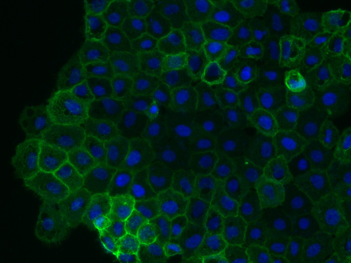
Immunocytochemistry/Immunofluorescence: CD44 Antibody (MEM-263) [NB500-481] - analysis of CD44 in 4% paraformaldehyde fixed Caov-3 cells (adherent human ovarian cancer cells) using anti-Cd44 antibody. The primary antibody was used at a dilution of 1:1000 in 2% goat serum in PBS and incubated for 2 hours at room temperature. Image from verified customer review.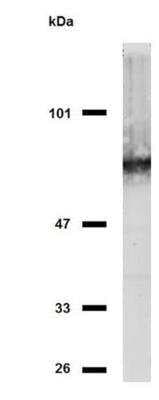
CD44 Antibody (MEM-263) [NB500-481] - Analysis of CD44 in HeLa cells using anti-CD44 (MEM-263) purified; non-reducing conditions.
![Flow Cytometry: CD44 Antibody (MEM-263) - BSA Free [NB500-481]](https://resources.rndsystems.com/images/products/CD44-Antibody-MEM-263-Flow-Cytometry-NB500-481-img0003.jpg "Flow Cytometry: CD44 Antibody (MEM-263) - BSA Free [NB500-481]")
Flow Cytometry: CD44 Antibody (MEM-263) - BSA Free [NB500-481]
Flow Cytometry: CD44 Antibody (MEM-263) [NB500-481] - A surface stain was performed on hPBMCs with CD44 (MEM-263) antibody NB500-481AF647 (blue) and a matched isotype control NBP2-27287AF647 (orange). Cells were incubated in an antibody dilution of 1 ug/mL for 20 minutes at room temperature. Both antibodies were conjugated to Alexa Fluor 647.![Flow Cytometry: CD44 Antibody (MEM-263) - BSA Free [NB500-481]](https://resources.rndsystems.com/images/products/CD44-Antibody-MEM-263-Flow-Cytometry-NB500-481-img0007.jpg "Flow Cytometry: CD44 Antibody (MEM-263) - BSA Free [NB500-481]")
Flow Cytometry: CD44 Antibody (MEM-263) - BSA Free [NB500-481]
Flow Cytometry: CD44 Antibody (MEM-263) [NB500-481] - Staining of human peripheral blood using anti-CD44 (MEM-263) APC conjugate.![Western Blot: CD44 Antibody (MEM-263)BSA Free [NB500-481]](https://resources.rndsystems.com/images/products/CD44-Antibody-MEM-263-Western-Blot-NB500-481-img0008.jpg "Western Blot: CD44 Antibody (MEM-263)BSA Free [NB500-481]")
Western Blot: CD44 Antibody (MEM-263)BSA Free [NB500-481]
CD44-Antibody-MEM-263-Western-Blot-NB500-481-img0008.jpg![Western Blot: CD44 Antibody (MEM-263)BSA Free [NB500-481]](https://resources.rndsystems.com/images/products/CD44-Antibody-MEM-263-Western-Blot-NB500-481-img0002.jpg "Western Blot: CD44 Antibody (MEM-263)BSA Free [NB500-481]")
Western Blot: CD44 Antibody (MEM-263)BSA Free [NB500-481]
Western Blot: CD44 Antibody (MEM-263) [NB500-481] - Fig. 1. Western Blotting analysis (non-reducing conditions) of isolated peripheral blood lymphocytes of various species using anti-CD44 (MEM-263). Lane 1: lysate of human PBL Lane 2: lysate of canine PBL Lane 3: lysate of porcine PBL.![Flow Cytometry: CD44 Antibody (MEM-263) - BSA Free [NB500-481]](https://resources.rndsystems.com/images/products/CD44-Antibody-MEM-263-Flow-Cytometry-NB500-481-img0004.jpg "Flow Cytometry: CD44 Antibody (MEM-263) - BSA Free [NB500-481]")
Flow Cytometry: CD44 Antibody (MEM-263) - BSA Free [NB500-481]
Flow Cytometry: CD44 Antibody (MEM-263) [NB500-481] - Analysis of human peripheral blood (lymphocyte gate) using anti-CD44 (MEM-263) PE conjugate.![Flow Cytometry: CD44 Antibody (MEM-263) - BSA Free [NB500-481]](https://resources.rndsystems.com/images/products/CD44-Antibody-MEM-263-Flow-Cytometry-NB500-481-img0005.jpg "Flow Cytometry: CD44 Antibody (MEM-263) - BSA Free [NB500-481]")
Flow Cytometry: CD44 Antibody (MEM-263) - BSA Free [NB500-481]
Flow Cytometry: CD44 Antibody (MEM-263) [NB500-481] - Staining of human peripheral blood using anti-CD44 (MEM-263) FITC conjugate. - BSA Free [NB500-481] -")
Western Blot: CD44 Antibody (MEM-263) - BSA Free [NB500-481] -
Expression of CD44 and CK18 in multiple clones of ovarian cancer cells. (a)-(b). CD44+ cells also express CK18 as determined by flow cytometry (a) and western blot analysis (b). Flow cytometer is representative of the eight evaluated clones. EOC stem cells, determined by either CD44 (c and d) or Ck18 (e and f) expression, are found in clusters surrounded by CD44− or Ck18− negative cancer cells.Applications for CD44 Antibody (MEM-263) - BSA Free
Application
Recommended Usage
Flow Cytometry
4 ug/ml
Immunocytochemistry/ Immunofluorescence
1:10-1:500
Immunohistochemistry
1:10-1:500
Immunohistochemistry-Paraffin
10 ug/ml
Immunoprecipitation
1:50
Western Blot
2 ug/ml
Application Notes
Western Blot - Sample preparation: Resuspend approx. 50 mil. cells in 1 ml cold Lysis buffer (1% laurylmaltoside in 20 mM Tris/Cl, 100 mM NaCl pH 8.2, 50 mM NaF including Protease inhibitor Cocktail). Incubate 60 min on ice. Centrifuge to remove cell debris. Mix lysate with non-reducing SDS-PAGE sample buffer. Application note: Non-reducing conditions. SDS-PAGE (6% separating gel). Use in Immunocytochemistry/immunofluorescence reported in scientific literature (PMID 25481699). This antibody is CyTOF ready.
Reviewed Applications
Read 1 review rated 4 using NB500-481 in the following applications:
Flow Cytometry Panel Builder
Bio-Techne Knows Flow Cytometry
Save time and reduce costly mistakes by quickly finding compatible reagents using the Panel Builder Tool.
Advanced Features
- Spectra Viewer - Custom analysis of spectra from multiple fluorochromes
- Spillover Popups - Visualize the spectra of individual fluorochromes
- Antigen Density Selector - Match fluorochrome brightness with antigen density

Formulation, Preparation, and Storage
Purification
Protein A purified
Formulation
Phosphate buffered saline (PBS), pH 7.4
Format
BSA Free
Preservative
15mM Sodium Azide
Concentration
1.0 mg/ml
Shipping
The product is shipped with polar packs. Upon receipt, store it immediately at the temperature recommended below.
Stability & Storage
Store at 4C. Do not freeze.
Background: CD44
Alternate Names
CD44, ECMR-III, HCAM, HCELL, LHR, MDU2, MDU3, MIC4, MUTCH-I, Pgp1
Entrez Gene IDs
960 (Human)
Gene Symbol
CD44
UniProt
Additional CD44 Products
Product Documents for CD44 Antibody (MEM-263) - BSA Free
Certificate of Analysis
To download a Certificate of Analysis, please enter a lot or batch number in the search box below.
Product Specific Notices for CD44 Antibody (MEM-263) - BSA Free
This product is for research use only and is not approved for use in humans or in clinical diagnosis. Primary Antibodies are guaranteed for 1 year from date of receipt.
Related Research Areas
Citations for CD44 Antibody (MEM-263) - BSA Free
 Powered by Bioz
Customer Reviews for CD44 Antibody (MEM-263) - BSA Free (1)
4 out of 5
1 Customer Rating
Have you used CD44 Antibody (MEM-263) - BSA Free?
Submit a review and receive an Amazon gift card!
$25/€18/£15/$25CAN/¥2500 Yen for a review with an image
$10/€7/£6/$10CAN/¥1110 Yen for a review without an image
Submit a review
Customer Images
Showing
1
-
1 of
1 review
Showing All
Filter By:
-
Application: ImmunofluorescenceSample Tested: adherent human ovarian cancer cellsSpecies: HumanVerified Customer | Posted 12/16/2014Caov-3 ovarian carcinoma cells

There are no reviews that match your criteria.
Protocols
Find general support by application which include: protocols, troubleshooting, illustrated assays, videos and webinars.
- 7-Amino Actinomycin D (7-AAD) Cell Viability Flow Cytometry Protocol
- Antigen Retrieval Protocol (PIER)
- Antigen Retrieval for Frozen Sections Protocol
- Appropriate Fixation of IHC/ICC Samples
- Cellular Response to Hypoxia Protocols
- Chromogenic IHC Staining of Formalin-Fixed Paraffin-Embedded (FFPE) Tissue Protocol
- Chromogenic Immunohistochemistry Staining of Frozen Tissue
- ClariTSA™ Fluorophore Kits
- Detection & Visualization of Antibody Binding
- Extracellular Membrane Flow Cytometry Protocol
- Flow Cytometry Protocol for Cell Surface Markers
- Flow Cytometry Protocol for Staining Membrane Associated Proteins
- Flow Cytometry Staining Protocols
- Flow Cytometry Troubleshooting Guide
- Fluorescent IHC Staining of Frozen Tissue Protocol
- Graphic Protocol for Heat-induced Epitope Retrieval
- Graphic Protocol for the Preparation and Fluorescent IHC Staining of Frozen Tissue Sections
- Graphic Protocol for the Preparation and Fluorescent IHC Staining of Paraffin-embedded Tissue Sections
- Graphic Protocol for the Preparation of Gelatin-coated Slides for Histological Tissue Sections
- ICC Cell Smear Protocol for Suspension Cells
- ICC Immunocytochemistry Protocol Videos
- ICC for Adherent Cells
- IHC Sample Preparation (Frozen sections vs Paraffin)
- Immunocytochemistry (ICC) Protocol
- Immunocytochemistry Troubleshooting
- Immunofluorescence of Organoids Embedded in Cultrex Basement Membrane Extract
- Immunofluorescent IHC Staining of Formalin-Fixed Paraffin-Embedded (FFPE) Tissue Protocol
- Immunohistochemistry (IHC) and Immunocytochemistry (ICC) Protocols
- Immunohistochemistry Frozen Troubleshooting
- Immunohistochemistry Paraffin Troubleshooting
- Immunoprecipitation Protocol
- Intracellular Flow Cytometry Protocol Using Alcohol (Methanol)
- Intracellular Flow Cytometry Protocol Using Detergents
- Intracellular Nuclear Staining Flow Cytometry Protocol Using Detergents
- Intracellular Staining Flow Cytometry Protocol Using Alcohol Permeabilization
- Intracellular Staining Flow Cytometry Protocol Using Detergents to Permeabilize Cells
- Preparing Samples for IHC/ICC Experiments
- Preventing Non-Specific Staining (Non-Specific Binding)
- Primary Antibody Selection & Optimization
- Propidium Iodide Cell Viability Flow Cytometry Protocol
- Protocol for Heat-Induced Epitope Retrieval (HIER)
- Protocol for Liperfluo
- Protocol for Making a 4% Formaldehyde Solution in PBS
- Protocol for VisUCyte™ HRP Polymer Detection Reagent
- Protocol for the Characterization of Human Th22 Cells
- Protocol for the Characterization of Human Th9 Cells
- Protocol for the Fluorescent ICC Staining of Cell Smears - Graphic
- Protocol for the Fluorescent ICC Staining of Cultured Cells on Coverslips - Graphic
- Protocol for the Preparation & Fixation of Cells on Coverslips
- Protocol for the Preparation and Chromogenic IHC Staining of Frozen Tissue Sections
- Protocol for the Preparation and Chromogenic IHC Staining of Frozen Tissue Sections - Graphic
- Protocol for the Preparation and Chromogenic IHC Staining of Paraffin-embedded Tissue Sections
- Protocol for the Preparation and Chromogenic IHC Staining of Paraffin-embedded Tissue Sections - Graphic
- Protocol for the Preparation and Fluorescent ICC Staining of Cells on Coverslips
- Protocol for the Preparation and Fluorescent ICC Staining of Non-adherent Cells
- Protocol for the Preparation and Fluorescent ICC Staining of Stem Cells on Coverslips
- Protocol for the Preparation and Fluorescent IHC Staining of Frozen Tissue Sections
- Protocol for the Preparation and Fluorescent IHC Staining of Paraffin-embedded Tissue Sections
- Protocol for the Preparation of Gelatin-coated Slides for Histological Tissue Sections
- Protocol for the Preparation of a Cell Smear for Non-adherent Cell ICC - Graphic
- Protocol: Annexin V and PI Staining by Flow Cytometry
- Protocol: Annexin V and PI Staining for Apoptosis by Flow Cytometry
- R&D Systems Quality Control Western Blot Protocol
- TUNEL and Active Caspase-3 Detection by IHC/ICC Protocol
- The Importance of IHC/ICC Controls
- Troubleshooting Guide: Fluorokine Flow Cytometry Kits
- Troubleshooting Guide: Immunohistochemistry
- Troubleshooting Guide: Western Blot Figures
- Western Blot Conditions
- Western Blot Protocol
- Western Blot Protocol for Cell Lysates
- Western Blot Troubleshooting
- Western Blot Troubleshooting Guide
- View all Protocols, Troubleshooting, Illustrated assays and Webinars
Loading...
Associated Pathways


