Claudin-1 Antibody - BSA Free
Novus Biologicals | Catalog # NBP1-77036
![Western Blot: Claudin-1 AntibodyBSA Free [NBP1-77036]](https://resources.rndsystems.com/images/products/Claudin-1-Antibody-Western-Blot-NBP1-77036-img0012.jpg "Western Blot: Claudin-1 AntibodyBSA Free [NBP1-77036]")
Key Product Details
Validated by
Independent Antibodies, Biological Validation
Species Reactivity
Validated:
Human, Mouse, Rat, Porcine, Canine
Cited:
Human, Mouse, Rat, Canine
Predicted:
Bovine (100%). Backed by our 100% Guarantee.
Applications
Validated:
Immunohistochemistry, Western Blot, ELISA, Immunocytochemistry/ Immunofluorescence
Cited:
Immunohistochemistry-Paraffin, Western Blot, Immunocytochemistry/ Immunofluorescence, IF/IHC
Label
Unconjugated
Antibody Source
Polyclonal Rabbit IgG
Format
BSA Free
Loading...
Product Specifications
Immunogen
Antibody was raised against a peptide corresponding to 20 amino acids near the carboxy terminus of human Claudin-1. The immunogen is located within the last 50 amino acids of Claudin-1.
Reactivity Notes
Porcine reactivity per customer review. Mouse reactivity reported in (PMID: 33392105), Canine reactivity reported in (PMID: 32419902).
Specificity
Human Claudin-1 has one isoform (211aa, 23kD). Mouse Claudin-1 has one isoform (211aa, 23kD) and Rat Claudin-1 also has one isoform (211aa, 23kD). NBP1-77036 can detect human, mouse and rat.
Clonality
Polyclonal
Host
Rabbit
Isotype
IgG
Theoretical MW
23 kDa.
Disclaimer note: The observed molecular weight of the protein may vary from the listed predicted molecular weight due to post translational modifications, post translation cleavages, relative charges, and other experimental factors.
Disclaimer note: The observed molecular weight of the protein may vary from the listed predicted molecular weight due to post translational modifications, post translation cleavages, relative charges, and other experimental factors.
Scientific Data Images for Claudin-1 Antibody - BSA Free
![Western Blot: Claudin-1 AntibodyBSA Free [NBP1-77036]](https://resources.rndsystems.com/images/products/Claudin-1-Antibody-Western-Blot-NBP1-77036-img0007.jpg "Western Blot: Claudin-1 AntibodyBSA Free [NBP1-77036]")
Western Blot: Claudin-1 AntibodyBSA Free [NBP1-77036]
Western Blot: Claudin-1 Antibody - BSA Free [NBP1-77036] - Claudin-1 Antibody [NBP1-77036] - Validation in Human Tissue Lysates Loading: 15 ug of lysates per lane.Antibodies: CLDN1, (1 ug/mL), 1h incubation at RT in 5% NFDM/TBST.Secondary: Goat anti-rabbit IgG HRP conjugate at 1:10000 dilution.![Western Blot: Claudin-1 AntibodyBSA Free [NBP1-77036]](https://resources.rndsystems.com/images/products/Claudin-1-Antibody-Western-Blot-NBP1-77036-img0009.jpg "Western Blot: Claudin-1 AntibodyBSA Free [NBP1-77036]")
Western Blot: Claudin-1 AntibodyBSA Free [NBP1-77036]
Western Blot: Claudin-1 Antibody - BSA Free [NBP1-77036] - Claudin-1 Antibody [NBP1-77036] - Validation in Human HepG2 Cell Lysate Loading: 15 ug of lysates per lane.Antibodies: CLDN1, 5187 (A: 1 gu/mL and B: 2 ug/mL), 1h incubation at RT in 5% NFDM/TBST.Secondary: Goat anti-rabbit IgG HRP conjugate at 1:10000 dilution.![Western Blot: Claudin-1 AntibodyBSA Free [NBP1-77036]](https://resources.rndsystems.com/images/products/Claudin-1-Antibody-NBP1-77036-img0011.jpg "Western Blot: Claudin-1 AntibodyBSA Free [NBP1-77036]")
Western Blot: Claudin-1 AntibodyBSA Free [NBP1-77036]
Western Blot: Claudin-1 Antibody - BSA Free [NBP1-77036] - Claudin-1 Antibody [NBP1-77036] - Independent Antibody Validation (IAV) via Protein Expression Profile in Human Cell Lines. Loading: 15 ug of lysates per lane. Antibodies: CLDN1, NBP1-77036 (1 ug/mL), CLDN1, (2 ug/mL), and beta-actin (1 ug/mL), 1h incubation at RT in 5% NFDM/TBST. Secondary: Goat anti-rabbit IgG HRP conjugate at 1:10000 dilution.![Western Blot: Claudin-1 AntibodyBSA Free [NBP1-77036]](https://resources.rndsystems.com/images/products/Claudin-1-Antibody-Western-Blot-NBP1-77036-img0008.jpg "Western Blot: Claudin-1 AntibodyBSA Free [NBP1-77036]")
Western Blot: Claudin-1 AntibodyBSA Free [NBP1-77036]
Western Blot: Claudin-1 Antibody - BSA Free [NBP1-77036] - Claudin-1 Antibody [NBP1-77036] - Validation in Human Tissue Lysates Loading: 15 ug of lysates per lane.Antibodies: CLDN1,(1 ug/mL), 1h incubation at RT in 5% NFDM/TBST.Secondary: Goat anti-rabbit IgG HRP conjugate at 1:10000 dilution.![Immunocytochemistry/ Immunofluorescence: Claudin-1 Antibody - BSA Free [NBP1-77036]](https://resources.rndsystems.com/images/products/Claudin-1-Antibody-Immunocytochemistry-NBP1-77036-img0004.jpg "Immunocytochemistry/ Immunofluorescence: Claudin-1 Antibody - BSA Free [NBP1-77036]")
Immunocytochemistry/ Immunofluorescence: Claudin-1 Antibody - BSA Free [NBP1-77036]
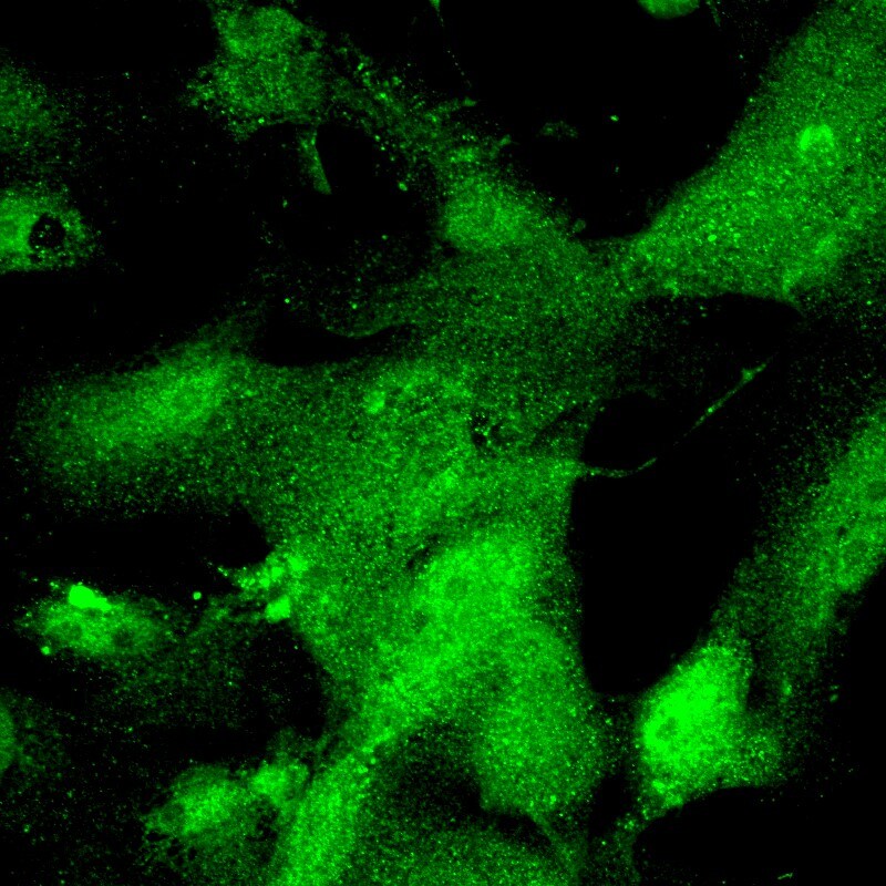
Immunocytochemistry/Immunofluorescence: Claudin-1 Antibody - BSA Free [NBP1-77036] - Claudin-1 Antibody [NBP1-77036] - Claudin-1 labeled pig cultured cornea cells visualized with Alexa488. This image was submitted via customer Review.![Immunocytochemistry/ Immunofluorescence: Claudin-1 Antibody - BSA Free [NBP1-77036]](https://resources.rndsystems.com/images/products/Claudin-1-Antibody-Immunocytochemistry-NBP1-77036-img0005.jpg "Immunocytochemistry/ Immunofluorescence: Claudin-1 Antibody - BSA Free [NBP1-77036]")
Immunocytochemistry/ Immunofluorescence: Claudin-1 Antibody - BSA Free [NBP1-77036]
Immunocytochemistry/Immunofluorescence: Claudin-1 Antibody - BSA Free [NBP1-77036] - Claudin-1 Antibody [NBP1-77036] - Claudin-1 labeled pig Sc cells visualized with Alexa488. This image was submitted via customer Review.
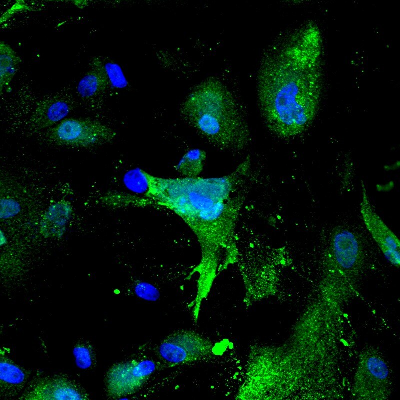
Immunocytochemistry/ Immunofluorescence: Claudin-1 Antibody - BSA Free [NBP1-77036] -
Immunocytochemistry/ Immunofluorescence: Claudin-1 Antibody - BSA Free [NBP1-77036] - Validation of Claudin-1 in HepG2 Cells. Immunofluorescent analysis of 4% paraformaldehyde-fixed HepG2 cells labeling Claudin-1 with at 20 ug/mL, followed by goat anti-rabbit IgG secondary antibody at 1/500 dilution (green) and DAPI staining (blue).Applications for Claudin-1 Antibody - BSA Free
Application
Recommended Usage
ELISA
1:100-1:2000
Immunocytochemistry/ Immunofluorescence
5-20 ug/mL
Western Blot
1-2 ug/ml
Application Notes
Use in ICC/IF reported in multiple pieces of scientific literature. Use in IHC reported in scientific literature (PMID:32067099).
Reviewed Applications
Read 2 reviews rated 3.5 using NBP1-77036 in the following applications:
Formulation, Preparation, and Storage
Purification
Peptide affinity purified
Formulation
PBS
Format
BSA Free
Preservative
0.02% Sodium Azide
Concentration
1 mg/ml
Shipping
The product is shipped with polar packs. Upon receipt, store it immediately at the temperature recommended below.
Stability & Storage
Store at 4C short term. Aliquot and store at -20C long term. Avoid freeze-thaw cycles.
Background: Claudin-1
Alternate Names
Claudin1, CLD1, CLDN1, SEMP1
Gene Symbol
CLDN1
UniProt
Additional Claudin-1 Products
Product Documents for Claudin-1 Antibody - BSA Free
Certificate of Analysis
To download a Certificate of Analysis, please enter a lot or batch number in the search box below.
Product Specific Notices for Claudin-1 Antibody - BSA Free
This product is for research use only and is not approved for use in humans or in clinical diagnosis. Primary Antibodies are guaranteed for 1 year from date of receipt.
Citations for Claudin-1 Antibody - BSA Free
 Powered by Bioz
Customer Reviews for Claudin-1 Antibody - BSA Free (2)
3.5 out of 5
2 Customer Ratings
Have you used Claudin-1 Antibody - BSA Free?
Submit a review and receive an Amazon gift card!
$25/€18/£15/$25CAN/¥2500 Yen for a review with an image
$10/€7/£6/$10CAN/¥1110 Yen for a review without an image
Submit a review
Customer Images
Showing
1
-
2 of
2 reviews
Showing All
Filter By:
-
Application: ImmunocytochemistrySample Tested: pig Sc cellsSpecies: PigVerified Customer | Posted 06/07/2017Claudin-1 Antibody (NBP1-77036) labeling pig Sc cells with Alexa488

-
Application: ImmunocytochemistrySample Tested: pig cultured cornea cellsSpecies: PigVerified Customer | Posted 06/07/2017Claudin-1 Antibody (NBP1-77036) labeling pig cultured cornea cells with Alexa488
There are no reviews that match your criteria.
Protocols
Find general support by application which include: protocols, troubleshooting, illustrated assays, videos and webinars.
- Antigen Retrieval Protocol (PIER)
- Antigen Retrieval for Frozen Sections Protocol
- Appropriate Fixation of IHC/ICC Samples
- Cellular Response to Hypoxia Protocols
- Chromogenic IHC Staining of Formalin-Fixed Paraffin-Embedded (FFPE) Tissue Protocol
- Chromogenic Immunohistochemistry Staining of Frozen Tissue
- ClariTSA™ Fluorophore Kits
- Detection & Visualization of Antibody Binding
- ELISA Sample Preparation & Collection Guide
- ELISA Troubleshooting Guide
- Fluorescent IHC Staining of Frozen Tissue Protocol
- Graphic Protocol for Heat-induced Epitope Retrieval
- Graphic Protocol for the Preparation and Fluorescent IHC Staining of Frozen Tissue Sections
- Graphic Protocol for the Preparation and Fluorescent IHC Staining of Paraffin-embedded Tissue Sections
- Graphic Protocol for the Preparation of Gelatin-coated Slides for Histological Tissue Sections
- How to Run an R&D Systems DuoSet ELISA
- How to Run an R&D Systems Quantikine ELISA
- How to Run an R&D Systems Quantikine™ QuicKit™ ELISA
- ICC Cell Smear Protocol for Suspension Cells
- ICC Immunocytochemistry Protocol Videos
- ICC for Adherent Cells
- IHC Sample Preparation (Frozen sections vs Paraffin)
- Immunocytochemistry (ICC) Protocol
- Immunocytochemistry Troubleshooting
- Immunofluorescence of Organoids Embedded in Cultrex Basement Membrane Extract
- Immunofluorescent IHC Staining of Formalin-Fixed Paraffin-Embedded (FFPE) Tissue Protocol
- Immunohistochemistry (IHC) and Immunocytochemistry (ICC) Protocols
- Immunohistochemistry Frozen Troubleshooting
- Immunohistochemistry Paraffin Troubleshooting
- Preparing Samples for IHC/ICC Experiments
- Preventing Non-Specific Staining (Non-Specific Binding)
- Primary Antibody Selection & Optimization
- Protocol for Heat-Induced Epitope Retrieval (HIER)
- Protocol for Making a 4% Formaldehyde Solution in PBS
- Protocol for VisUCyte™ HRP Polymer Detection Reagent
- Protocol for the Fluorescent ICC Staining of Cell Smears - Graphic
- Protocol for the Fluorescent ICC Staining of Cultured Cells on Coverslips - Graphic
- Protocol for the Preparation & Fixation of Cells on Coverslips
- Protocol for the Preparation and Chromogenic IHC Staining of Frozen Tissue Sections
- Protocol for the Preparation and Chromogenic IHC Staining of Frozen Tissue Sections - Graphic
- Protocol for the Preparation and Chromogenic IHC Staining of Paraffin-embedded Tissue Sections
- Protocol for the Preparation and Chromogenic IHC Staining of Paraffin-embedded Tissue Sections - Graphic
- Protocol for the Preparation and Fluorescent ICC Staining of Cells on Coverslips
- Protocol for the Preparation and Fluorescent ICC Staining of Non-adherent Cells
- Protocol for the Preparation and Fluorescent ICC Staining of Stem Cells on Coverslips
- Protocol for the Preparation and Fluorescent IHC Staining of Frozen Tissue Sections
- Protocol for the Preparation and Fluorescent IHC Staining of Paraffin-embedded Tissue Sections
- Protocol for the Preparation of Gelatin-coated Slides for Histological Tissue Sections
- Protocol for the Preparation of a Cell Smear for Non-adherent Cell ICC - Graphic
- Quantikine HS ELISA Kit Assay Principle, Alkaline Phosphatase
- Quantikine HS ELISA Kit Principle, Streptavidin-HRP Polymer
- R&D Systems Quality Control Western Blot Protocol
- Sandwich ELISA (Colorimetric) – Biotin/Streptavidin Detection Protocol
- Sandwich ELISA (Colorimetric) – Direct Detection Protocol
- TUNEL and Active Caspase-3 Detection by IHC/ICC Protocol
- The Importance of IHC/ICC Controls
- Troubleshooting Guide: ELISA
- Troubleshooting Guide: Immunohistochemistry
- Troubleshooting Guide: Western Blot Figures
- Western Blot Conditions
- Western Blot Protocol
- Western Blot Protocol for Cell Lysates
- Western Blot Troubleshooting
- Western Blot Troubleshooting Guide
- View all Protocols, Troubleshooting, Illustrated assays and Webinars
Loading...
Associated Pathways


