Collagen I alpha 1 Antibody (COL-1)
Novus Biologicals | Catalog # NB600-450
![Immunohistochemistry-Frozen: Collagen I alpha 1 Antibody (COL-1) [NB600-450]](https://resources.rndsystems.com/images/products/Collagen-I-alpha-1-Antibody-COL-1-Immunohistochemistry-Frozen-NB600-450-img0003.jpg "Immunohistochemistry-Frozen: Collagen I alpha 1 Antibody (COL-1) [NB600-450]")
Loading...
Key Product Details
Validated by
Biological Validation
Species Reactivity
Validated:
Human, Mouse, Rat, Porcine, Bovine, Equine, Feline, Mammal, Rabbit
Cited:
Human, Mouse, Rat, Equine, Feline, Rabbit
Applications
Validated:
Immunohistochemistry, Immunohistochemistry-Paraffin, Immunohistochemistry-Frozen, Western Blot, ELISA, Immunocytochemistry/ Immunofluorescence, Dot Blot
Cited:
Immunohistochemistry, Immunohistochemistry-Paraffin, Immunohistochemistry-Frozen, Western Blot, ELISA, Immunocytochemistry/ Immunofluorescence, IF/IHC, Electron Microscopy
Label
Unconjugated
Antibody Source
Monoclonal Mouse IgG1 Clone # COL-1
Loading...
Product Specifications
Immunogen
This Collagen I alpha 1 antibody (COL-1) was raised against full length bovine native protein (purified).
Epitope
The epitope recognized by the antibody may be sensitive to routine formalin fixation and paraffin embedding.
Reactivity Notes
Reported reactivity in deer. Mouse reactivity reported in scientific literature (PMID: 25287675). Goat reactivity reported by customer review. Please note that this antibody is reactive to Mouse and derived from the same host, Mouse. Mouse-On-Mouse blocking reagent may be needed for IHC and ICC experiments to reduce high background signal. You can find these reagents under catalog numbers PK-2200-NB and MP-2400-NB. Please contact Technical Support if you have any questions. Feline reactivity reported in scientific literature (PMID:33091431).
Localization
Extracellular matrix
Specificity
This Collagen I alpha 1 antibody (COL-1) recognizes the native (helical) form of collagen type I using ELISA and dot-blot. It does not react with the thermally-denatured molecule, and shows no cross-reactivity with collagen types II, III, IV, V, VI, VII, IX, X and XI.
Clonality
Monoclonal
Host
Mouse
Isotype
IgG1
Theoretical MW
139 kDa.
Disclaimer note: The observed molecular weight of the protein may vary from the listed predicted molecular weight due to post translational modifications, post translation cleavages, relative charges, and other experimental factors.
Disclaimer note: The observed molecular weight of the protein may vary from the listed predicted molecular weight due to post translational modifications, post translation cleavages, relative charges, and other experimental factors.
Scientific Data Images for Collagen I alpha 1 Antibody (COL-1)
Immunohistochemistry-Frozen: Collagen I alpha 1 Antibody (COL-1) [NB600-450]
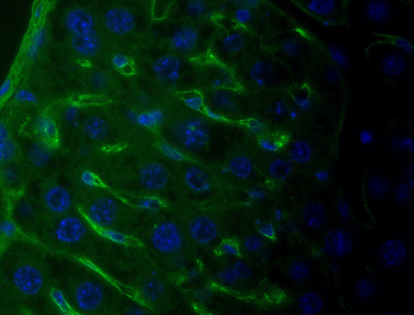
Immunohistochemistry-Frozen: Collagen I alpha 1 Antibody (COL-1) [NB600-450] - Staining of acetone-fixed, frozen sections of pig tongue at a dilution of 1:4000![Immunohistochemistry: Collagen I alpha 1 Antibody (COL-1) [NB600-450]](https://resources.rndsystems.com/images/products/Collagen-I-alpha-1-Antibody-COL-1-Immunohistochemistry-NB600-450-img0006.jpg "Immunohistochemistry: Collagen I alpha 1 Antibody (COL-1) [NB600-450]")
Immunohistochemistry: Collagen I alpha 1 Antibody (COL-1) [NB600-450]
Immunohistochemistry: Collagen I alpha 1 Antibody (COL-1) [NB600-450] - Analysis of an FFPE ligament section. Enzymatic antigen retrieval and blocking using 1% BSA for 30 min at 20C was performed. Sample was incubated for 1 hour with primary antibody at a dilution of 1:400 at 20C.![Immunocytochemistry/ Immunofluorescence: Collagen I alpha 1 Antibody (COL-1) [NB600-450]](https://resources.rndsystems.com/images/products/Collagen-I-alpha-1-Antibody-COL-1-Immunocytochemistry-Immunofluorescence-NB600-450-img0014.jpg "Immunocytochemistry/ Immunofluorescence: Collagen I alpha 1 Antibody (COL-1) [NB600-450]")
![Immunohistochemistry: Collagen I alpha 1 Antibody (COL-1) [NB600-450]](https://resources.rndsystems.com/images/products/Collagen-I-alpha-1-Antibody-COL-1-Immunohistochemistry-NB600-450-img0017.jpg "Immunohistochemistry: Collagen I alpha 1 Antibody (COL-1) [NB600-450]")
Immunohistochemistry: Collagen I alpha 1 Antibody (COL-1) [NB600-450]
Immunohistochemistry: Collagen I alpha 1 Antibody (COL-1) [NB600-450] - Staining of acetone-fixed, frozen sections of human tongue at a dilution of 1:2000.![Immunocytochemistry/ Immunofluorescence: Collagen I alpha 1 Antibody (COL-1) [NB600-450]](https://resources.rndsystems.com/images/products/Collagen-I-alpha-1-Antibody-COL-1-Immunocytochemistry-Immunofluorescence-NB600-450-img0010.jpg "Immunocytochemistry/ Immunofluorescence: Collagen I alpha 1 Antibody (COL-1) [NB600-450]")
Immunocytochemistry/ Immunofluorescence: Collagen I alpha 1 Antibody (COL-1) [NB600-450]
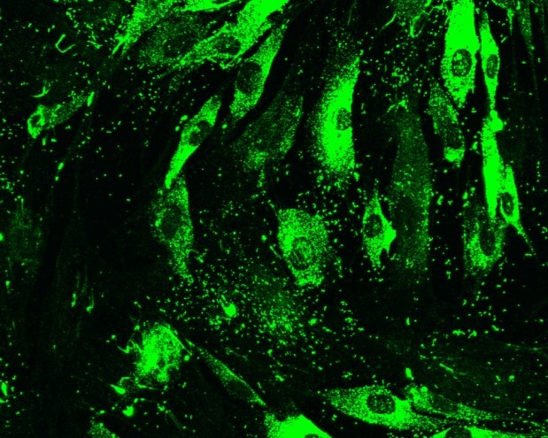
Immunocytochemistry/Immunofluorescence: Collagen I alpha 1 Antibody (COL-1) [NB600-450] - Analysis of Collagen I alpha 1 in 4% PFA fixed equine tenocytes using anti-Collagen I alpha 1 antibody. ICC/IF image submitted by a verified customer review.![Immunocytochemistry/ Immunofluorescence: Collagen I alpha 1 Antibody (COL-1) [NB600-450]](https://resources.rndsystems.com/images/products/Collagen-I-alpha-1-Antibody-COL-1-Immunocytochemistry-Immunofluorescence-NB600-450-img0012.jpg "Immunocytochemistry/ Immunofluorescence: Collagen I alpha 1 Antibody (COL-1) [NB600-450]")
Immunocytochemistry/ Immunofluorescence: Collagen I alpha 1 Antibody (COL-1) [NB600-450]
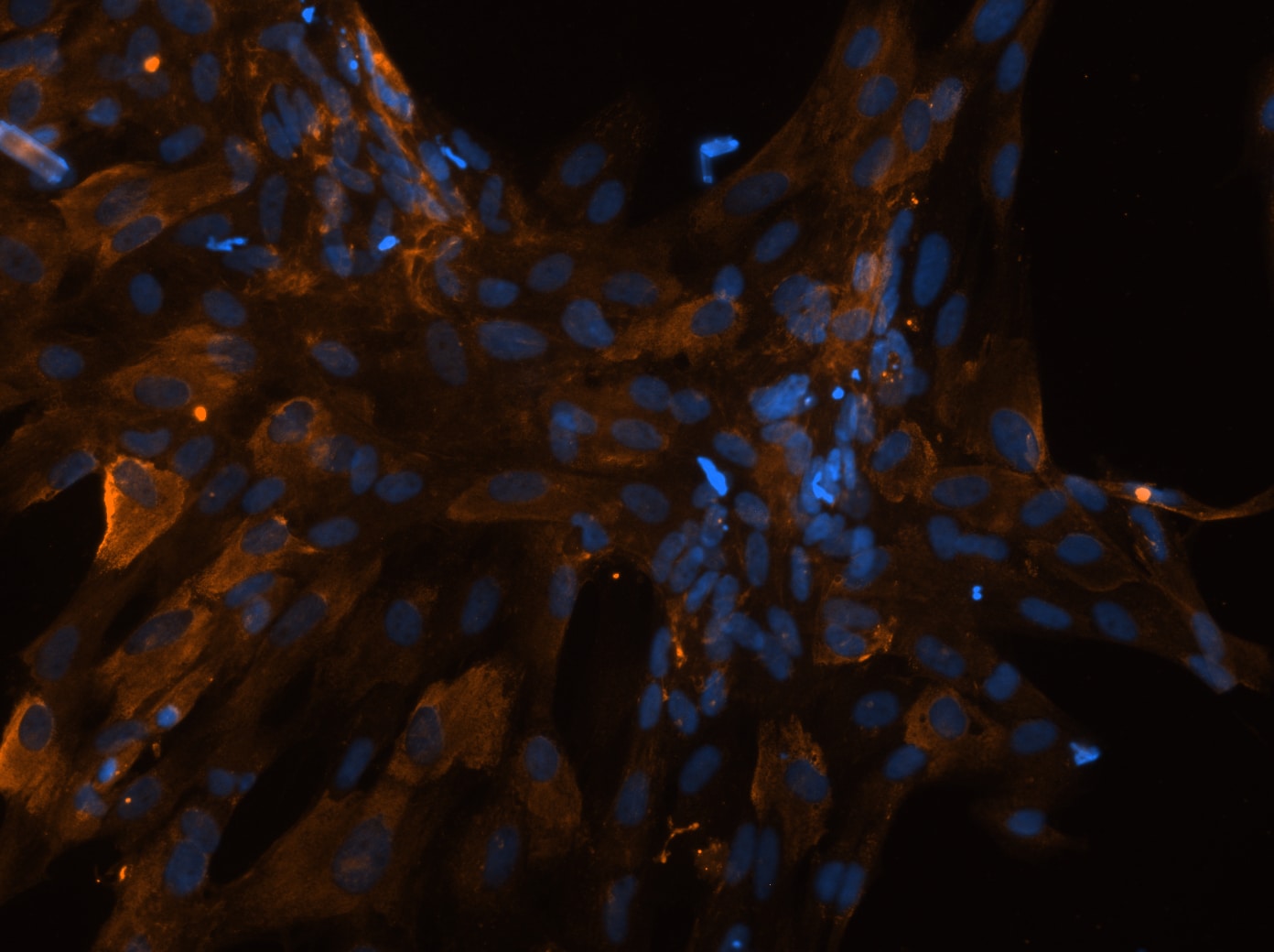
Immunocytochemistry/Immunofluorescence: Collagen I alpha 1 Antibody (COL-1) [NB600-450] - Collagen I alpha1 labeled pig trabecular meshwork cells indirectly visualized with Alexa Fluor 488 conjugated secondary antibody. ICC/IF image submitted by a verified customer review.![Immunohistochemistry: Collagen I alpha 1 Antibody (COL-1) [NB600-450]](https://resources.rndsystems.com/images/products/Collagen-I-alpha-1-Antibody-COL-1-Immunohistochemistry-NB600-450-img0008.jpg "Immunohistochemistry: Collagen I alpha 1 Antibody (COL-1) [NB600-450]")
Immunohistochemistry: Collagen I alpha 1 Antibody (COL-1) [NB600-450]
Immunohistochemistry: Collagen I alpha 1 Antibody (COL-1) [NB600-450] - Analysis of formalin fixed frozen porcine skin tissue sections. Blocking: 5% serum for 1 hr at 20C. Sample was incubated for 16 hours at 4C and used at a dilution of 1:500.![Immunohistochemistry: Collagen I alpha 1 Antibody (COL-1) [NB600-450]](https://resources.rndsystems.com/images/products/Collagen-I-alpha-1-Antibody-COL-1-Immunohistochemistry-NB600-450-img0013.jpg "Immunohistochemistry: Collagen I alpha 1 Antibody (COL-1) [NB600-450]")
Immunohistochemistry: Collagen I alpha 1 Antibody (COL-1) [NB600-450]
Collagen-I-alpha-1-Antibody-COL-1-Immunohistochemistry-NB600-450-img0013.jpg![Immunohistochemistry-Paraffin: Collagen I alpha 1 Antibody (COL-1) [NB600-450]](https://resources.rndsystems.com/images/products/Collagen-I-alpha-1-Antibody-COL-1-Immunohistochemistry-Paraffin-NB600-450-img0015.jpg "Immunohistochemistry-Paraffin: Collagen I alpha 1 Antibody (COL-1) [NB600-450]")
Immunohistochemistry-Paraffin: Collagen I alpha 1 Antibody (COL-1) [NB600-450]
Immunohistochemistry-Paraffin: Collagen I alpha 1 Antibody (COL-1) [NB600-450] - Mouse healthy liver. Anibody at 1:200. Enzymatic/proteolytic antigen retrieval. No permeabilization. IHC-P image submitted by a verified customer review. [NB600-450] -")
Immunohistochemistry: Collagen I alpha 1 Antibody (COL-1) [NB600-450] -
(a) Histological section of keloid stained with picrosirius seen in ordinary light (20x). (b) Histological section of keloid stained with picrosirius seen in polarized light (20x). (c) Immunohistochemistry for type I collagen in keloid (20x). (d) Histological section of keloid stained with picrosirius seen in ordinary light (20x). (e) Histological section of keloid stained with picrosirius seen in polarized light (20x). (f) Immunohistochemistry for type III collagen in keloid (20x). [NB600-450] -")
Immunohistochemistry: Collagen I alpha 1 Antibody (COL-1) [NB600-450] -
Immunohistochemistry: Collagen I alpha 1 Antibody (COL-1) [NB600-450] - FSP‐1–positive cells only partially responsible for collagen I & III production in irradiated bladders. Bladder strips from irradiated mice were costained for collagen I or for collagen III mRNA (pink) & for fibroblasts (FSP‐1, brown). U: urothelium; LP: lamina propria. Black arrow: FSP‐1 positive cells. Gray arrow: collagen only positive cells. Scale bar = 50 µm Image collected & cropped by CiteAb from the following publication (https://pubmed.ncbi.nlm.nih.gov/32109348), licensed under a CC-BY license. Not internally tested by Novus Biologicals. [NB600-450] -")
Immunocytochemistry/ Immunofluorescence: Collagen I alpha 1 Antibody (COL-1) [NB600-450] -
Collagen I and collagen III accumulate in C57BL/6 mice in response to radiation. Bladder strips from irradiated and control mice were stained for collagen I or for collagen III and imaged using fluorescent microscopy. (a) Representative images (100 x 100 um) of each mouse strain and treatment group are provided for collagen I and collagen III immunofluorescence. (b–c) Percentage of tissue‐positive for collagen I or collagen III staining. Collagen I and III density is significantly elevated in C57BL/6 mice, but not in the C3H or BALB/c strains. Results are mean +/- SD of n = 3–6 mice. Dashed line: ANOVA; Full line: multiple t‐test. **p <.01, *** p <.001 Image collected and cropped by CiteAb from the following open publication (https://pubmed.ncbi.nlm.nih.gov/32109348), licensed under a CC-BY license. Not internally tested by Novus Biologicals.Applications for Collagen I alpha 1 Antibody (COL-1)
Application
Recommended Usage
Dot Blot
1:100-1:2000
ELISA
1:100-1:2000
Immunohistochemistry
1:10-1:500
Immunohistochemistry-Frozen
1:2000
Immunohistochemistry-Paraffin
1:10-1:500
Western Blot
1:100-1:2000
Reviewed Applications
Read 5 reviews rated 4.6 using NB600-450 in the following applications:
Formulation, Preparation, and Storage
Purification
Unpurified
Formulation
Ascites
Preservative
15mM Sodium Azide
Concentration
This product is unpurified. The exact concentration of antibody is not quantifiable.
Shipping
The product is shipped with polar packs. Upon receipt, store it immediately at the temperature recommended below.
Stability & Storage
Store at 4C short term. Aliquot and store at -20C long term. Avoid freeze-thaw cycles.
Background: Collagen I alpha 1
Type I collagen is a fibril-forming collagen found in most connective tissues and is abundant in bone, cornea, dermis and tendon tissue. Collagens are fibrous, extracellular matrix proteins with high tensile strength and are the major components of connective tissue. Several collagens play a role in cell adhesion, responsible for maintaining normal tissue architecture and function. All collagens contain a triple helix domain and frequently show lateral self-association in order to form complex connective tissues. Post-Golgi LH3 trafficking is essential for collagen homeostasis and for the development and function of multiple organs and tissues (1).
The COL1A1 gene encodes the pro-alpha1 chains of type I collagen protein, whose triple helix is comprised of two alpha1 chains and one alpha2 chain. Mutations in the encoding COL1A1 gene are associated with brittle bone disease (Osteogenesis Imperfecta), cortical hyperostosis (Caffey disease) and disorders that affect the connective tissues (Ehlers-Danlos syndrome) (2). Studies have found that HIF-1 transcription regulation of collagen prolyl hydroxylases regulates collagen deposition, promoting cancer cell alignment along collagen fibers, which enhances invasion and metastasis to lymph nodes and lung tissue by breast cancer cells (3).
References
1. Banushi, B., Forneris, F., Straatman-Iwanowska, A., Strange, A., Lyne, A. M., Rogerson, C.,... Gissen, P. (2016). Regulation of post-Golgi LH3 trafficking is essential for collagen homeostasis. Nat Commun, 7, 12111. doi:10.1038/ncomms12111
2. Lu, Y., Zhang, S., Wang, Y., Ren, X., & Han, J. (2019). Molecular mechanisms and clinical manifestations of rare genetic disorders associated with type I collagen. Intractable Rare Dis Res, 8(2), 98-107. doi:10.5582/irdr.2019.01064
3. Gilkes, D. M., Chaturvedi, P., Bajpai, S., Wong, C. C., Wei, H., Pitcairn, S.,... Semenza, G. L. (2013). Collagen prolyl hydroxylases are essential for breast cancer metastasis. Cancer Res, 73(11), 3285-3296. doi:10.1158/0008-5472.Can-12-3963
Additional Collagen I alpha 1 Products
Product Documents for Collagen I alpha 1 Antibody (COL-1)
Certificate of Analysis
To download a Certificate of Analysis, please enter a lot or batch number in the search box below.
Product Specific Notices for Collagen I alpha 1 Antibody (COL-1)
This product is for research use only and is not approved for use in humans or in clinical diagnosis. Primary Antibodies are guaranteed for 1 year from date of receipt.
Related Research Areas
Citations for Collagen I alpha 1 Antibody (COL-1)
 Powered by Bioz
Customer Reviews for Collagen I alpha 1 Antibody (COL-1) (5)
4.6 out of 5
5 Customer Ratings
Have you used Collagen I alpha 1 Antibody (COL-1)?
Submit a review and receive an Amazon gift card!
$25/€18/£15/$25CAN/¥2500 Yen for a review with an image
$10/€7/£6/$10CAN/¥1110 Yen for a review without an image
Submit a review
Customer Images

Showing
1
-
5 of
5 reviews
Showing All
Filter By:
-
Application: Immunohistochemistry-ParaffinSample Tested: LiverSpecies: MouseVerified Customer | Posted 06/24/2021Mouse healthy liverPrimary antibody dilution 1:200 Enzymatic/proteolytic antigen retrieval No Permeabilization

-
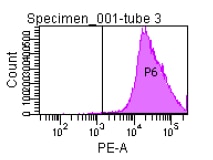
Application: Flow CytometrySample Tested: ChondrocyteSpecies: GoatVerified Customer | Posted 01/22/2019
-
Application: Immunohistochemistry-FrozenSample Tested: ScleraSpecies: FelineVerified Customer | Posted 09/15/2018NB600-450 was secondary immunolabelied shown as red. DAPI (blue). The image was captured using epifluorescent microscope.
-
Application: ImmunocytochemistrySample Tested: Pig trabecular meshwork cellsSpecies: PigVerified Customer | Posted 06/07/2017NB600-450 Collagen I alpha1 antibody labeling pig trabecular meshwork cells with Alexa488
-
Application: ImmunofluorescenceSample Tested: Equine TenocytesSpecies: OtherVerified Customer | Posted 04/21/2015Collagen I-positive equine tenocytes
There are no reviews that match your criteria.
Protocols
View specific protocols for Collagen I alpha 1 Antibody (COL-1) (NB600-450):
Sample
Human Tissue sections (Normal Skin and Basal Cell Carcinoma/Solar Elastos)
Application
Immunohistochemistry (Formalin-fixed paraffin-embedded sections)
Blocking step:
None
Antigen retrieval step:
Heat mediated
Incubation time:
30 minute(s)
Dilution: 1/800
Additional notes
The tissue was FFPE (formalin fixed, paraffin embedded tissue sectioned at 3-4 microns). The antigen retreival method was a heat-induced antigen retreival (HEIR) using a vegetable steamer. Sections were incubated in the steamer for 20 minutes in 1 mM EDTA buffer, pH 8.0. Sections were then rinsed in H2O and TBS buffer prior to starting the staining protocol.
The staining appeared to be very specific and robust, with very little to no non-specific background staining.
Staining Summary & Notes:
1mM EDTA Buffer, pH 8.0, HIER for 20 minutes in steamer
Rinse with TBS containing 0.05% Tween 20
10 minute blocking in 3% Peroxide
Rinse with TBS containing 0.05% Tween 20
30 minutes in Primary Antibody @ RT
Rinse with TBS containing 0.05% Tween 20
30 minutes in Secondary Antibody @ RT
Rinse with TBS containing 0.05% Tween 20
10 minutes in DAB+ (DakoCytomation) @ RT
Water Rinse
Counterstain for 6 minutes in Mayer's Hematoxylin (DakoCytomation) @ RT
Secondary antibody
Name:
DakoCytomation Envision+ Anti-Mouse/HRP (RTU)
Conjugation: HRP
Dilution: 1/1
Find general support by application which include: protocols, troubleshooting, illustrated assays, videos and webinars.
- Antigen Retrieval Protocol (PIER)
- Antigen Retrieval for Frozen Sections Protocol
- Appropriate Fixation of IHC/ICC Samples
- Cellular Response to Hypoxia Protocols
- Chromogenic IHC Staining of Formalin-Fixed Paraffin-Embedded (FFPE) Tissue Protocol
- Chromogenic Immunohistochemistry Staining of Frozen Tissue
- ClariTSA™ Fluorophore Kits
- Detection & Visualization of Antibody Binding
- ELISA Sample Preparation & Collection Guide
- ELISA Troubleshooting Guide
- Fluorescent IHC Staining of Frozen Tissue Protocol
- Graphic Protocol for Heat-induced Epitope Retrieval
- Graphic Protocol for the Preparation and Fluorescent IHC Staining of Frozen Tissue Sections
- Graphic Protocol for the Preparation and Fluorescent IHC Staining of Paraffin-embedded Tissue Sections
- Graphic Protocol for the Preparation of Gelatin-coated Slides for Histological Tissue Sections
- How to Run an R&D Systems DuoSet ELISA
- How to Run an R&D Systems Quantikine ELISA
- How to Run an R&D Systems Quantikine™ QuicKit™ ELISA
- ICC Cell Smear Protocol for Suspension Cells
- ICC Immunocytochemistry Protocol Videos
- ICC for Adherent Cells
- IHC Sample Preparation (Frozen sections vs Paraffin)
- Immunocytochemistry (ICC) Protocol
- Immunocytochemistry Troubleshooting
- Immunofluorescence of Organoids Embedded in Cultrex Basement Membrane Extract
- Immunofluorescent IHC Staining of Formalin-Fixed Paraffin-Embedded (FFPE) Tissue Protocol
- Immunohistochemistry (IHC) and Immunocytochemistry (ICC) Protocols
- Immunohistochemistry Frozen Troubleshooting
- Immunohistochemistry Paraffin Troubleshooting
- Preparing Samples for IHC/ICC Experiments
- Preventing Non-Specific Staining (Non-Specific Binding)
- Primary Antibody Selection & Optimization
- Protocol for Heat-Induced Epitope Retrieval (HIER)
- Protocol for Making a 4% Formaldehyde Solution in PBS
- Protocol for VisUCyte™ HRP Polymer Detection Reagent
- Protocol for the Fluorescent ICC Staining of Cell Smears - Graphic
- Protocol for the Fluorescent ICC Staining of Cultured Cells on Coverslips - Graphic
- Protocol for the Preparation & Fixation of Cells on Coverslips
- Protocol for the Preparation and Chromogenic IHC Staining of Frozen Tissue Sections
- Protocol for the Preparation and Chromogenic IHC Staining of Frozen Tissue Sections - Graphic
- Protocol for the Preparation and Chromogenic IHC Staining of Paraffin-embedded Tissue Sections
- Protocol for the Preparation and Chromogenic IHC Staining of Paraffin-embedded Tissue Sections - Graphic
- Protocol for the Preparation and Fluorescent ICC Staining of Cells on Coverslips
- Protocol for the Preparation and Fluorescent ICC Staining of Non-adherent Cells
- Protocol for the Preparation and Fluorescent ICC Staining of Stem Cells on Coverslips
- Protocol for the Preparation and Fluorescent IHC Staining of Frozen Tissue Sections
- Protocol for the Preparation and Fluorescent IHC Staining of Paraffin-embedded Tissue Sections
- Protocol for the Preparation of Gelatin-coated Slides for Histological Tissue Sections
- Protocol for the Preparation of a Cell Smear for Non-adherent Cell ICC - Graphic
- Quantikine HS ELISA Kit Assay Principle, Alkaline Phosphatase
- Quantikine HS ELISA Kit Principle, Streptavidin-HRP Polymer
- R&D Systems Quality Control Western Blot Protocol
- Sandwich ELISA (Colorimetric) – Biotin/Streptavidin Detection Protocol
- Sandwich ELISA (Colorimetric) – Direct Detection Protocol
- TUNEL and Active Caspase-3 Detection by IHC/ICC Protocol
- The Importance of IHC/ICC Controls
- Troubleshooting Guide: ELISA
- Troubleshooting Guide: Immunohistochemistry
- Troubleshooting Guide: Western Blot Figures
- Western Blot Conditions
- Western Blot Protocol
- Western Blot Protocol for Cell Lysates
- Western Blot Troubleshooting
- Western Blot Troubleshooting Guide
- View all Protocols, Troubleshooting, Illustrated assays and Webinars
FAQs for Collagen I alpha 1 Antibody (COL-1)
Showing
1
-
5 of
5 FAQs
Showing All
-
Q: How long does delivery take for your collagen 1 alpha 1 antibody?
A: Our most popular collagen I alpha 1 antibody (NB600-408) is usually in stock and ships priority overnight for next business day delivery in the U.S.
-
Q: I am looking for anti collagen 1 antibody, I see your product calls it collagen I alpha 1 is there a difference?
A: Collagen I alpha 1 is the most common protein for the collagen 1 protein. There are other subunits as well (such as collagen 1 alpha 2…etc.).
-
Q: The predicted molecular weight says 150kDA but the band on the gel appears at 130kDA why is that?
A: Differences between the predicted and observed MW can be affected by a number of factors including PTMs, relative charges and experimental factors. These cumulative effect of these differences can be quite substantial with larger molecules such as collagen I alpha 1.
-
Q: We are looking for an anti-Collagen antibody for Flow Cytometry (FACS) analysis in Swine sample. Would you please provide some references if there is any available as well?
A:
We currently have one antibody for porcine Collagen validated in flow cytometry (catalog # NBP1-94202).
-
Q: What is the dilution factor for IHC for starting? Because the range of dilution factor on the datasheet is so broad, I am confused which dilution factor is suitable for starting.
A: I can recommend the following dilution to start. Keep in mind that these will all vary greatly by sample type, so these are just suggestions. You may need to use more or less antibody. For catalog number NB600-450, the dilution range is 1:1000-1:2000.
-
Q: How long does delivery take for your collagen 1 alpha 1 antibody?
A: Our most popular collagen I alpha 1 antibody (NB600-408) is usually in stock and ships priority overnight for next business day delivery in the U.S.
-
Q: I am looking for anti collagen 1 antibody, I see your product calls it collagen I alpha 1 is there a difference?
A: Collagen I alpha 1 is the most common protein for the collagen 1 protein. There are other subunits as well (such as collagen 1 alpha 2…etc.).
-
Q: The predicted molecular weight says 150kDA but the band on the gel appears at 130kDA why is that?
A: Differences between the predicted and observed MW can be affected by a number of factors including PTMs, relative charges and experimental factors. These cumulative effect of these differences can be quite substantial with larger molecules such as collagen I alpha 1.
-
Q: We are looking for an anti-Collagen antibody for Flow Cytometry (FACS) analysis in Swine sample. Would you please provide some references if there is any available as well?
A:
We currently have one antibody for porcine Collagen validated in flow cytometry (catalog # NBP1-94202).
-
Q: What is the dilution factor for IHC for starting? Because the range of dilution factor on the datasheet is so broad, I am confused which dilution factor is suitable for starting.
A: I can recommend the following dilution to start. Keep in mind that these will all vary greatly by sample type, so these are just suggestions. You may need to use more or less antibody. For catalog number NB600-450, the dilution range is 1:1000-1:2000.
Loading...