EpCAM/TROP1 Antibody (AUA1) - BSA Free
Novus Biologicals | Catalog # NB600-1182
Key Product Details
Species Reactivity
Validated:
Human
Cited:
Human
Applications
Validated:
Immunohistochemistry, Immunohistochemistry-Paraffin, Western Blot, ELISA, Flow Cytometry, Immunocytochemistry/ Immunofluorescence, CyTOF-ready
Cited:
Western Blot, Flow Cytometry, Immunocytochemistry/ Immunofluorescence, IF/IHC
Label
Unconjugated
Antibody Source
Monoclonal Mouse IgG1 Clone # AUA1
Format
BSA Free
Loading...
Product Specifications
Immunogen
LoVo cell line preparation (Human).
Localization
Cell Membrane
Clonality
Monoclonal
Host
Mouse
Isotype
IgG1
Theoretical MW
35 kDa.
Disclaimer note: The observed molecular weight of the protein may vary from the listed predicted molecular weight due to post translational modifications, post translation cleavages, relative charges, and other experimental factors.
Disclaimer note: The observed molecular weight of the protein may vary from the listed predicted molecular weight due to post translational modifications, post translation cleavages, relative charges, and other experimental factors.
Scientific Data Images for EpCAM/TROP1 Antibody (AUA1) - BSA Free
![Immunohistochemistry-Paraffin: EpCAM/TROP1 Antibody (AUA1) - BSA Free [NB600-1182]](https://resources.rndsystems.com/images/products/EpCAM-TROP1-Antibody-AUA1-Immunohistochemistry-Paraffin-NB600-1182-img0009.jpg "Immunohistochemistry-Paraffin: EpCAM/TROP1 Antibody (AUA1) - BSA Free [NB600-1182]")
Immunohistochemistry-Paraffin: EpCAM/TROP1 Antibody (AUA1) - BSA Free [NB600-1182]
Immunohistochemistry-Paraffin: EpCAM/TROP1 Antibody (AUA1) [NB600-1182] - Analysis of a FFPE human breast carcinoma tissue section using 1:100 dilution of EpCAM/TROP1 antibody (clone AUA1) on a Bond Rx autostainer (Leica Biosystems). The assay involved 20 minutes of heat induced antigen retrieval (HIER) with 10mM sodium citrate buffer (pH 6.0) and endogenous peroxidase quenching using peroxide block. The sections were incubated with primary antibody for 30 minutes. Bond Polymer Refine Detection (Leica Biosystems) and DAB were used for signal detection which followed counterstaining with hematoxylin. Whole slide scanning and capturing of representative images (20X) were performed using Aperio AT2 (Leica Biosystems). This EpCAM antibody generated an expected immunostaining of EpCAM (CD326) protein in the membranes of the cancer cells and the inter-cellular spaces. The tumor stroma and the stromal cells did not show EpCAM immunopositivity.![Western Blot: EpCAM/TROP1 Antibody (AUA1)BSA Free [NB600-1182]](https://resources.rndsystems.com/images/products/EpCAM-TROP1-Antibody-AUA1-Western-Blot-NB600-1182-img0015.jpg "Western Blot: EpCAM/TROP1 Antibody (AUA1)BSA Free [NB600-1182]")
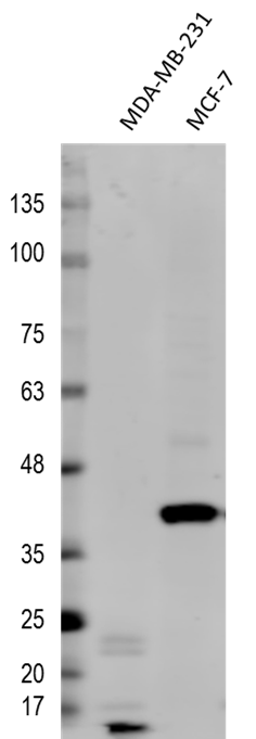
Western Blot: EpCAM/TROP1 Antibody (AUA1)BSA Free [NB600-1182]
Western Blot: EpCAM/TROP1 Antibody (AUA1) [NB600-1182] - Detection of EpCAM/CD326 in two Human mammary tumor cell lines, MDA-MB-231 (mesenchyme-like) and MCF-7 (epithelium-like). Dilution: 1:500 in PBS with 5% BSA. Secondary Ab: anti-Mouse IgG 1:5,000. This image was submitted via customer Review.![Immunocytochemistry/ Immunofluorescence: EpCAM/TROP1 Antibody (AUA1) - BSA Free [NB600-1182]](https://resources.rndsystems.com/images/products/EpCAM-TROP1-Antibody-AUA1-Immunocytochemistry-Immunofluorescence-NB600-1182-img0016.jpg "Immunocytochemistry/ Immunofluorescence: EpCAM/TROP1 Antibody (AUA1) - BSA Free [NB600-1182]")
Immunocytochemistry/ Immunofluorescence: EpCAM/TROP1 Antibody (AUA1) - BSA Free [NB600-1182]
Immunocytochemistry/Immunofluorescence: EpCAM/TROP1 Antibody (AUA1) [NB600-1182] - A431 cells were fixed for 10 minutes using 10% formalin and then permeabilized for 5 minutes using 1X PBS + 0.05% Triton X-100. The cells were incubated with anti-EpCAM/TROP1 (AUA1) conjugated to Alexa Fluor 488 [NB600-1182AF488] at 10ug/ml for 1 hour at room temperature. Nuclei were counterstained with DAPI (blue). Cells were imaged using a 40X objective.![Immunohistochemistry-Paraffin: EpCAM/TROP1 Antibody (AUA1) - BSA Free [NB600-1182]](https://resources.rndsystems.com/images/products/EpCAM-TROP1-Antibody-AUA1-Immunohistochemistry-Paraffin-NB600-1182-img0014.jpg "Immunohistochemistry-Paraffin: EpCAM/TROP1 Antibody (AUA1) - BSA Free [NB600-1182]")
Immunohistochemistry-Paraffin: EpCAM/TROP1 Antibody (AUA1) - BSA Free [NB600-1182]
Immunohistochemistry-Paraffin: EpCAM/TROP1 Antibody (AUA1) [NB600-1182] - Analysis of a FFPE human breast carcinoma tissue with 1:500 dilution of EpCAM/TROP1 antibody (clone AUA1) on a Bond Rx autostainer (Leica Biosystems). The assay involved 20 minutes of heat induced antigen retrieval (HIER) with 10mM sodium citrate buffer (pH 6.0) and endogenous peroxidase quenching using peroxide block. The sections were incubated with primary antibody for 30 minutes. Bond Polymer Refine Detection (Leica Biosystems) and DAB were used for signal detection which followed counterstaining with hematoxylin. Whole slide scanning and capturing of representative images (20X) were performed using Aperio AT2 (Leica Biosystems). The staining was primarily localized to the membranes and inter-cellular spaces of the cancer cells. Staining intensity for 1:500 dilution was lower than what was seen in sections tested with 1:100 dilution. Staining was performed by Histowiz.![Flow Cytometry: EpCAM/TROP1 Antibody (AUA1) - BSA Free [NB600-1182]](https://resources.rndsystems.com/images/products/EpCAM-TROP1-Antibody-AUA1-Flow-Cytometry-NB600-1182-img0008.jpg "Flow Cytometry: EpCAM/TROP1 Antibody (AUA1) - BSA Free [NB600-1182]")
Flow Cytometry: EpCAM/TROP1 Antibody (AUA1) - BSA Free [NB600-1182]
Flow Cytometry: EpCAM/TROP1 Antibody (AUA1) [NB600-1182] - Using the PE direct conjugate A cell surface stain was performed on HT-29 cells with EpCAM (AUA1) antibody NB600-1182PE (blue) and a matched isotype control NBP2-27287PE (orange). Cells were incubated in an antibody dilution of 5 ug/mL for 20 minutes at room temperature. Both antibodies were conjugated to phycoerythrin.![Western Blot: EpCAM/TROP1 Antibody (AUA1)BSA Free [NB600-1182]](https://resources.rndsystems.com/images/products/EpCAM-TROP1-Antibody-AUA1-Western-Blot-NB600-1182-img0001.jpg "Western Blot: EpCAM/TROP1 Antibody (AUA1)BSA Free [NB600-1182]")
Western Blot: EpCAM/TROP1 Antibody (AUA1)BSA Free [NB600-1182]
Western Blot: EpCAM/TROP1 Antibody (AUA1) [NB600-1182] - Analysis of A431 lysate (A) and Hek293 lysate (B) using EpCAM/CD326 antibody (AUA1) at 2 ug/ml. Non-reduced lysate was used in each lane.![Flow Cytometry: EpCAM/TROP1 Antibody (AUA1) - BSA Free [NB600-1182]](https://resources.rndsystems.com/images/products/EpCAM-TROP1-Antibody-AUA1-Flow-Cytometry-NB600-1182-img0007.jpg "Flow Cytometry: EpCAM/TROP1 Antibody (AUA1) - BSA Free [NB600-1182]")
Flow Cytometry: EpCAM/TROP1 Antibody (AUA1) - BSA Free [NB600-1182]
Flow Cytometry: EpCAM/TROP1 Antibody (AUA1) [NB600-1182] - Intracellular flow cytometric staining of 1 x 10^6 CHO (A) and HeLa (B) cells using EpCAM/CD326 antibody (blue) and matched Isotype control (orange). An antibody concentration of 1 ug/1x10^6 cells was used.Applications for EpCAM/TROP1 Antibody (AUA1) - BSA Free
Application
Recommended Usage
ELISA
1:100-1:2000
Flow Cytometry
1 ug per million cells
Immunocytochemistry/ Immunofluorescence
1:10-1:500. Use reported in scientific literature (PMID 21763624)
Immunohistochemistry
1:10-1:500
Immunohistochemistry-Paraffin
1:10-1:500
Western Blot
1:500
Application Notes
This antibody is CyTOF ready. Flow Cytometry reported by Alvarez et al - Conference on Retroviruses and Opportunistic Infections - CROI 2014 (Abstract: The NCOR2-Nurr1-CoREST Transrepression Axis Impairs HIV Reactivation in Latently Infected Microglial Cells)
Reviewed Applications
Read 1 review rated 5 using NB600-1182 in the following applications:
Flow Cytometry Panel Builder
Bio-Techne Knows Flow Cytometry
Save time and reduce costly mistakes by quickly finding compatible reagents using the Panel Builder Tool.
Advanced Features
- Spectra Viewer - Custom analysis of spectra from multiple fluorochromes
- Spillover Popups - Visualize the spectra of individual fluorochromes
- Antigen Density Selector - Match fluorochrome brightness with antigen density

Formulation, Preparation, and Storage
Purification
Protein G purified
Formulation
PBS
Format
BSA Free
Preservative
0.05% Sodium Azide
Concentration
1.0 mg/ml
Shipping
The product is shipped with polar packs. Upon receipt, store it immediately at the temperature recommended below.
Stability & Storage
Store at 4C short term. Aliquot and store at -20C long term. Avoid freeze-thaw cycles.
Background: EpCAM/TROP1
EpCAM functions as an intracellular signaling molecule and contributes to regulation of epithelial-to-mesenchymal (EMT) transition (4,5). EpCAM is cleaved during the process of regulated intramembrane proteolysis (RIP) (4,5). Initial cleavage occurs at the membrane via a disintegrin and metalloprotease 17 (ADAM17) which releases EpCAM's extracellular domain (EpEx) (4,5). A secondary cleavage is mediated by presenilin 2 (PSEN2) which releases EpCAM's cytoplasmic trail (EpICD) (4,5). EpICD translocates to the nucleus where it associates with beta-catenin, FHL-2, and LEF-1 and induces transcription of genes related to EMT and tumor growth (4,5). EpCAM has also been shown to regulate structure and functionality of the apical junction complex of cells through direct interaction with claudin-7 and association with E-cadherin (2,5). Loss of EpCAM disrupts adherins junction and tight junction structure and function (2).
References
1. Mohtar MA, Syafruddin SE, Nasir SN, Low TY. Revisiting the Roles of Pro-Metastatic EpCAM in Cancer. Biomolecules. 2020; 10(2):255. https://doi.org/10.3390/biom10020255
2. Huang L, Yang Y, Yang F, et al. Functions of EpCAM in physiological processes and diseases (Review). Int J Mol Med. 2018; 42(4):1771-1785. https://doi.org/10.3892/ijmm.2018.3764
3. Brown TC, Sankpal NV, Gillanders WE. Functional Implications of the Dynamic Regulation of EpCAM during Epithelial-to-Mesenchymal Transition. Biomolecules. 2021; 11(7):956. https://doi.org/10.3390/biom11070956
4. Uniprot (P16422)
5. Schnell U, Cirulli V, Giepmans BN. EpCAM: structure and function in health and disease. Biochim Biophys Acta. 2013; 1828(8):1989-2001. https://doi.org/10.1016/j.bbamem.2013.04.018
Long Name
Epithelial Cell Adhesion Molecule
Alternate Names
17-1A, CD326, GA733-2, gp40, KS1/4, M4S1, TACSTD1, TROP1
Entrez Gene IDs
4072 (Human)
Gene Symbol
EPCAM
Additional EpCAM/TROP1 Products
Product Documents for EpCAM/TROP1 Antibody (AUA1) - BSA Free
Certificate of Analysis
To download a Certificate of Analysis, please enter a lot or batch number in the search box below.
Product Specific Notices for EpCAM/TROP1 Antibody (AUA1) - BSA Free
This product is for research use only and is not approved for use in humans or in clinical diagnosis. Primary Antibodies are guaranteed for 1 year from date of receipt.
Citations for EpCAM/TROP1 Antibody (AUA1) - BSA Free
 Powered by Bioz
Customer Reviews for EpCAM/TROP1 Antibody (AUA1) - BSA Free (1)
5 out of 5
1 Customer Rating
Have you used EpCAM/TROP1 Antibody (AUA1) - BSA Free?
Submit a review and receive an Amazon gift card!
$25/€18/£15/$25CAN/¥2500 Yen for a review with an image
$10/€7/£6/$10CAN/¥1110 Yen for a review without an image
Submit a review
Customer Images
Showing
1
-
1 of
1 review
Showing All
Filter By:
-
Application: Western BlotSample Tested: Human breast cancer cell linesSpecies: HumanVerified Customer | Posted 03/06/2017Detection of EpCAM/CD326 in two Human mammary tumor cell lines, MDA-MB-231 (mesenchyme-like) and MCF-7 (epithelium-like). Dilution: 1:500 in PBS with 5% BSA. Secondary Ab: anti-Mouse IgG 1:5,000.

There are no reviews that match your criteria.
Protocols
Find general support by application which include: protocols, troubleshooting, illustrated assays, videos and webinars.
- 7-Amino Actinomycin D (7-AAD) Cell Viability Flow Cytometry Protocol
- Antigen Retrieval Protocol (PIER)
- Antigen Retrieval for Frozen Sections Protocol
- Appropriate Fixation of IHC/ICC Samples
- Cellular Response to Hypoxia Protocols
- Chromogenic IHC Staining of Formalin-Fixed Paraffin-Embedded (FFPE) Tissue Protocol
- Chromogenic Immunohistochemistry Staining of Frozen Tissue
- ClariTSA™ Fluorophore Kits
- Detection & Visualization of Antibody Binding
- ELISA Sample Preparation & Collection Guide
- ELISA Troubleshooting Guide
- Extracellular Membrane Flow Cytometry Protocol
- Flow Cytometry Protocol for Cell Surface Markers
- Flow Cytometry Protocol for Staining Membrane Associated Proteins
- Flow Cytometry Staining Protocols
- Flow Cytometry Troubleshooting Guide
- Fluorescent IHC Staining of Frozen Tissue Protocol
- Graphic Protocol for Heat-induced Epitope Retrieval
- Graphic Protocol for the Preparation and Fluorescent IHC Staining of Frozen Tissue Sections
- Graphic Protocol for the Preparation and Fluorescent IHC Staining of Paraffin-embedded Tissue Sections
- Graphic Protocol for the Preparation of Gelatin-coated Slides for Histological Tissue Sections
- How to Run an R&D Systems DuoSet ELISA
- How to Run an R&D Systems Quantikine ELISA
- How to Run an R&D Systems Quantikine™ QuicKit™ ELISA
- ICC Cell Smear Protocol for Suspension Cells
- ICC Immunocytochemistry Protocol Videos
- ICC for Adherent Cells
- IHC Sample Preparation (Frozen sections vs Paraffin)
- Immunocytochemistry (ICC) Protocol
- Immunocytochemistry Troubleshooting
- Immunofluorescence of Organoids Embedded in Cultrex Basement Membrane Extract
- Immunofluorescent IHC Staining of Formalin-Fixed Paraffin-Embedded (FFPE) Tissue Protocol
- Immunohistochemistry (IHC) and Immunocytochemistry (ICC) Protocols
- Immunohistochemistry Frozen Troubleshooting
- Immunohistochemistry Paraffin Troubleshooting
- Intracellular Flow Cytometry Protocol Using Alcohol (Methanol)
- Intracellular Flow Cytometry Protocol Using Detergents
- Intracellular Nuclear Staining Flow Cytometry Protocol Using Detergents
- Intracellular Staining Flow Cytometry Protocol Using Alcohol Permeabilization
- Intracellular Staining Flow Cytometry Protocol Using Detergents to Permeabilize Cells
- Preparing Samples for IHC/ICC Experiments
- Preventing Non-Specific Staining (Non-Specific Binding)
- Primary Antibody Selection & Optimization
- Propidium Iodide Cell Viability Flow Cytometry Protocol
- Protocol for Heat-Induced Epitope Retrieval (HIER)
- Protocol for Liperfluo
- Protocol for Making a 4% Formaldehyde Solution in PBS
- Protocol for VisUCyte™ HRP Polymer Detection Reagent
- Protocol for the Characterization of Human Th22 Cells
- Protocol for the Characterization of Human Th9 Cells
- Protocol for the Fluorescent ICC Staining of Cell Smears - Graphic
- Protocol for the Fluorescent ICC Staining of Cultured Cells on Coverslips - Graphic
- Protocol for the Preparation & Fixation of Cells on Coverslips
- Protocol for the Preparation and Chromogenic IHC Staining of Frozen Tissue Sections
- Protocol for the Preparation and Chromogenic IHC Staining of Frozen Tissue Sections - Graphic
- Protocol for the Preparation and Chromogenic IHC Staining of Paraffin-embedded Tissue Sections
- Protocol for the Preparation and Chromogenic IHC Staining of Paraffin-embedded Tissue Sections - Graphic
- Protocol for the Preparation and Fluorescent ICC Staining of Cells on Coverslips
- Protocol for the Preparation and Fluorescent ICC Staining of Non-adherent Cells
- Protocol for the Preparation and Fluorescent ICC Staining of Stem Cells on Coverslips
- Protocol for the Preparation and Fluorescent IHC Staining of Frozen Tissue Sections
- Protocol for the Preparation and Fluorescent IHC Staining of Paraffin-embedded Tissue Sections
- Protocol for the Preparation of Gelatin-coated Slides for Histological Tissue Sections
- Protocol for the Preparation of a Cell Smear for Non-adherent Cell ICC - Graphic
- Protocol: Annexin V and PI Staining by Flow Cytometry
- Protocol: Annexin V and PI Staining for Apoptosis by Flow Cytometry
- Quantikine HS ELISA Kit Assay Principle, Alkaline Phosphatase
- Quantikine HS ELISA Kit Principle, Streptavidin-HRP Polymer
- R&D Systems Quality Control Western Blot Protocol
- Sandwich ELISA (Colorimetric) – Biotin/Streptavidin Detection Protocol
- Sandwich ELISA (Colorimetric) – Direct Detection Protocol
- TUNEL and Active Caspase-3 Detection by IHC/ICC Protocol
- The Importance of IHC/ICC Controls
- Troubleshooting Guide: ELISA
- Troubleshooting Guide: Fluorokine Flow Cytometry Kits
- Troubleshooting Guide: Immunohistochemistry
- Troubleshooting Guide: Western Blot Figures
- Western Blot Conditions
- Western Blot Protocol
- Western Blot Protocol for Cell Lysates
- Western Blot Troubleshooting
- Western Blot Troubleshooting Guide
- View all Protocols, Troubleshooting, Illustrated assays and Webinars
FAQs for EpCAM/TROP1 Antibody (AUA1) - BSA Free
Showing
1
-
1 of
1 FAQ
Showing All
-
Q: I'm looking for EpCAM antibody for rat. Do you have any antibody which can use FACS?
A:
We unfortunately do not have any EpCAM antibodies validated in FACS for detection of the rat protein. I am sorry for any inconvenience. I do have three products to EpCAM that can detect the rat protein. These products have been validated to detect the native rat protein, so they may work for FACS however they have not yet been tested in that particular application. If you would like to try them in your experiment you would qualify for our Innovator's Reward Program. This program was designed to help scientists with the cost of their antibodies in return for valuable information. If you decide to test an antibody in a previously untested species or application we will issue you a credit for the purchase price of the antibody in exchange for a review of your experiment using our product.

