ERCC1 Antibody (8F1) - BSA Free
Novus Biologicals | Catalog # NB500-704
Key Product Details
Species Reactivity
Validated:
Human, Rat, Chinese Hamster
Cited:
Human, Hamster - Cricetulus (Chinese Hamster)
Applications
Validated:
Immunohistochemistry, Immunohistochemistry-Paraffin, Western Blot, Immunocytochemistry/ Immunofluorescence
Cited:
Immunohistochemistry-Paraffin, Western Blot, IF/IHC
Label
Unconjugated
Antibody Source
Monoclonal Mouse IgG2B Clone # 8F1
Format
BSA Free
Loading...
Product Specifications
Immunogen
Full length recombinant human ERCC1. [UniProt# P07992]
Reactivity Notes
Human, rat. Chinese Hamster reactivity reported in scientific literature (PMID: 17962301) Not yet tested in other species.
Localization
Nuclear.
Clonality
Monoclonal
Host
Mouse
Isotype
IgG2B
Scientific Data Images for ERCC1 Antibody (8F1) - BSA Free
![Immunohistochemistry-Paraffin: ERCC1 Antibody (8F1) - BSA Free [NB500-704]](https://resources.rndsystems.com/images/products/ERCC1-Antibody-8F1-Immunohistochemistry-Paraffin-NB500-704-img0004.jpg "Immunohistochemistry-Paraffin: ERCC1 Antibody (8F1) - BSA Free [NB500-704]")
Immunohistochemistry-Paraffin: ERCC1 Antibody (8F1) - BSA Free [NB500-704]
ERCC1-Antibody-8F1-Immunohistochemistry-Paraffin-NB500-704-img0004.jpg![Immunohistochemistry-Paraffin: ERCC1 Antibody (8F1) - BSA Free [NB500-704]](https://resources.rndsystems.com/images/products/ERCC1-Antibody-8F1-Immunohistochemistry-Paraffin-NB500-704-img0002.jpg "Immunohistochemistry-Paraffin: ERCC1 Antibody (8F1) - BSA Free [NB500-704]")
Immunohistochemistry-Paraffin: ERCC1 Antibody (8F1) - BSA Free [NB500-704]
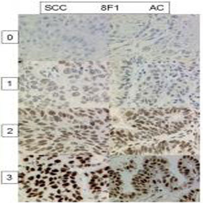
Immunohistochemistry-Paraffin: ERCC1 Antibody (8F1) [NB500-704] - Analysis of ERCC1 in human non-small cell lung cancer. Image courtesy of product review submitted by Alex Soltermann.![Immunohistochemistry-Paraffin: ERCC1 Antibody (8F1) - BSA Free [NB500-704]](https://resources.rndsystems.com/images/products/ERCC1-Antibody-8F1-Immunohistochemistry-Paraffin-NB500-704-img0003.jpg "Immunohistochemistry-Paraffin: ERCC1 Antibody (8F1) - BSA Free [NB500-704]")
Immunohistochemistry-Paraffin: ERCC1 Antibody (8F1) - BSA Free [NB500-704]
Immunohistochemistry-Paraffin: ERCC1 Antibody (8F1) [NB500-704] - Formalin fixed paraffin embedded human tonsil stained with ERCC1 antibody.![Immunohistochemistry-Paraffin: ERCC1 Antibody (8F1) - BSA Free [NB500-704]](https://resources.rndsystems.com/images/products/ERCC1-Antibody-8F1-Immunohistochemistry-Paraffin-NB500-704-img0005.jpg "Immunohistochemistry-Paraffin: ERCC1 Antibody (8F1) - BSA Free [NB500-704]")
Immunohistochemistry-Paraffin: ERCC1 Antibody (8F1) - BSA Free [NB500-704]
ERCC1-Antibody-8F1-Immunohistochemistry-Paraffin-NB500-704-img0005.jpg - BSA Free [NB500-704] -")
Immunohistochemistry: ERCC1 Antibody (8F1) - BSA Free [NB500-704] -
Immunohistochemistry: ERCC1 Antibody (8F1) - BSA Free [NB500-704] - Anti-ERCC1 immunocytochemistry on cell block core of malignant pleural mesothelioma, using Mab 8F1 & D-10 with CC1-mono & H2-60 protocols. Arrow: Surrounding non-tumoral cells, including lymphocytes, macrophages & neutrophil granulocytes. Arrowhead: Unspecific plasma membrane staining with D-10. 200 × original magnification. Inset lower left: Staining of intracellular mucin vacuoles of a mucinous adenocarcinoma of unknown origin. 400 × original magnification. Image collected & cropped by CiteAb from the following publication (http://jclinbioinformatics.biomedcentral.com/articles/10.1186/2043-9113…), licensed under a CC-BY license. Not internally tested by Novus Biologicals. - BSA Free [NB500-704] -")
Western Blot: ERCC1 Antibody (8F1) - BSA Free [NB500-704] -
DHO hydrophilic extract increases the Single Strand Annealing activity in response to CPT treatment (A) Chromatin enhriched purification of HeLa cells pretreated or not with the DHO extract at 1 mg/ml followed by 1 μM CPT treatment for additional two hours. Cells were then lysed to obtain a soluble (S) and a chromatin-enriched (P, as pellet) fraction. Western blotting was performed to analyse the loading onto chromatin of the Poly (ADP-ribose) polymerase (PARP1) protein, involved in the cell response to DNA damage. Total RPA32 and Lamin A/C were used as controls of the supernatant or the chromatin-enriched fraction, respectively. (B) Chromatin enhriched purification of HeLa cells performed as previously for the analysis of RAD52 chromatin loading. (C) Chromatin enhiched purification of HeLa cells was performed as previously described followed by incubation with ERCC1 antibody. (D) HeLa hprtSAGFP cells were transfected with the plasmid encoding the SceI restriction enzyme followed by incubation with 1 mg/ml of DHO extract or vehicle (DMSO) for 48 hours followed by FACS analysis measurement of GFP levels to calculate %SSA frequency compared with control cells which were set as 100%. Data represent the mean % +/- SD. obtained from three independent experiments. Statistically significant differences are indicated with: ***significant (P < 0.001). Image collected and cropped by CiteAb from the following open publication (https://pubmed.ncbi.nlm.nih.gov/37409248), licensed under a CC-BY license. Not internally tested by Novus Biologicals.Applications for ERCC1 Antibody (8F1) - BSA Free
Application
Recommended Usage
Immunocytochemistry/ Immunofluorescence
1:10-1:500
Immunohistochemistry
1:10-1:500
Immunohistochemistry-Paraffin
1:100-1:200
Western Blot
1:100-1:2000
Application Notes
IHC: In an ABC method, we suggest an incubation period of 30 minutes at room temperature. However, depending upon the fixation conditions and the staining system employed, optimal incubation conditions and antibody dilutions should be determined by the user. Formalin fixed paraffin embedded tissue sections require high temperature antigen unmasking with 10 mM citrate buffer, pH 6.0 prior to immunostaining. Antibody can be used in Immunocytochemistry/Immunoflourescence as reported in the literature (PMID: 21961533)Western Blot was reported in scientific literature.
Reviewed Applications
Read 1 review rated 5 using NB500-704 in the following applications:
Formulation, Preparation, and Storage
Purification
Protein G purified
Formulation
PBS
Format
BSA Free
Preservative
0.02% Sodium Azide
Concentration
1.0 mg/ml
Shipping
The product is shipped with polar packs. Upon receipt, store it immediately at the temperature recommended below.
Stability & Storage
Store at 4C short term. Aliquot and store at -20C long term. Avoid freeze-thaw cycles.
Background: ERCC1
Long Name
Excision Repair Cross-complementing 1
Alternate Names
COFS4, DNA excision repair protein ERCC-1, excision repair cross-complementing rodent repair deficiency, complementationgroup 1 (includes overlapping antisense sequence), RAD10, UV20
Gene Symbol
ERCC1
UniProt
Additional ERCC1 Products
Product Documents for ERCC1 Antibody (8F1) - BSA Free
Certificate of Analysis
To download a Certificate of Analysis, please enter a lot or batch number in the search box below.
Product Specific Notices for ERCC1 Antibody (8F1) - BSA Free
This product is for research use only and is not approved for use in humans or in clinical diagnosis. Primary Antibodies are guaranteed for 1 year from date of receipt.
Related Research Areas
Citations for ERCC1 Antibody (8F1) - BSA Free
 Powered by Bioz
Customer Reviews for ERCC1 Antibody (8F1) - BSA Free (1)
5 out of 5
1 Customer Rating
Have you used ERCC1 Antibody (8F1) - BSA Free?
Submit a review and receive an Amazon gift card!
$25/€18/£15/$25CAN/¥2500 Yen for a review with an image
$10/€7/£6/$10CAN/¥1110 Yen for a review without an image
Submit a review
Customer Images
Showing
1
-
1 of
1 review
Showing All
Filter By:
-
Application: Immunohistochemistry-ParaffinSample Tested: Non-small cell lung cancerSpecies: HumanVerified Customer | Posted 12/14/2011

There are no reviews that match your criteria.
Protocols
Find general support by application which include: protocols, troubleshooting, illustrated assays, videos and webinars.
- Antigen Retrieval Protocol (PIER)
- Antigen Retrieval for Frozen Sections Protocol
- Appropriate Fixation of IHC/ICC Samples
- Cellular Response to Hypoxia Protocols
- Chromogenic IHC Staining of Formalin-Fixed Paraffin-Embedded (FFPE) Tissue Protocol
- Chromogenic Immunohistochemistry Staining of Frozen Tissue
- ClariTSA™ Fluorophore Kits
- Detection & Visualization of Antibody Binding
- Fluorescent IHC Staining of Frozen Tissue Protocol
- Graphic Protocol for Heat-induced Epitope Retrieval
- Graphic Protocol for the Preparation and Fluorescent IHC Staining of Frozen Tissue Sections
- Graphic Protocol for the Preparation and Fluorescent IHC Staining of Paraffin-embedded Tissue Sections
- Graphic Protocol for the Preparation of Gelatin-coated Slides for Histological Tissue Sections
- ICC Cell Smear Protocol for Suspension Cells
- ICC Immunocytochemistry Protocol Videos
- ICC for Adherent Cells
- IHC Sample Preparation (Frozen sections vs Paraffin)
- Immunocytochemistry (ICC) Protocol
- Immunocytochemistry Troubleshooting
- Immunofluorescence of Organoids Embedded in Cultrex Basement Membrane Extract
- Immunofluorescent IHC Staining of Formalin-Fixed Paraffin-Embedded (FFPE) Tissue Protocol
- Immunohistochemistry (IHC) and Immunocytochemistry (ICC) Protocols
- Immunohistochemistry Frozen Troubleshooting
- Immunohistochemistry Paraffin Troubleshooting
- Preparing Samples for IHC/ICC Experiments
- Preventing Non-Specific Staining (Non-Specific Binding)
- Primary Antibody Selection & Optimization
- Protocol for Heat-Induced Epitope Retrieval (HIER)
- Protocol for Making a 4% Formaldehyde Solution in PBS
- Protocol for VisUCyte™ HRP Polymer Detection Reagent
- Protocol for the Fluorescent ICC Staining of Cell Smears - Graphic
- Protocol for the Fluorescent ICC Staining of Cultured Cells on Coverslips - Graphic
- Protocol for the Preparation & Fixation of Cells on Coverslips
- Protocol for the Preparation and Chromogenic IHC Staining of Frozen Tissue Sections
- Protocol for the Preparation and Chromogenic IHC Staining of Frozen Tissue Sections - Graphic
- Protocol for the Preparation and Chromogenic IHC Staining of Paraffin-embedded Tissue Sections
- Protocol for the Preparation and Chromogenic IHC Staining of Paraffin-embedded Tissue Sections - Graphic
- Protocol for the Preparation and Fluorescent ICC Staining of Cells on Coverslips
- Protocol for the Preparation and Fluorescent ICC Staining of Non-adherent Cells
- Protocol for the Preparation and Fluorescent ICC Staining of Stem Cells on Coverslips
- Protocol for the Preparation and Fluorescent IHC Staining of Frozen Tissue Sections
- Protocol for the Preparation and Fluorescent IHC Staining of Paraffin-embedded Tissue Sections
- Protocol for the Preparation of Gelatin-coated Slides for Histological Tissue Sections
- Protocol for the Preparation of a Cell Smear for Non-adherent Cell ICC - Graphic
- R&D Systems Quality Control Western Blot Protocol
- TUNEL and Active Caspase-3 Detection by IHC/ICC Protocol
- The Importance of IHC/ICC Controls
- Troubleshooting Guide: Immunohistochemistry
- Troubleshooting Guide: Western Blot Figures
- Western Blot Conditions
- Western Blot Protocol
- Western Blot Protocol for Cell Lysates
- Western Blot Troubleshooting
- Western Blot Troubleshooting Guide
- View all Protocols, Troubleshooting, Illustrated assays and Webinars
Loading...