Fibronectin Antibody - BSA Free
Novus Biologicals | Catalog # NBP1-91258
![Western Blot: Fibronectin AntibodyBSA Free [NBP1-91258]](https://resources.rndsystems.com/images/products/Fibronectin-Antibody---BSA-Free-Western-Blot-NBP1-91258-img0020.jpg "Western Blot: Fibronectin AntibodyBSA Free [NBP1-91258]")
Key Product Details
Validated by
Species Reactivity
Validated:
Cited:
Applications
Validated:
Cited:
Label
Antibody Source
Format
Product Specifications
Immunogen
Reactivity Notes
Localization
Marker
Clonality
Host
Isotype
Scientific Data Images for Fibronectin Antibody - BSA Free
Western Blot: Fibronectin AntibodyBSA Free [NBP1-91258]
Fibronectin-Antibody---BSA-Free-Western-Blot-NBP1-91258-img0020.jpg![Immunocytochemistry/ Immunofluorescence: Fibronectin Antibody - BSA Free [NBP1-91258]](https://resources.rndsystems.com/images/products/Fibronectin-Antibody---BSA-Free-Immunocytochemistry-Immunofluorescence-NBP1-91258-img0021.jpg "Immunocytochemistry/ Immunofluorescence: Fibronectin Antibody - BSA Free [NBP1-91258]")
Immunocytochemistry/ Immunofluorescence: Fibronectin Antibody - BSA Free [NBP1-91258]
Immunocytochemistry/Immunofluorescence: Fibronectin Antibody - BSA Free [NBP1-91258] - NIH3T3 cells were fixed in 4% paraformaldehyde for 10 minutes and permeabilized in 0.05% Triton X-100 in PBS for 5 minutes. The cells were incubated with anti- NBP1-91258 at 1 ug/ml overnight at 4C and detected with an anti-rabbit Dylight 488 (Green) at a 1:1000 dilution for 60 minutes. Nuclei were counterstained with DAPI (Blue). Cells were imaged using a 100X objective and digitally deconvolved.![Immunohistochemistry-Frozen: Fibronectin Antibody - BSA Free [NBP1-91258]](https://resources.rndsystems.com/images/products/Fibronectin-Antibody---BSA-Free-Immunohistochemistry-Frozen-NBP1-91258-img0019.jpg "Immunohistochemistry-Frozen: Fibronectin Antibody - BSA Free [NBP1-91258]")
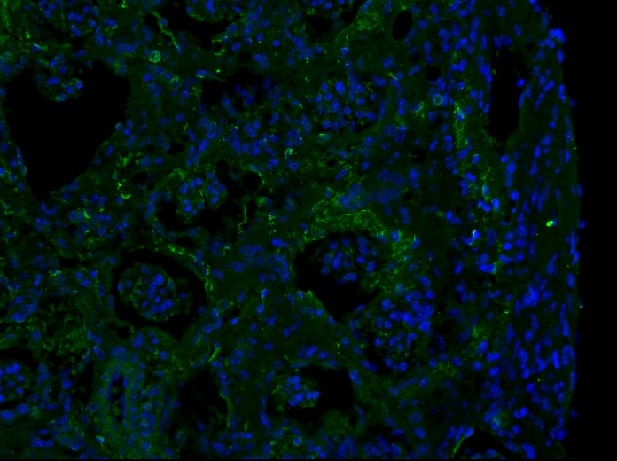
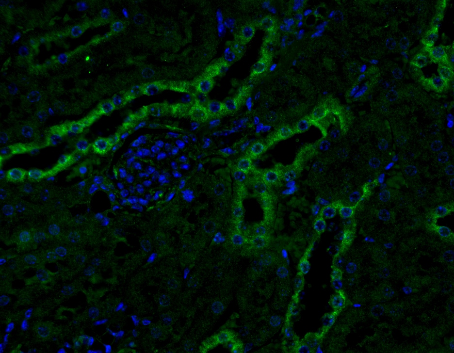
Immunohistochemistry-Frozen: Fibronectin Antibody - BSA Free [NBP1-91258]
Immunohistochemistry-Frozen: Fibronectin Antibody - BSA Free [NBP1-91258] - Analysis in a 6 month old Alport mouse kidney. Image from a verified customer review.![Western Blot: Fibronectin AntibodyBSA Free [NBP1-91258]](https://resources.rndsystems.com/images/products/Fibronectin-Antibody---BSA-Free-Western-Blot-NBP1-91258-img0003.jpg "Western Blot: Fibronectin AntibodyBSA Free [NBP1-91258]")
Western Blot: Fibronectin AntibodyBSA Free [NBP1-91258]
Western Blot: Fibronectin Antibody - BSA Free [NBP1-91258] - Analysis of Fibronectin in HepG2 cell lysate.![Western Blot: Fibronectin AntibodyBSA Free [NBP1-91258]](https://resources.rndsystems.com/images/products/Fibronectin-Antibody---BSA-Free-Western-Blot-NBP1-91258-img0004.jpg "Western Blot: Fibronectin AntibodyBSA Free [NBP1-91258]")
Western Blot: Fibronectin AntibodyBSA Free [NBP1-91258]
Western Blot: Fibronectin Antibody - BSA Free [NBP1-91258] - Analysis of Fibronectin in NIH 3T3 cell lysate.![Immunocytochemistry/ Immunofluorescence: Fibronectin Antibody - BSA Free [NBP1-91258]](https://resources.rndsystems.com/images/products/Fibronectin-Antibody---BSA-Free-Immunocytochemistry-Immunofluorescence-NBP1-91258-img0008.jpg "Immunocytochemistry/ Immunofluorescence: Fibronectin Antibody - BSA Free [NBP1-91258]")
Immunocytochemistry/ Immunofluorescence: Fibronectin Antibody - BSA Free [NBP1-91258]
Immunocytochemistry/Immunofluorescence: Fibronectin Antibody - BSA Free [NBP1-91258] - Fibronectin antibody was tested in HeLa cells with DyLight 488 (Green). Nuclei and alpha-tubulin were counterstained with DAPI (Blue) and DyLight 550 (Red).![Immunocytochemistry/ Immunofluorescence: Fibronectin Antibody - BSA Free [NBP1-91258]](https://resources.rndsystems.com/images/products/Fibronectin-Antibody---BSA-Free-Immunocytochemistry-Immunofluorescence-NBP1-91258-img0012.jpg "Immunocytochemistry/ Immunofluorescence: Fibronectin Antibody - BSA Free [NBP1-91258]")
Immunocytochemistry/ Immunofluorescence: Fibronectin Antibody - BSA Free [NBP1-91258]
Immunocytochemistry/Immunofluorescence: Fibronectin Antibody - BSA Free [NBP1-91258] - NIH-3T3 cells were fixed for 10 minutes using 10% formalin and then permeabilized for 5 minutes using 1X TBS + 0.5% Triton X-100. The cells were incubated with anti-Fibronectin at 2 ug/mL overnight at 4C and detected with an anti-rabbit DyLight 488 (Green) at 1:500. Alpha tubulin (DM1A) NB100-690 was used as a co-stain at 1:1000 and detected with an anti-mouse DyLight 550 (Red) at a 1:500 dilution. Nuclei were counterstained with DAPI (Blue). Cells were imaged using a 40X objective.![Immunohistochemistry: Fibronectin Antibody - BSA Free [NBP1-91258]](https://resources.rndsystems.com/images/products/Fibronectin-Antibody---BSA-Free-Immunohistochemistry-NBP1-91258-img0007.jpg "Immunohistochemistry: Fibronectin Antibody - BSA Free [NBP1-91258]")
Immunohistochemistry: Fibronectin Antibody - BSA Free [NBP1-91258]
Immunohistochemistry: Fibronectin Antibody - BSA Free [NBP1-91258] - Analysis of Fibronectin in human renal cancer using DAB with hematoxylin counterstain.![Immunohistochemistry-Paraffin: Fibronectin Antibody - BSA Free [NBP1-91258]](https://resources.rndsystems.com/images/products/Fibronectin-Antibody---BSA-Free-Immunohistochemistry-Paraffin-NBP1-91258-img0010.jpg "Immunohistochemistry-Paraffin: Fibronectin Antibody - BSA Free [NBP1-91258]")
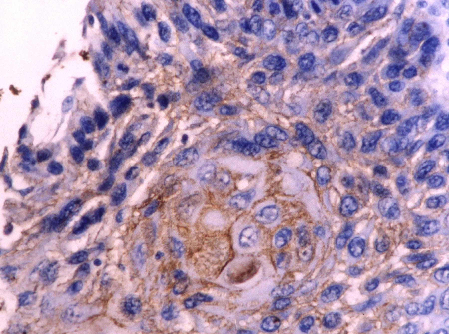
Immunohistochemistry-Paraffin: Fibronectin Antibody - BSA Free [NBP1-91258]
Immunohistochemistry-Paraffin: Fibronectin Antibody - BSA Free [NBP1-91258] - Analysis of Fibronectin in mouse kidney tissue section using anti-Fibronectin antibody. Image from verified customer review.![Immunohistochemistry-Paraffin: Fibronectin Antibody - BSA Free [NBP1-91258]](https://resources.rndsystems.com/images/products/Fibronectin-Antibody---BSA-Free-Immunohistochemistry-Paraffin-NBP1-91258-img0011.jpg "Immunohistochemistry-Paraffin: Fibronectin Antibody - BSA Free [NBP1-91258]")
Immunohistochemistry-Paraffin: Fibronectin Antibody - BSA Free [NBP1-91258]
Immunohistochemistry-Paraffin: Fibronectin Antibody - BSA Free [NBP1-91258] - Analysis of Fibronectin in human oesophageal cancer tissue using anti-Fibronectin antibody. Image from verified customer review.![Immunohistochemistry-Frozen: Fibronectin Antibody - BSA Free [NBP1-91258]](https://resources.rndsystems.com/images/products/Fibronectin-Antibody---BSA-Free-Immunohistochemistry-Frozen-NBP1-91258-img0013.jpg "Immunohistochemistry-Frozen: Fibronectin Antibody - BSA Free [NBP1-91258]")
Immunohistochemistry-Frozen: Fibronectin Antibody - BSA Free [NBP1-91258]
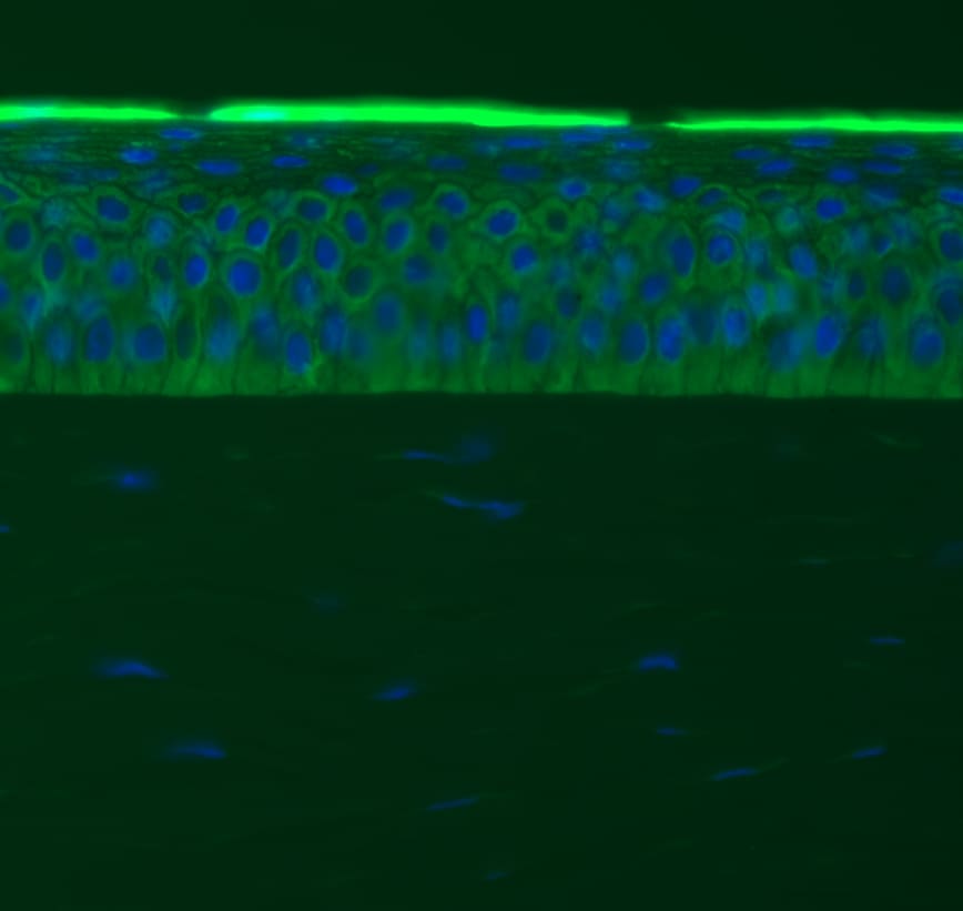
Immunohistochemistry-Frozen: Fibronectin Antibody - BSA Free [NBP1-91258] - Feline corneal stromal cells. IHC-Fr and IF (Alexa Fluor488) were performed using NBP1-91258 at 1:400. Image captured by epifluorescent microscopy. Image from verified customer review.![Immunohistochemistry-Frozen: Fibronectin Antibody - BSA Free [NBP1-91258]](https://resources.rndsystems.com/images/products/Fibronectin-Antibody---BSA-Free-Immunohistochemistry-Frozen-NBP1-91258-img0016.jpg "Immunohistochemistry-Frozen: Fibronectin Antibody - BSA Free [NBP1-91258]")
Immunohistochemistry-Frozen: Fibronectin Antibody - BSA Free [NBP1-91258]
Immunohistochemistry-Frozen: Fibronectin Antibody - BSA Free [NBP1-91258] - Trabecular meshwork (TM) region of pig eyes. NBP1-91258 Fibronectin antibody was labelled with Alexa Fluor 488 conjugated secondary antibody (Green). DAPI shown as blue. Image from verified customer review.![Immunohistochemistry-Frozen: Fibronectin Antibody - BSA Free [NBP1-91258]](https://resources.rndsystems.com/images/products/Fibronectin-Antibody---BSA-Free-Immunohistochemistry-Frozen-NBP1-91258-img0017.jpg "Immunohistochemistry-Frozen: Fibronectin Antibody - BSA Free [NBP1-91258]")
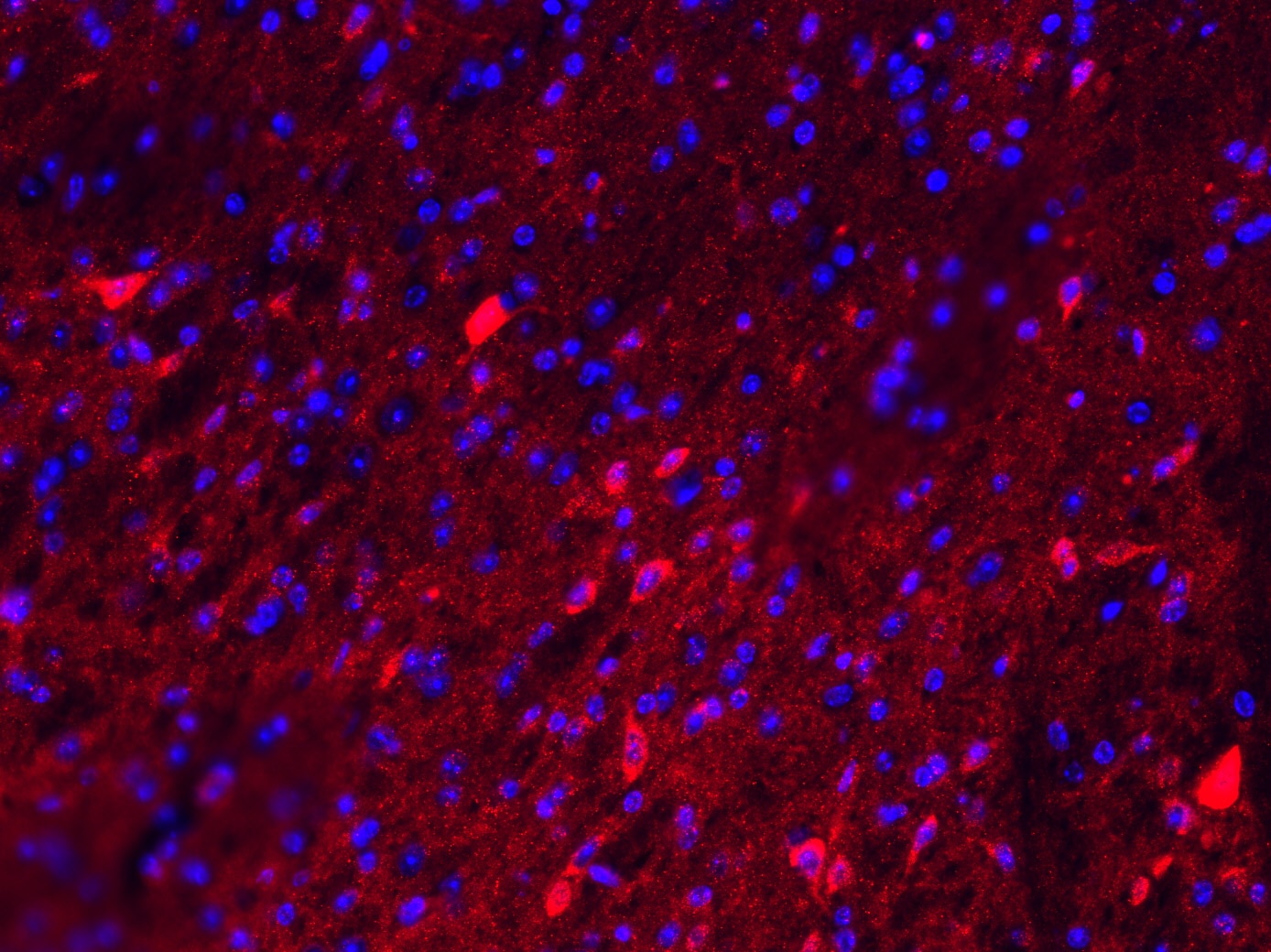
Immunohistochemistry-Frozen: Fibronectin Antibody - BSA Free [NBP1-91258]
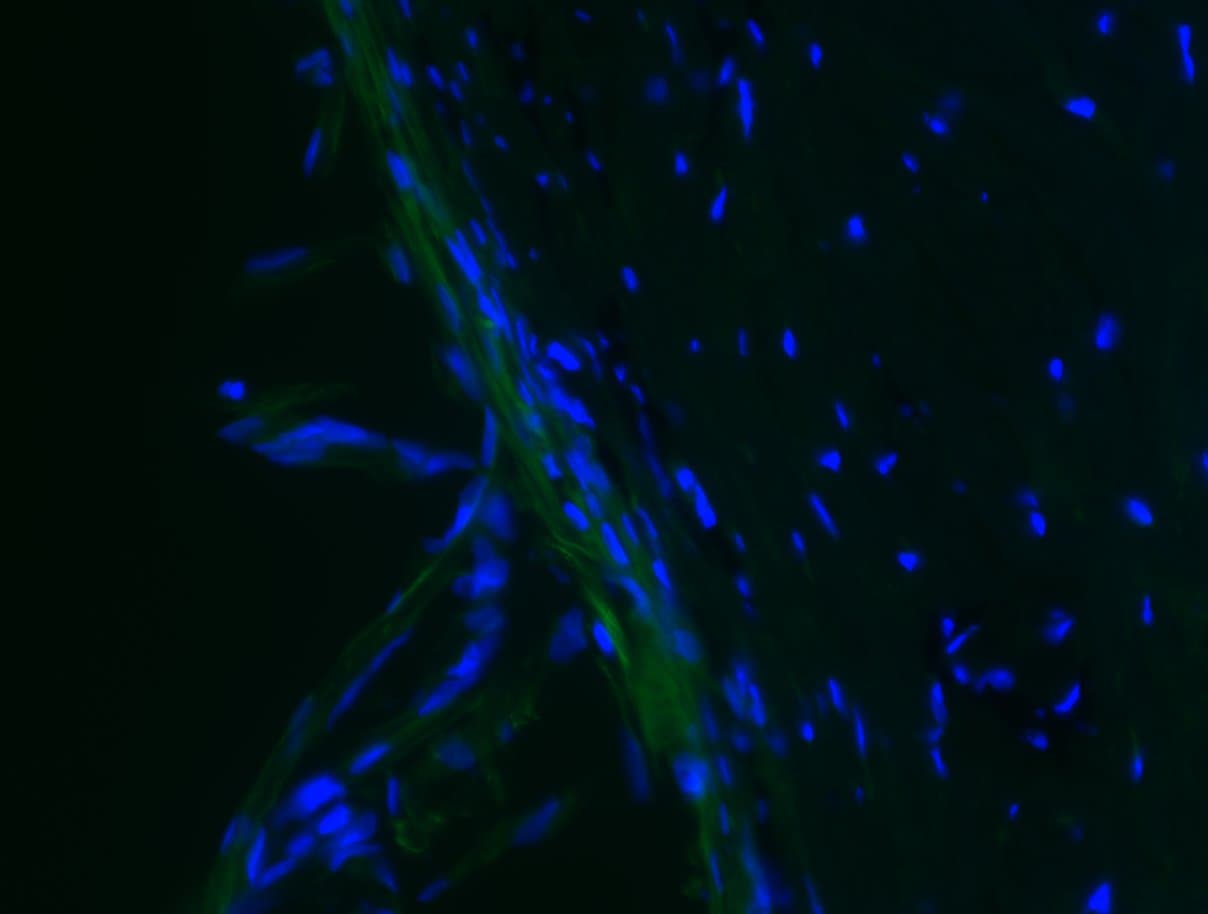
Immunohistochemistry-Frozen: Fibronectin Antibody - BSA Free [NBP1-91258] - Mouse brain cryosections were stained with Fibronectin and anti-rabbit Alexa Fluor 555. Magnification 20x. Image from vrified customer review.![Immunohistochemistry-Frozen: Fibronectin Antibody - BSA Free [NBP1-91258]](https://resources.rndsystems.com/images/products/Fibronectin-Antibody---BSA-Free-Immunohistochemistry-Frozen-NBP1-91258-img0018.jpg "Immunohistochemistry-Frozen: Fibronectin Antibody - BSA Free [NBP1-91258]")
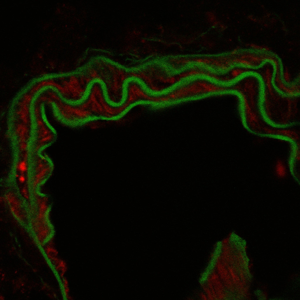
Immunohistochemistry-Frozen: Fibronectin Antibody - BSA Free [NBP1-91258]
Immunohistochemistry-Frozen: Fibronectin Antibody - BSA Free [NBP1-91258] - Staining in mouse carotid artery. Image from a verified customer review.![Simple Western: Fibronectin AntibodyBSA Free [NBP1-91258]](https://resources.rndsystems.com/images/products/Fibronectin-Antibody---BSA-Free-Simple-Western-NBP1-91258-img0009.jpg "Simple Western: Fibronectin AntibodyBSA Free [NBP1-91258]")
Simple Western: Fibronectin AntibodyBSA Free [NBP1-91258]
Simple Western: Fibronectin Antibody - BSA Free [NBP1-91258] - Lane view shows a specific band for Fibronectin in 0.5 mg/ml of HepG2 lysate. This experiment was performed under reducing conditions using the 12-230 kDa separation system.
Western Blot: Fibronectin Antibody - BSA Free [NBP1-91258] -
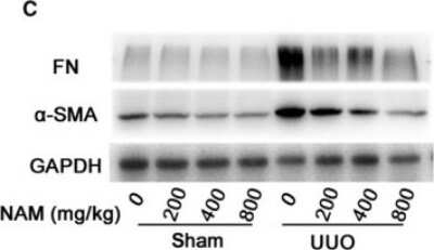
Western blot analysis of the effects of IP6, Ins, IP6 + Ins & normal saline on the levels of collagen IV, Lamininand Fibronectin. IP6 or Ins treatment decreased the protein expression of collagen IV, LN & FN, & the combined IP6 + Ins treatment resulted in significantly greater effects compared with treatment with either compound alone. The samples were probed with antibodies against p-collagen IV, p-LN, & p-FN. The Western blot membranes were stripped & reprobed for beta -actin as an internal control to confirm equal loading. (A) representative blots from one of three separate experiments; (B) relative band intensities based on densitometry. The results are expressed as the mean ± standard deviation from three independent experiments. * p < 0.05 compared to the IP6 + Ins group; #p < 0.05 compared to the normal saline group. Image collected & cropped by CiteAb from the following publication (http://www.mdpi.com/2072-6643/8/5/286), licensed under a CC-BY license. Not internally tested by Novus Biologicals.
Immunocytochemistry/ Immunofluorescence: Fibronectin Antibody - BSA Free [NBP1-91258] -
Immunocytochemistry/ Immunofluorescence: Fibronectin Antibody - BSA Free [NBP1-91258] - LC depressed both the protein & mRNA level of fibronectin via depletion of peritoneal M2. Values were expressed as the mean ± SD. (A) The overexpression of fibronectin induced by Lactate-4.25% dialysate was evidently downregulated by LC treatment measured by Western blotting; (B) The relative protein level of fibronectin normalized by GAPDH; (C) The mRNA level of fibronectin was depressed by LC treatment; (D) Immunofluorescence staining of fibronectin in the four groups. Blue corresponds to nuclear staining, & red corresponds to fibronectin staining. #p < 0.05 vs. NC & LC group. Image collected & cropped by CiteAb from the following publication (https://pubmed.ncbi.nlm.nih.gov/23685870), licensed under a CC-BY license. Not internally tested by Novus Biologicals.
Western Blot: Fibronectin Antibody - BSA Free [NBP1-91258] -
Western Blot: Fibronectin Antibody - BSA Free [NBP1-91258] - EMT in cancer cells is regulated by m6A levels of mRNAs. a HeLa & HepG2 cells were treated with or without 10 ng/ml TGF-beta for 3 days, the m6A/A ratio of the total mRNA were determined by LC–MS/MS. b Wound healing of wild-type (control) or Mettl3Mut/− cells was recorded (left) & quantitatively analyzed (right). c Wild-type or Mettl3Mut/− cells were allowed to invade for 24 h & tested by CytoSelect™ 24-well Cell Invasion assay kits (8 µm, colorimetric format); d, e mRNA (d) & protein (e) expressions of MMP2, FN, & E-Cad in wild-type & Mettl3Mut/− HeLa cells were measured by qRT-PCR & western blot analysis, respectively. f HeLa cells were transfected with pcDNA/ALKBH5 or a vector control for 48 h, protein expression was determined by western blot analysis (left) & quantitatively analyzed (right). g Wild-type or Mettl3Mut/− cells were treated with or without 10 ng/ml TGF-beta for 3 days, protein expression was determined by western blot analysis (left) & quantitatively analyzed (right). h The expression of METTL3 in liver cancer & its matched adjacent normal tissues of 50 patients from TCGA database. i Correlation between METTL3 & CDH1 in liver cancer patients (n = 364) from TCGA database. j HeLa cells were pretreated with or without Smad2/3 inhibitor SB431542 (10 μM) & then further treated with 10 ng/ml TGF-beta for 3 days, the m6A/A ratio of the total mRNA were determined by LC–MS/MS. Data are presented as means ± SD from three independent experiments. *p < 0.05, **p < 0.01, NS, no significant, by Student’s t test. Red bar = 200 μm Image collected & cropped by CiteAb from the following publication (https://pubmed.ncbi.nlm.nih.gov/31061416), licensed under a CC-BY license. Not internally tested by Novus Biologicals.
Western Blot: Fibronectin Antibody - BSA Free [NBP1-91258] -
Western Blot: Fibronectin Antibody - BSA Free [NBP1-91258] - Snail is involved in m6A-regulated EMT in cancer cells. a Overlapping of 2.0-fold m6A expression changes in EMT cells & EMT-related functional genes. b m6A peaks were enriched in CDS & 3′UTRs of SNAI1 genes from m6A RIP-seq data. Squares marked increases of m6A peaks in cancer cells undergoing EMT; c m6A RIP-qPCR analysis of SNAI1 mRNA in the control & EMT undergoing HeLa cells. d Protein expression of Snail in Mettl3Mut/− or AKLBH5 transfected (24 h) HeLa cells & the control. e The wound healing of wild-type or Mettl3Mut/− HeLa cells transfected with or without pcDNA/Snail for 48 h were recorded (left) & quantitatively analyzed (right). f Wild-type or Mettl3Mut/− HeLa cells were transfected with or without pcDNA/Snail for 48 h, expression of Snail, FN & E-Cad were measured by western blot analysis. Data are presented as means ± SD from three independent experiments. *p < 0.05, NS, no significant, by Student’s t test. Red bar = 200μm Image collected & cropped by CiteAb from the following publication (https://pubmed.ncbi.nlm.nih.gov/31061416), licensed under a CC-BY license. Not internally tested by Novus Biologicals.
Western Blot: Fibronectin Antibody - BSA Free [NBP1-91258] -
Western Blot: Fibronectin Antibody - BSA Free [NBP1-91258] - EMT in cancer cells is regulated by m6A levels of mRNAs. a HeLa & HepG2 cells were treated with or without 10 ng/ml TGF-beta for 3 days, the m6A/A ratio of the total mRNA were determined by LC–MS/MS. b Wound healing of wild-type (control) or Mettl3Mut/− cells was recorded (left) & quantitatively analyzed (right). c Wild-type or Mettl3Mut/− cells were allowed to invade for 24 h & tested by CytoSelect™ 24-well Cell Invasion assay kits (8 µm, colorimetric format); d, e mRNA (d) & protein (e) expressions of MMP2, FN, & E-Cad in wild-type & Mettl3Mut/− HeLa cells were measured by qRT-PCR & western blot analysis, respectively. f HeLa cells were transfected with pcDNA/ALKBH5 or a vector control for 48 h, protein expression was determined by western blot analysis (left) & quantitatively analyzed (right). g Wild-type or Mettl3Mut/− cells were treated with or without 10 ng/ml TGF-beta for 3 days, protein expression was determined by western blot analysis (left) & quantitatively analyzed (right). h The expression of METTL3 in liver cancer & its matched adjacent normal tissues of 50 patients from TCGA database. i Correlation between METTL3 & CDH1 in liver cancer patients (n = 364) from TCGA database. j HeLa cells were pretreated with or without Smad2/3 inhibitor SB431542 (10 μM) & then further treated with 10 ng/ml TGF-beta for 3 days, the m6A/A ratio of the total mRNA were determined by LC–MS/MS. Data are presented as means ± SD from three independent experiments. *p < 0.05, **p < 0.01, NS, no significant, by Student’s t test. Red bar = 200 μm Image collected & cropped by CiteAb from the following publication (https://pubmed.ncbi.nlm.nih.gov/31061416), licensed under a CC-BY license. Not internally tested by Novus Biologicals.
Fibronectin in HepG2 Human Cell Line.
Fibronectin was detected in immersion fixed HepG2 human hepatocellular carcinoma cell line using Rabbit anti-Fibronectin Affinity Purified Polyclonal Antibody conjugated to Alexa Fluor® 647 (Catalog # NBP1-91258AF647) (light blue) at 10 µg/mL overnight at 4C. Cells were counterstained with DAPI (dark blue). Cells were imaged using a 100X objective and digitally deconvolved.
Fibronectin in HepG2 Human Cell Line.
Fibronectin was detected in immersion fixed HepG2 human hepatocellular carcinoma cell line using Rabbit anti-Fibronectin Affinity Purified Polyclonal Antibody conjugated to FITC (Catalog # NBP1-91258F) (green) at 10 µg/mL overnight at 4C. Cells were counterstained with DAPI (dark blue). Cells were imaged using a 100X objective and digitally deconvolved.Applications for Fibronectin Antibody - BSA Free
Immunocytochemistry/ Immunofluorescence
Immunohistochemistry
Immunohistochemistry-Frozen
Immunohistochemistry-Paraffin
Simple Western
Western Blot
See Simple Western Antibody Database for Simple Western validation: Tested in HepG2 lysate 0.5 mg/mL, separated by Size, antibody dilution of 1:100. Separated by Size-Wes, Sally Sue/Peggy Sue. The 12-230kDa separation system and EZ Standard Pack 5 are recommended for detecting human Fibronectin using Simple Western.
Reviewed Applications
Read 11 reviews rated 4.2 using NBP1-91258 in the following applications:
Formulation, Preparation, and Storage
Purification
Formulation
Format
Preservative
Concentration
Shipping
Stability & Storage
Background: Fibronectin
Alternate Names
Entrez Gene IDs
Gene Symbol
UniProt
Additional Fibronectin Products
Product Documents for Fibronectin Antibody - BSA Free
Certificate of Analysis
To download a Certificate of Analysis, please enter a lot or batch number in the search box below.
Product Specific Notices for Fibronectin Antibody - BSA Free
This product is for research use only and is not approved for use in humans or in clinical diagnosis. Primary Antibodies are guaranteed for 1 year from date of receipt.
Citations for Fibronectin Antibody - BSA Free
 Powered by Bioz
Customer Reviews for Fibronectin Antibody - BSA Free (11)
Have you used Fibronectin Antibody - BSA Free?
Submit a review and receive an Amazon gift card!
$25/€18/£15/$25CAN/¥2500 Yen for a review with an image
$10/€7/£6/$10CAN/¥1110 Yen for a review without an image
Submit a review
Customer Images

-
Application: Western BlotSample Tested: Mouse PancreasSpecies: MouseVerified Customer | Posted 10/02/2020Caerulein dosed mouse pancreas

-
Application: Western BlotSample Tested: PancreasSpecies: MouseVerified Customer | Posted 10/02/2020Caerulein dosed mouse pancreas
-
Application: Western BlotSample Tested: SVG-A whole cell lysateSpecies: HumanVerified Customer | Posted 06/17/2019SVG-A immortalized astrocytes were stimulated with different cytokines (unstimulated control: right lane) for 24 h; 15 µg per lane ptorein were seperated by SDS page, blotted and detected using 1:250 anti-fibronectin.
-
Application: Immunohistochemistry-FrozenSample Tested: Mouse brainSpecies: MouseVerified Customer | Posted 05/09/2019Mouse brain cryosections were stained with Fibronectin and anti-rabbit Alexa Fluor 555. Magnification 20x.
-
Application: Immunohistochemistry-FrozenSample Tested: trabecular meshwork (TM) region of pig eyesSpecies: PigVerified Customer | Posted 12/16/2018NBP1-91258 Fibronectin was labelled with Alexa Fluor 488 conjugated secondary antibody - green. DAPI shown as blue.
-
Application: Immunohistochemistry-FrozenSample Tested: corneal stromal cellsSpecies: FelineVerified Customer | Posted 04/27/2018IF was performed using NBP1-91258(1:400) and Alexa fluor 488 and the image was captured by epifluorescent microscope.
-
Application: ImmunofluorescenceSample Tested: Mouse Kidneys with Alport DiseaseSpecies: MouseVerified Customer | Posted 07/22/20166 month Kidney from Alport Mouse
-
Application: Western BlotSample Tested: Mouse Atherosclerotic PlaqueSpecies: MouseVerified Customer | Posted 04/07/2016Dot Blot of Protein Standards
-
Application: ImmunocytochemistrySample Tested:Species: MouseVerified Customer | Posted 06/06/2015Mouse Carotid Artery
-
Application: ImmunocytochemistrySample Tested:Species: HumanVerified Customer | Posted 04/30/2015fibronectin antibody (NBP1-91258)
-
Application: Immunohistochemistry-ParaffinSample Tested: Mouse kidneySpecies: MouseVerified Customer | Posted 03/02/2015Fibronectin expression in mouse kidney
There are no reviews that match your criteria.
Protocols
View specific protocols for Fibronectin Antibody - BSA Free (NBP1-91258):
Immunocytochemistry Protocol
Culture cells to appropriate density in 35 mm culture dishes or 6-well plates.
1. Remove culture medium and add 10% formalin to the dish. Fix at room temperature for 30 minutes.
2. Remove the formalin and add ice cold methanol. Incubate for 5-10 minutes.
3. Remove methanol and add washing solution (i.e. PBS). Be sure to not let the specimen dry out. Wash three times for 10 minutes.
4. To block nonspecific antibody binding incubate in 10% normal goat serum from 1 hour to overnight at room temperature.
5. Add primary antibody at appropriate dilution and incubate at room temperature from 2 hours to overnight at room temperature.
6. Remove primary antibody and replace with washing solution. Wash three times for 10 minutes.
7. Add secondary antibody at appropriate dilution. Incubate for 1 hour at room temperature.
8. Remove antibody and replace with wash solution, then wash for 10 minutes. Add Hoechst 33258 to wash solution at 1:25,0000 and incubate for 10 minutes. Wash a third time for 10 minutes.
9. Cells can be viewed directly after washing. The plates can also be stored in PBS containing Azide covered in Parafilm (TM). Cells can also be cover-slipped using Fluoromount, with appropriate sealing.
*The above information is only intended as a guide. The researcher should determine what protocol best meets their needs. Please follow safe laboratory procedures.
Immunohistochemistry-Paraffin Embedded Sections
Antigen Unmasking:
Bring slides to a boil in 10 mM sodium citrate buffer (pH 6.0) then maintain at a sub-boiling temperature for 10 minutes. Cool slides on bench-top for 30 minutes.
Staining:
1. Wash sections in deionized water three times for 5 minutes each.
2. Wash sections in wash buffer for 5 minutes.
3. Block each section with 100-400 ul blocking solution for 1 hour at room temperature.
4. Remove blocking solution and add 100-400 ul diluted primary antibody. Incubate overnight at 4 C.
5. Remove antibody solution and wash sections in wash buffer three times for 5 minutes each.
6. Add 100-400 ul biotinylated diluted secondary antibody. Incubate 30 minutes at room temperature.
7. Remove secondary antibody solution and wash sections three times with wash buffer for 5 minutes each.
8. Add 100-400 ul Streptavidin-HRP reagent to each section and incubate for 30 minutes at room temperature.
9. Wash sections three times in wash buffer for 5 minutes each.
10. Add 100-400 ul DAB substrate to each section and monitor staining closely.
11. As soon as the sections develop, immerse slides in deionized water.
12. Counterstain sections in hematoxylin.
13. Wash sections in deionized water two times for 5 minutes each.
14. Dehydrate sections.
15. Mount coverslips.
*The above information is only intended as a guide. The researcher should determine what protocol best meets their needs. Please follow safe laboratory procedures.
Western Blot Protocol
1. Perform SDS-PAGE on samples to be analyzed, loading 40 ug of total protein per lane.
2. Transfer proteins to membrane according to the instructions provided by the manufacturer of the membrane and transfer apparatus.
3. Stain according to standard Ponceau S procedure (or similar product) to assess transfer success, and mark molecular weight standards where appropriate.
4. Rinse the blot.
5. Block the membrane using standard blocking buffer for at least 1 hour.
6. Wash the membrane in wash buffer three times for 10 minutes each.
7. Dilute primary antibody in blocking buffer and incubate 1 hour at room temperature.
8. Wash the membrane in wash buffer three times for 10 minutes each.
9. Apply the diluted HRP conjugated secondary antibody in blocking buffer (as per manufacturers instructions) and incubate 1 hour at room temperature.
10. Wash the blot in wash buffer three times for 10 minutes each (this step can be repeated as required to reduce background).
11. Apply the detection reagent of choice in accordance with the manufacturers instructions.
Note: Tween-20 can be added to the blocking or antibody dilution buffer at a final concentration of 0.05-0.2%.
Immunocytochemistry Protocol
Culture cells to appropriate density in 35 mm culture dishes or 6-well plates.
1. Remove culture medium and add 10% formalin to the dish. Fix at room temperature for 30 minutes.
2. Remove the formalin and add ice cold methanol. Incubate for 5-10 minutes.
3. Remove methanol and add washing solution (i.e. PBS). Be sure to not let the specimen dry out. Wash three times for 10 minutes.
4. To block nonspecific antibody binding incubate in 10% normal goat serum from 1 hour to overnight at room temperature.
5. Add primary antibody at appropriate dilution and incubate at room temperature from 2 hours to overnight at room temperature.
6. Remove primary antibody and replace with washing solution. Wash three times for 10 minutes.
7. Add secondary antibody at appropriate dilution. Incubate for 1 hour at room temperature.
8. Remove antibody and replace with wash solution, then wash for 10 minutes. Add Hoechst 33258 to wash solution at 1:25,0000 and incubate for 10 minutes. Wash a third time for 10 minutes.
9. Cells can be viewed directly after washing. The plates can also be stored in PBS containing Azide covered in Parafilm (TM). Cells can also be cover-slipped using Fluoromount, with appropriate sealing.
*The above information is only intended as a guide. The researcher should determine what protocol best meets their needs. Please follow safe laboratory procedures.
Find general support by application which include: protocols, troubleshooting, illustrated assays, videos and webinars.
- Antigen Retrieval Protocol (PIER)
- Antigen Retrieval for Frozen Sections Protocol
- Appropriate Fixation of IHC/ICC Samples
- Cellular Response to Hypoxia Protocols
- Chromogenic IHC Staining of Formalin-Fixed Paraffin-Embedded (FFPE) Tissue Protocol
- Chromogenic Immunohistochemistry Staining of Frozen Tissue
- ClariTSA™ Fluorophore Kits
- Detection & Visualization of Antibody Binding
- Fluorescent IHC Staining of Frozen Tissue Protocol
- Graphic Protocol for Heat-induced Epitope Retrieval
- Graphic Protocol for the Preparation and Fluorescent IHC Staining of Frozen Tissue Sections
- Graphic Protocol for the Preparation and Fluorescent IHC Staining of Paraffin-embedded Tissue Sections
- Graphic Protocol for the Preparation of Gelatin-coated Slides for Histological Tissue Sections
- ICC Cell Smear Protocol for Suspension Cells
- ICC Immunocytochemistry Protocol Videos
- ICC for Adherent Cells
- IHC Sample Preparation (Frozen sections vs Paraffin)
- Immunocytochemistry (ICC) Protocol
- Immunocytochemistry Troubleshooting
- Immunofluorescence of Organoids Embedded in Cultrex Basement Membrane Extract
- Immunofluorescent IHC Staining of Formalin-Fixed Paraffin-Embedded (FFPE) Tissue Protocol
- Immunohistochemistry (IHC) and Immunocytochemistry (ICC) Protocols
- Immunohistochemistry Frozen Troubleshooting
- Immunohistochemistry Paraffin Troubleshooting
- Preparing Samples for IHC/ICC Experiments
- Preventing Non-Specific Staining (Non-Specific Binding)
- Primary Antibody Selection & Optimization
- Protocol for Heat-Induced Epitope Retrieval (HIER)
- Protocol for Making a 4% Formaldehyde Solution in PBS
- Protocol for VisUCyte™ HRP Polymer Detection Reagent
- Protocol for the Fluorescent ICC Staining of Cell Smears - Graphic
- Protocol for the Fluorescent ICC Staining of Cultured Cells on Coverslips - Graphic
- Protocol for the Preparation & Fixation of Cells on Coverslips
- Protocol for the Preparation and Chromogenic IHC Staining of Frozen Tissue Sections
- Protocol for the Preparation and Chromogenic IHC Staining of Frozen Tissue Sections - Graphic
- Protocol for the Preparation and Chromogenic IHC Staining of Paraffin-embedded Tissue Sections
- Protocol for the Preparation and Chromogenic IHC Staining of Paraffin-embedded Tissue Sections - Graphic
- Protocol for the Preparation and Fluorescent ICC Staining of Cells on Coverslips
- Protocol for the Preparation and Fluorescent ICC Staining of Non-adherent Cells
- Protocol for the Preparation and Fluorescent ICC Staining of Stem Cells on Coverslips
- Protocol for the Preparation and Fluorescent IHC Staining of Frozen Tissue Sections
- Protocol for the Preparation and Fluorescent IHC Staining of Paraffin-embedded Tissue Sections
- Protocol for the Preparation of Gelatin-coated Slides for Histological Tissue Sections
- Protocol for the Preparation of a Cell Smear for Non-adherent Cell ICC - Graphic
- R&D Systems Quality Control Western Blot Protocol
- TUNEL and Active Caspase-3 Detection by IHC/ICC Protocol
- The Importance of IHC/ICC Controls
- Troubleshooting Guide: Immunohistochemistry
- Troubleshooting Guide: Western Blot Figures
- Western Blot Conditions
- Western Blot Protocol
- Western Blot Protocol for Cell Lysates
- Western Blot Troubleshooting
- Western Blot Troubleshooting Guide
- View all Protocols, Troubleshooting, Illustrated assays and Webinars
FAQs for Fibronectin Antibody - BSA Free
-
Q: What is the difference between Bovine Fibronectin Protein, CF (Catalog # 1030-FN) and Human Fibronectin Protein, CF (Catalog # 1918-FN)?
A: Only the source species for the protein is different. Both carrier-free proteins have been validated to be bioactive with the same assay protocols listed and recommended concentrations fall within the same range, although customers should determine their optimal concentrations for their particular cell type and application.


