FIH-1/HIF-1AN Antibody - BSA Free
Novus Biologicals | Catalog # NB100-428
![Western Blot: FIH-1/HIF-1AN Antibody [NB100-428]](https://resources.rndsystems.com/images/products/FIH-1-HIF-1AN-Antibody-Western-Blot-NB100-428-img0007.jpg "Western Blot: FIH-1/HIF-1AN Antibody [NB100-428]")
Key Product Details
Validated by
Knockout/Knockdown, Biological Validation
Species Reactivity
Validated:
Human, Mouse, Rat
Cited:
Human, Mouse, Rat
Applications
Validated:
Knockout Validated, Immunohistochemistry, Immunohistochemistry-Paraffin, Immunohistochemistry-Frozen, Western Blot, Immunocytochemistry/ Immunofluorescence, Immunoprecipitation, Electron Microscopy, Knockdown Validated
Cited:
Knockout Validated, Immunohistochemistry-Paraffin, Western Blot, Immunocytochemistry/ Immunofluorescence, Immunoprecipitation, Electron Microscopy
Label
Unconjugated
Antibody Source
Polyclonal Rabbit IgG
Format
BSA Free
Loading...
Product Specifications
Immunogen
A synthetic peptide made to the C-terminal region of human Factor Inhibiting HIF-1 (between residues 300 and the C-terminus). [UniProt# Q9NWT6]
Reactivity Notes
Human, rat and mouse (PMID 19720742).
Localization
Nuclear
Clonality
Polyclonal
Host
Rabbit
Isotype
IgG
Theoretical MW
40.3 kDa.
Disclaimer note: The observed molecular weight of the protein may vary from the listed predicted molecular weight due to post translational modifications, post translation cleavages, relative charges, and other experimental factors.
Disclaimer note: The observed molecular weight of the protein may vary from the listed predicted molecular weight due to post translational modifications, post translation cleavages, relative charges, and other experimental factors.
Scientific Data Images for FIH-1/HIF-1AN Antibody - BSA Free
Western Blot: FIH-1/HIF-1AN Antibody [NB100-428]
Western Blot: FIH-1/HIF-1AN Antibody [NB100-428] - Analysis of astrocytes lysate from Rat brain using FIH-1/HIF-1AN antibody at 1:500 dilution with ECL detection. The antibody detected a major band at ~40 kDa position representing FIH-1/HIF-1AN protein.![Western Blot: FIH-1/HIF-1AN Antibody [NB100-428]](https://resources.rndsystems.com/images/products/FIH-1-HIF-1AN-Antibody-Western-Blot-NB100-428-img0010.jpg "Western Blot: FIH-1/HIF-1AN Antibody [NB100-428]")
Western Blot: FIH-1/HIF-1AN Antibody [NB100-428]
FIH-1-HIF-1AN-Antibody-Western-Blot-NB100-428-img0010.jpg![Immunohistochemistry: FIH-1/HIF-1AN Antibody [NB100-428]](https://resources.rndsystems.com/images/products/FIH-1-HIF-1AN-Antibody-Immunohistochemistry-NB100-428-img0008.jpg "Immunohistochemistry: FIH-1/HIF-1AN Antibody [NB100-428]")
Immunohistochemistry: FIH-1/HIF-1AN Antibody [NB100-428]
Immunohistochemistry: FIH-1/HIF-1AN Antibody [NB100-428] - Detection of FIH-1 in rat brain tissue.![Western Blot: FIH-1/HIF-1AN Antibody [NB100-428]](https://resources.rndsystems.com/images/products/FIH-1-HIF-1AN-Antibody-Western-Blot-NB100-428-img0009.jpg "Western Blot: FIH-1/HIF-1AN Antibody [NB100-428]")
![Knockdown Validated: FIH-1/HIF-1AN Antibody [NB100-428]](https://resources.rndsystems.com/images/products/FIH-1-HIF-1AN%20Antibody-Knockdown%20Validated-NB100-428-img0011.jpg "Western Blot: FIH-1/HIF-1AN Antibody [NB100-428]")

Immunohistochemistry: FIH-1/HIF-1AN Antibody [NB100-428] -
Immunohistochemistry: FIH-1/HIF-1AN Antibody [NB100-428] - 3D target protein expression in multicellular tumor spheroids3D expression analysis was carried out in the MCF-7, MDA-MB-231 & HBL-100 cell lines using multicellular tumor spheroids. The spheroids were cultured in spinner flasks for 1 week before fixation. Hypoxyprobe was used to examine the formation of hypoxic areas within the spheroids. NHE1, CAIX, MMP14 & FIH-1 expression was also analyzed. Incubation for an hour with PBS instead of primary antibody acted as a control. Image collected & cropped by CiteAb from the following publication (https://www.oncotarget.com/lookup/doi/10.18632/oncotarget.17143), licensed under a CC-BY license. Not internally tested by Novus Biologicals.Applications for FIH-1/HIF-1AN Antibody - BSA Free
Application
Recommended Usage
Immunohistochemistry
1:100
Immunohistochemistry-Frozen
1:50-1:200
Immunohistochemistry-Paraffin
1:50-1:200
Immunoprecipitation
1:10-1:500
Knockout Validated
reported in scientific literature (PMID 31937588)
Western Blot
1:1000-1:5000
Reviewed Applications
Read 3 reviews rated 4.3 using NB100-428 in the following applications:
Formulation, Preparation, and Storage
Purification
Immunogen affinity purified
Formulation
Tris-Citrate/Phosphate (pH 7.0 - 8.0)
Format
BSA Free
Preservative
0.1% Sodium Azide
Concentration
1.0 mg/ml
Shipping
The product is shipped with polar packs. Upon receipt, store it immediately at the temperature recommended below.
Stability & Storage
Store at 4C. Do not freeze.
Background: FIH-1/HIF-1AN
Long Name
Factor Inhibiting HIF-1
Alternate Names
FIH1, HIF-1AN, HIF1AN, Peptide-aspartate beta-dioxygenase
Gene Symbol
HIF1AN
UniProt
Additional FIH-1/HIF-1AN Products
Product Documents for FIH-1/HIF-1AN Antibody - BSA Free
Certificate of Analysis
To download a Certificate of Analysis, please enter a lot or batch number in the search box below.
Product Specific Notices for FIH-1/HIF-1AN Antibody - BSA Free
This product is for research use only and is not approved for use in humans or in clinical diagnosis. Primary Antibodies are guaranteed for 1 year from date of receipt.
Related Research Areas
Citations for FIH-1/HIF-1AN Antibody - BSA Free
 Powered by Bioz
Customer Reviews for FIH-1/HIF-1AN Antibody - BSA Free (3)
4.3 out of 5
3 Customer Ratings
Have you used FIH-1/HIF-1AN Antibody - BSA Free?
Submit a review and receive an Amazon gift card!
$25/€18/£15/$25CAN/¥2500 Yen for a review with an image
$10/€7/£6/$10CAN/¥1110 Yen for a review without an image
Submit a review
Customer Images
Showing
1
-
3 of
3 reviews
Showing All
Filter By:
-
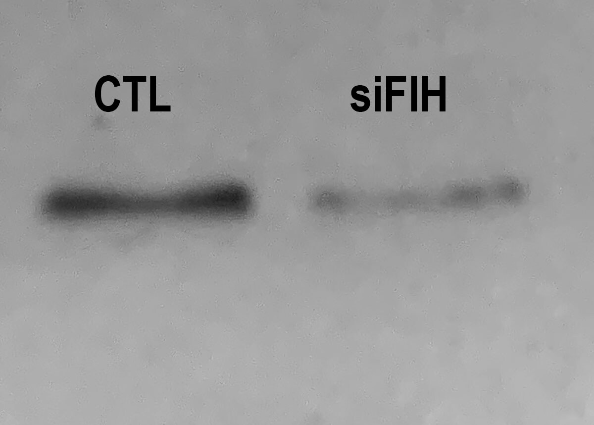
Application: Western BlotSample Tested: Vascular Smooth Muscle Cells and Whole cell lysateSpecies: RatVerified Customer | Posted 06/05/2020Rat VSMC cells treated (siFIH) or not (CTL) with a specific siRNA against FIH-1.

-
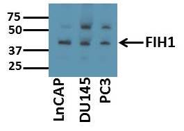
Application: Western BlotSample Tested: Cell lysates from prostate cancer cell linesSpecies: HumanVerified Customer | Posted 11/12/2017Sample: Whole cell lysates of prostate cancer cell lines (20ug) Blocking: 5% nonfat dry milk reconstituted in TBS-T for 1 hour at room temperature Primary Antibody diluted (1:500) in 5% nonfat dry milk reconstituted in TBS-T overnight at 4deg C Secondary Antibody: Goat Anti-Rabbit IgG H&L (HRP) (ab97051) at 1/10000 dilution in TBS-T
-
Application: Western BlotSample Tested: See PMID 22277691Species: HumanVerified Customer | Posted 12/12/2014
There are no reviews that match your criteria.
Protocols
View specific protocols for FIH-1/HIF-1AN Antibody - BSA Free (NB100-428):
1. Prepared 18 micron frozen rat brain tissue sections (post fixed with 10% formalin for 30 minutes).
2. Dilute the primary antibody 1:100 and incubated with diluted FIH antibody (cat# NB 100-428) at 4C, overnight.
3. Diluted the secondary antibody, biotinylated anti-rabbit IgG, according to the manufacturer's guidelines and incubated overnight at 4C.
4. Diluted the tertiary antibody, HRP-conjugated streptavidin, according to the manufacturer's guidelines and incubated overnight at 4C.
5. Visualized with DAB oxidation by HRP.
1. (30-40ug) of protein were separated via gel electrophoresis (10-20%) under denaturing conditions and transferred to nitrocellulose membrane.
2. Membrane was blocked overnight (4C) in TBS/Tween-20 (TBST) + 5% milk (blocking buffer).
3. Membrane was incubated with primary anti-FIH [cat# NB 100-428] (1:500) in blocking buffer for 2 hours at room temp.
4. Membrane was washed 3 times with TBST. 10 minutes each wash.
5. Membrane was incubated with HRP conjugated Donkey anti-rabbit secondary in blocking buffer for 2 hours at room temp.
6. Membrane was washed 3 times with TBST. 10 minutes each wash.
7. Membranes were developed for visualization using Super Signal(R) West Pico Chemiluminescent Substrate and captured on Hyperfilm ECL (Amersham Biosciences).
Find general support by application which include: protocols, troubleshooting, illustrated assays, videos and webinars.
- Antigen Retrieval Protocol (PIER)
- Antigen Retrieval for Frozen Sections Protocol
- Appropriate Fixation of IHC/ICC Samples
- Cellular Response to Hypoxia Protocols
- Chromogenic IHC Staining of Formalin-Fixed Paraffin-Embedded (FFPE) Tissue Protocol
- Chromogenic Immunohistochemistry Staining of Frozen Tissue
- ClariTSA™ Fluorophore Kits
- Detection & Visualization of Antibody Binding
- Fluorescent IHC Staining of Frozen Tissue Protocol
- Graphic Protocol for Heat-induced Epitope Retrieval
- Graphic Protocol for the Preparation and Fluorescent IHC Staining of Frozen Tissue Sections
- Graphic Protocol for the Preparation and Fluorescent IHC Staining of Paraffin-embedded Tissue Sections
- Graphic Protocol for the Preparation of Gelatin-coated Slides for Histological Tissue Sections
- ICC Cell Smear Protocol for Suspension Cells
- ICC Immunocytochemistry Protocol Videos
- ICC for Adherent Cells
- IHC Sample Preparation (Frozen sections vs Paraffin)
- Immunocytochemistry (ICC) Protocol
- Immunocytochemistry Troubleshooting
- Immunofluorescence of Organoids Embedded in Cultrex Basement Membrane Extract
- Immunofluorescent IHC Staining of Formalin-Fixed Paraffin-Embedded (FFPE) Tissue Protocol
- Immunohistochemistry (IHC) and Immunocytochemistry (ICC) Protocols
- Immunohistochemistry Frozen Troubleshooting
- Immunohistochemistry Paraffin Troubleshooting
- Immunoprecipitation Protocol
- Preparing Samples for IHC/ICC Experiments
- Preventing Non-Specific Staining (Non-Specific Binding)
- Primary Antibody Selection & Optimization
- Protocol for Heat-Induced Epitope Retrieval (HIER)
- Protocol for Making a 4% Formaldehyde Solution in PBS
- Protocol for VisUCyte™ HRP Polymer Detection Reagent
- Protocol for the Fluorescent ICC Staining of Cell Smears - Graphic
- Protocol for the Fluorescent ICC Staining of Cultured Cells on Coverslips - Graphic
- Protocol for the Preparation & Fixation of Cells on Coverslips
- Protocol for the Preparation and Chromogenic IHC Staining of Frozen Tissue Sections
- Protocol for the Preparation and Chromogenic IHC Staining of Frozen Tissue Sections - Graphic
- Protocol for the Preparation and Chromogenic IHC Staining of Paraffin-embedded Tissue Sections
- Protocol for the Preparation and Chromogenic IHC Staining of Paraffin-embedded Tissue Sections - Graphic
- Protocol for the Preparation and Fluorescent ICC Staining of Cells on Coverslips
- Protocol for the Preparation and Fluorescent ICC Staining of Non-adherent Cells
- Protocol for the Preparation and Fluorescent ICC Staining of Stem Cells on Coverslips
- Protocol for the Preparation and Fluorescent IHC Staining of Frozen Tissue Sections
- Protocol for the Preparation and Fluorescent IHC Staining of Paraffin-embedded Tissue Sections
- Protocol for the Preparation of Gelatin-coated Slides for Histological Tissue Sections
- Protocol for the Preparation of a Cell Smear for Non-adherent Cell ICC - Graphic
- R&D Systems Quality Control Western Blot Protocol
- TUNEL and Active Caspase-3 Detection by IHC/ICC Protocol
- The Importance of IHC/ICC Controls
- Troubleshooting Guide: Immunohistochemistry
- Troubleshooting Guide: Western Blot Figures
- Western Blot Conditions
- Western Blot Protocol
- Western Blot Protocol for Cell Lysates
- Western Blot Troubleshooting
- Western Blot Troubleshooting Guide
- View all Protocols, Troubleshooting, Illustrated assays and Webinars
Loading...
Associated Pathways
