GAD1/GAD67 Antibody (GAD1/2391) [PE]
Novus Biologicals | Catalog # NBP3-08276PE

Key Product Details
Species Reactivity
Human
Applications
Immunohistochemistry-Paraffin, Western Blot, ELISA, Protein Array
Label
PE (Excitation = 488 nm, Emission = 575 nm)
Antibody Source
Monoclonal Mouse IgG1 kappa Clone # GAD1/2391
Loading...
Product Specifications
Immunogen
Recombinant human GAD1/GAD67 protein fragment (around aa 72-135) (exact sequence is proprietary) (Uniprot: Q99259)
Localization
Cytoplasmic
Marker
GABAergic Neuronal Marker
Specificity
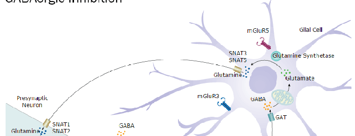
This monoclonal antibody recognizes a protein of 67kDa, which is identified as glutamic acid decarboxylase 1 (GDA1). There are two forms of glutamic acid decarboxylases (GADs) that are found in the brain: GAD65 (also known as GAD2) and GAD67 (also known as GAD1. GAD65 and GAD67 are members of the group II decarboxylase family of proteins and are responsible for catalyzing the rate-limiting step in the production of GABA (-aminobutyric acid) from L-glutamic acid. Although both GAD s are found in the brain, GAD65 localizes to synaptic vesicle membranes in nerve terminals, while GAD67 is distributed throughout the cell. GAD67 is responsible for the basal levels of GABA synthesis. In the case of a heightened demand for GABA in neurotransmission, GAD65 will transiently activate to assist in GABA production. The loss of GAD65 is detrimental and can impair GABA neurotransmission, however the loss of GAD67 is lethal.
Clonality
Monoclonal
Host
Mouse
Isotype
IgG1 kappa
Applications for GAD1/GAD67 Antibody (GAD1/2391) [PE]
Application
Recommended Usage
ELISA
Optimal dilutions of this antibody should be experimentally determined.
Immunohistochemistry-Paraffin
Optimal dilutions of this antibody should be experimentally determined.
Protein Array
Optimal dilutions of this antibody should be experimentally determined.
Western Blot
Optimal dilutions of this antibody should be experimentally determined.
Application Notes
Optimal dilution of this antibody should be experimentally determined.
Formulation, Preparation, and Storage
Purification
Protein A or G purified
Formulation
PBS
Preservative
0.05% Sodium Azide
Concentration
Please see the vial label for concentration. If unlisted please contact technical services.
Shipping
The product is shipped with polar packs. Upon receipt, store it immediately at the temperature recommended below.
Stability & Storage
Store at 4C in the dark.
Background: GAD1/GAD67
Long Name
Glutamate Decarboxylase 1 (Brain, 67kDa)
Alternate Names
GAD67
Gene Symbol
GAD1
Additional GAD1/GAD67 Products
Product Documents for GAD1/GAD67 Antibody (GAD1/2391) [PE]
Certificate of Analysis
To download a Certificate of Analysis, please enter a lot or batch number in the search box below.
Product Specific Notices for GAD1/GAD67 Antibody (GAD1/2391) [PE]
This product is for research use only and is not approved for use in humans or in clinical diagnosis. Primary Antibodies are guaranteed for 1 year from date of receipt.
Customer Reviews for GAD1/GAD67 Antibody (GAD1/2391) [PE]
There are currently no reviews for this product. Be the first to review GAD1/GAD67 Antibody (GAD1/2391) [PE] and earn rewards!
Have you used GAD1/GAD67 Antibody (GAD1/2391) [PE]?
Submit a review and receive an Amazon gift card!
$25/€18/£15/$25CAN/¥2500 Yen for a review with an image
$10/€7/£6/$10CAN/¥1110 Yen for a review without an image
Submit a review
Protocols
Find general support by application which include: protocols, troubleshooting, illustrated assays, videos and webinars.
- Antigen Retrieval Protocol (PIER)
- Antigen Retrieval for Frozen Sections Protocol
- Appropriate Fixation of IHC/ICC Samples
- Cellular Response to Hypoxia Protocols
- Chromogenic IHC Staining of Formalin-Fixed Paraffin-Embedded (FFPE) Tissue Protocol
- Chromogenic Immunohistochemistry Staining of Frozen Tissue
- ClariTSA™ Fluorophore Kits
- Detection & Visualization of Antibody Binding
- ELISA Sample Preparation & Collection Guide
- ELISA Troubleshooting Guide
- Fluorescent IHC Staining of Frozen Tissue Protocol
- Graphic Protocol for Heat-induced Epitope Retrieval
- Graphic Protocol for the Preparation and Fluorescent IHC Staining of Frozen Tissue Sections
- Graphic Protocol for the Preparation and Fluorescent IHC Staining of Paraffin-embedded Tissue Sections
- Graphic Protocol for the Preparation of Gelatin-coated Slides for Histological Tissue Sections
- How to Run an R&D Systems DuoSet ELISA
- How to Run an R&D Systems Quantikine ELISA
- How to Run an R&D Systems Quantikine™ QuicKit™ ELISA
- IHC Sample Preparation (Frozen sections vs Paraffin)
- Immunofluorescent IHC Staining of Formalin-Fixed Paraffin-Embedded (FFPE) Tissue Protocol
- Immunohistochemistry (IHC) and Immunocytochemistry (ICC) Protocols
- Immunohistochemistry Frozen Troubleshooting
- Immunohistochemistry Paraffin Troubleshooting
- Preparing Samples for IHC/ICC Experiments
- Preventing Non-Specific Staining (Non-Specific Binding)
- Primary Antibody Selection & Optimization
- Protocol for Heat-Induced Epitope Retrieval (HIER)
- Protocol for Making a 4% Formaldehyde Solution in PBS
- Protocol for VisUCyte™ HRP Polymer Detection Reagent
- Protocol for the Preparation & Fixation of Cells on Coverslips
- Protocol for the Preparation and Chromogenic IHC Staining of Frozen Tissue Sections
- Protocol for the Preparation and Chromogenic IHC Staining of Frozen Tissue Sections - Graphic
- Protocol for the Preparation and Chromogenic IHC Staining of Paraffin-embedded Tissue Sections
- Protocol for the Preparation and Chromogenic IHC Staining of Paraffin-embedded Tissue Sections - Graphic
- Protocol for the Preparation and Fluorescent IHC Staining of Frozen Tissue Sections
- Protocol for the Preparation and Fluorescent IHC Staining of Paraffin-embedded Tissue Sections
- Protocol for the Preparation of Gelatin-coated Slides for Histological Tissue Sections
- Quantikine HS ELISA Kit Assay Principle, Alkaline Phosphatase
- Quantikine HS ELISA Kit Principle, Streptavidin-HRP Polymer
- R&D Systems Quality Control Western Blot Protocol
- Sandwich ELISA (Colorimetric) – Biotin/Streptavidin Detection Protocol
- Sandwich ELISA (Colorimetric) – Direct Detection Protocol
- TUNEL and Active Caspase-3 Detection by IHC/ICC Protocol
- The Importance of IHC/ICC Controls
- Troubleshooting Guide: ELISA
- Troubleshooting Guide: Immunohistochemistry
- Troubleshooting Guide: Western Blot Figures
- Western Blot Conditions
- Western Blot Protocol
- Western Blot Protocol for Cell Lysates
- Western Blot Troubleshooting
- Western Blot Troubleshooting Guide
- View all Protocols, Troubleshooting, Illustrated assays and Webinars
Loading...
Associated Pathways