GADD153/CHOP Antibody (9C8) - BSA Free
Novus Biologicals | Catalog # NB600-1335
Key Product Details
Validated by
Biological Validation
Species Reactivity
Validated:
Human, Mouse, Rat, Primate
Cited:
Human, Mouse, Rat, Primate
Applications
Validated:
Immunohistochemistry, Immunohistochemistry-Paraffin, Western Blot, ELISA, Flow Cytometry, Immunocytochemistry/ Immunofluorescence, Simple Western, Immunoprecipitation, Chromatin Immunoprecipitation (ChIP), Gel Super Shift Assays, Knockdown Validated
Cited:
Immunohistochemistry-Paraffin, Western Blot, ELISA, Immunocytochemistry/ Immunofluorescence, Immunoprecipitation, Chemotaxis, Gel Supershift Assay
Label
Unconjugated
Antibody Source
Monoclonal Mouse IgG2b Kappa Clone # 9C8
Format
BSA Free
Loading...
Product Specifications
Immunogen
Full length mouse CHOP/GADD153 [Swiss-Prot# P35639]
Epitope
The N-terminal region of CHOP/GADD153.
Reactivity Notes
Mouse reactivity reported in scientific literature (PMID:32828953). Human, mouse, rat and primate.
Localization
Nuclear
Marker
ER Stress Marker
Clonality
Monoclonal
Host
Mouse
Isotype
IgG2b Kappa
Theoretical MW
19 kDa.
Disclaimer note: The observed molecular weight of the protein may vary from the listed predicted molecular weight due to post translational modifications, post translation cleavages, relative charges, and other experimental factors.
Disclaimer note: The observed molecular weight of the protein may vary from the listed predicted molecular weight due to post translational modifications, post translation cleavages, relative charges, and other experimental factors.
Scientific Data Images for GADD153/CHOP Antibody (9C8) - BSA Free
![Western Blot: GADD153/CHOP Antibody (9C8) [NB600-1335]](https://resources.rndsystems.com/images/products/GADD153-CHOP-Antibody-9C8-Western-Blot-NB600-1335-img0011.jpg "Western Blot: GADD153/CHOP Antibody (9C8) [NB600-1335]")
Western Blot: GADD153/CHOP Antibody (9C8) [NB600-1335]
Western Blot: GADD153/CHOP Antibody (9C8) [NB600-1335] - Ethanol feeding increases CHOP expression. Image from verified customer review.![Immunohistochemistry-Paraffin: GADD153/CHOP Antibody (9C8) [NB600-1335]](https://resources.rndsystems.com/images/products/GADD153-CHOP-Antibody-9C8-Immunohistochemistry-Paraffin-NB600-1335-img0010.jpg "Immunohistochemistry-Paraffin: GADD153/CHOP Antibody (9C8) [NB600-1335]")
Immunohistochemistry-Paraffin: GADD153/CHOP Antibody (9C8) [NB600-1335]
Immunohistochemistry-Paraffin: GADD153/CHOP Antibody (9C8) [NB600-1335] - FFPE tissue section of mouse brain using 1:100 dilution of GADD153/CHOP antibody. The signal was developed using HRP-DAB based detection method which followed counterstaining of the nuclei with hematoxylin. The antibody generated a cytoplasmic and nuclear staining of CHOP in various cell types in the tested section.![Flow Cytometry: GADD153/CHOP Antibody (9C8) [NB600-1335]](https://resources.rndsystems.com/images/products/GADD153-CHOP-Antibody-9C8-Flow-Cytometry-NB600-1335-img0012.jpg "Flow Cytometry: GADD153/CHOP Antibody (9C8) [NB600-1335]")
Flow Cytometry: GADD153/CHOP Antibody (9C8) [NB600-1335]
Flow Cytometry: GADD153/CHOP Antibody (9C8) [NB600-1335] - An intracellular stain was performed on SK-MEL-28 cells with GADD153/CHOP Antibody (9C8) NB600-1335 (blue) and a matched isotype control MAB004 (orange). Cells were fixed with 4% PFA and then permeabilized with 0.1% saponin. Cells were incubated in an antibody dilution of 2.5 ug/mL for 30 minutes at room temperature, followed by Mouse IgG (H+L) Cross-Adsorbed Secondary Antibody, Dylight 550 (84540, Thermo Fisher).![Western Blot: GADD153/CHOP Antibody (9C8) [NB600-1335]](https://resources.rndsystems.com/images/products/GADD153-CHOP-Antibody-9C8-Western-Blot-NB600-1335-img0001.jpg "Western Blot: GADD153/CHOP Antibody (9C8) [NB600-1335]")
![Western Blot: GADD153/CHOP Antibody (9C8) [NB600-1335]](https://resources.rndsystems.com/images/products/GADD153-CHOP-Antibody-9C8-Western-Blot-NB600-1335-img0004.jpg "Western Blot: GADD153/CHOP Antibody (9C8) [NB600-1335]")
![Western Blot: GADD153/CHOP Antibody (9C8) [NB600-1335]](https://resources.rndsystems.com/images/products/GADD153-CHOP-Antibody-9C8-Western-Blot-NB600-1335-img0005.jpg "Western Blot: GADD153/CHOP Antibody (9C8) [NB600-1335]")
Western Blot: GADD153/CHOP Antibody (9C8) [NB600-1335]
Western Blot: GADD153/CHOP Antibody (9C8) [NB600-1335] - Analysis of CHOP in rat heart tissue lysate. Image courtesy of product review submitted by Lee Hsiao-Wei.![Immunohistochemistry-Paraffin: GADD153/CHOP Antibody (9C8) [NB600-1335]](https://resources.rndsystems.com/images/products/GADD153-CHOP-Antibody-9C8-Immunohistochemistry-Paraffin-NB600-1335-img0008.jpg "Immunohistochemistry-Paraffin: GADD153/CHOP Antibody (9C8) [NB600-1335]")
Immunohistochemistry-Paraffin: GADD153/CHOP Antibody (9C8) [NB600-1335]
Immunohistochemistry-Paraffin: GADD153/CHOP Antibody (9C8) [NB600-1335] - FFPE tissue section of mouse brain using 1:100 dilution of GADD153/CHOP antibody. The signal was developed using HRP-DAB based detection method which followed counterstaining of the nuclei with hematoxylin. The antibody generated a cytoplasmic and nuclear staining of CHOP in various cell types in the tested section.![Simple Western: GADD153/CHOP Antibody (9C8) [NB600-1335]](https://resources.rndsystems.com/images/products/GADD153-CHOP-Antibody-9C8-Simple-Western-NB600-1335-img0007.jpg "Simple Western: GADD153/CHOP Antibody (9C8) [NB600-1335]")
Simple Western: GADD153/CHOP Antibody (9C8) [NB600-1335]
Simple Western: GADD153/CHOP Antibody (9C8) [NB600-1335] - Image shows a specific band for CHOP/GADD153 in 1.0 mg/mL of HeLa lysate. This experiment was performed under reducing conditions using the 12-230 kDa separation system. - BSA Free [NB600-1335] -")
Western Blot: GADD153/CHOP Antibody (9C8) - BSA Free [NB600-1335] -
The TUG1‐miR‐144‐3p‐Nrf2 axis regulates H/R‐induced cell apoptosis by adjusting oxidative stress and endoplasmic reticulum stress in vitro. A, B, TUNEL stain and quantitative analysis for H/R‐injured TCMK cells with different transfections (n = 3). C–J, The expression levels of Nrf2, H0‐1, CHOP, GRP78, Bax, Bcl‐2 and cleaved‐caspase3 in H/R‐injured TCMK cells with different transfections were measured by Western blot analysis. Representative protein bands are shown in F, and the quantitative analysis of protein expression is shown in C‐E, G‐J (n = 3). K, L, The levels of SOD and MDA in different transfection groups (n = 3). All data are expressed as the mean +/- SD; data comparisons between multiple groups were performed using one‐way analysis of variance (ANOVA) with Tukey's post hoc test. *p < 0.05, **p < 0.01 Image collected and cropped by CiteAb from the following open publication (https://pubmed.ncbi.nlm.nih.gov/34547172), licensed under a CC-BY license. Not internally tested by Novus Biologicals. - BSA Free [NB600-1335] -")
Western Blot: GADD153/CHOP Antibody (9C8) - BSA Free [NB600-1335] -
MiR‐144‐3p regulates H/R‐induced Nrf2‐HO‐1 signaling pathway activation, oxidative stress, mitochondria and endoplasmic reticulum functions in vitro. A–C, The expression levels of Nrf2 and HO‐1 in TCMK cells transfected with miR‐144‐3p mimics or inhibitor after H/R treatment. Representative bands are shown in A, and the quantitative analysis of protein expression level is shown in B, C (n = 3). D, E, The SOD and MDA levels in cellular supernatant of TCMK cells transfected with miR‐144‐3p mimics or inhibitor after H/R treatment (n = 3). F–I, The expression levels of CHOP, GRP78 and Cytochrome C in TCMK cells transfected with miR‐144‐3p mimics or inhibitor after H/R treatment. Representative bands are shown in F, and the quantitative analysis of protein expression level is shown in G–I (n = 3). All data are expressed as the mean +/- SD; data comparisons between multiple groups were performed using one‐way analysis of variance (ANOVA) with Tukey's post hoc test. *p < 0.05, **p < 0.01 Image collected and cropped by CiteAb from the following open publication (https://pubmed.ncbi.nlm.nih.gov/34547172), licensed under a CC-BY license. Not internally tested by Novus Biologicals. - BSA Free [NB600-1335] -")
Western Blot: GADD153/CHOP Antibody (9C8) - BSA Free [NB600-1335] -
UPR Western blotting in vitro (A). (B) ASC-CO and ASC-CM reduced the expression of BIP under exposure to CoCl2, incubations with tunicamycin, and CoCl2 were used as controls (C) ASC-CO and ASC-CM reduced the protein expression of ATF6 under CoCl2, incubations with tunicamycin, and CoCl2 were used as controls (D) ASC-CO 57 and ASC-CM reduced the protein expression of XBP1, incubations with tunicamycin, and CoCl2. (E) The protein expression of CHOP and tunicamycin remained unchanged with ASC-CO and ASC-CM as compared to controls. (n = 3, ANOVA ns, not significant; * p ≤ 0.05, ** p ≤ 0.01, *** p ≤ 0.001). Image collected and cropped by CiteAb from the following open publication (https://www.mdpi.com/1422-0067/24/24/17197), licensed under a CC-BY license. Not internally tested by Novus Biologicals. - BSA Free [NB600-1335] -")
Western Blot: GADD153/CHOP Antibody (9C8) - BSA Free [NB600-1335] -
Knocking down Nrf2 reversed the miR‐144‐3p inhibitor‐alleviated H/R‐induced apoptosis, oxidative stress, mitochondrial damage and endoplasmic reticulum stress in vitro. A–c, F–K, The protein expression level of Nrf2, HO‐1, cleaved‐caspase3, Bax, Bcl‐2, CHOP, GRP78 and Cytochrome C in H/R‐injured TCMK cells transfected with miR‐144‐3p inhibitor or si‐Nrf2. Representative bands are shown in A, and the quantitative analysis of protein expression is shown in B, C, F–K (n = 3). D, E, The SOD and MDA levels in cellular supernatants in H/R‐injured TCMK cells transfected with miR‐144‐3p inhibitor or si‐Nrf2 (n = 3). All data are expressed as the mean +/- SD; data comparisons between multiple groups were performed using one‐way analysis of variance (ANOVA) with Tukey's post hoc test. *p < 0.05, **p < 0.01, NS, no significant difference Image collected and cropped by CiteAb from the following open publication (https://pubmed.ncbi.nlm.nih.gov/34547172), licensed under a CC-BY license. Not internally tested by Novus Biologicals. - BSA Free [NB600-1335] -")
Western Blot: GADD153/CHOP Antibody (9C8) - BSA Free [NB600-1335] -
TUG1 plays an important role in H/R‐induced cell apoptosis possibly through regulating the Nrf2‐HO‐1 pathway, oxidative stress, mitochondrial damage and endoplasmic reticulum stress via targeting miR‐144‐3p. A, The expression of miR‐144‐3p in TUG1‐overexpressing or TUG1‐knockdown TCMK cells detected by RT‐qPCR (n = 3). B, C, The levels of SOD and MDA in cellular supernatant (n = 3). D–J, The relative expression levels of Nrf2, HO‐1, GRP78, CHOP, Cyt C and cleaved‐caspase3 were examined. Representative protein bands are shown in D, and the quantitative analysis of protein expression is shown in E‐J (n = 3). All data are expressed as the mean +/- SD; data comparisons between multiple groups were performed using one‐way analysis of variance (ANOVA) with Tukey's post hoc test. *p < 0.05, **p < 0.01 Image collected and cropped by CiteAb from the following open publication (https://pubmed.ncbi.nlm.nih.gov/34547172), licensed under a CC-BY license. Not internally tested by Novus Biologicals. - BSA Free [NB600-1335] -")
Western Blot: GADD153/CHOP Antibody (9C8) - BSA Free [NB600-1335] -
ASCs lead to changes in UPR expression in vivo (A) The comparison of the left and right legs—with DLFA- of the two groups of mice are shown (B) Ratio between BIP expression between left and right leg with and without ASCs (C) Ratio of ATF6 expression between left and right leg with and without ASCs (D) Ratio of CHOP expression between left and right leg with and without ASCs (E) Ratio of XBP1 expression between left and right leg with and without ASCs (n = 3, ANOVA, * p ≤ 0.05, ** p ≤ 0.01). Image collected and cropped by CiteAb from the following open publication (https://www.mdpi.com/1422-0067/24/24/17197), licensed under a CC-BY license. Not internally tested by Novus Biologicals. - BSA Free [NB600-1335] -")
Western Blot: GADD153/CHOP Antibody (9C8) - BSA Free [NB600-1335] -
Response to ER Stress Mediates the Reprogramming Facilitation by Surf4. (A) The RNA level of ER stress‐related genes on day 3 of reprogramming with or without exogenous Surf4. Relative expression of these genes relative to beta ‐actin (n = 3, average +/-SEM). (B) The protein level of ER stress‐related genes on day 3 of reprogramming with or without exogenous Surf4. (C) Kinetics of Oct4‐GFP+colony formation with or without exogenous Surf4 and sXbp1‐ delta DBD during reprogramming. (D) The number of Oct4‐GFP+ colonies and the percentage of Oct4‐GFP+ cells induced by OSKM plus Surf4 and sXbp1‐ delta DBD. (E) Morphology of the primary colonies induced by OSKM plus Surf4 and sXbp1‐ delta DBD. Scale bars, 1000 μm. Magnification: ×40. (F) AP staining of the primary iPS colonies. See also Figure S4 and Table S1 Image collected and cropped by CiteAb from the following open publication (https://pubmed.ncbi.nlm.nih.gov/34585448), licensed under a CC-BY license. Not internally tested by Novus Biologicals. - BSA Free [NB600-1335] -")
Western Blot: GADD153/CHOP Antibody (9C8) - BSA Free [NB600-1335] -
Response to ER Stress Mediates the Reprogramming Facilitation by Surf4. (A) The RNA level of ER stress‐related genes on day 3 of reprogramming with or without exogenous Surf4. Relative expression of these genes relative to beta ‐actin (n = 3, average +/-SEM). (B) The protein level of ER stress‐related genes on day 3 of reprogramming with or without exogenous Surf4. (C) Kinetics of Oct4‐GFP+colony formation with or without exogenous Surf4 and sXbp1‐ delta DBD during reprogramming. (D) The number of Oct4‐GFP+ colonies and the percentage of Oct4‐GFP+ cells induced by OSKM plus Surf4 and sXbp1‐ delta DBD. (E) Morphology of the primary colonies induced by OSKM plus Surf4 and sXbp1‐ delta DBD. Scale bars, 1000 μm. Magnification: ×40. (F) AP staining of the primary iPS colonies. See also Figure S4 and Table S1 Image collected and cropped by CiteAb from the following open publication (https://pubmed.ncbi.nlm.nih.gov/34585448), licensed under a CC-BY license. Not internally tested by Novus Biologicals.Applications for GADD153/CHOP Antibody (9C8) - BSA Free
Application
Recommended Usage
Immunocytochemistry/ Immunofluorescence
1:100
Immunohistochemistry
1:100
Immunohistochemistry-Paraffin
1:100
Immunoprecipitation
1:10 - 1:500
Simple Western
1:250
Application Notes
Use in Knockdown Validated reported in scientific literature (PMID:32828953)In Western blot a band can be seen at approx. 29 Knockdown Validateda. Gel Super Shift Assays was reported in scientific literature.
In Simple Western only 10 - 15 uL of the recommended dilution is used per data point.
See Simple Western Antibody Database for Simple Western validation: Tested in HeLa lysate 1.0 mg/mL, separated by Size, antibody dilution of 1:250, apparent MW was 34 kDa. Separated by Size-Wes, Sally Sue/Peggy Sue.
The observed molecular weight of the protein may vary from the listed predicted molecular weight due to post translational modifications, post translation cleavages, relative charges, and other experimental factors. Use in chromatin immunoprecipitation reported in scientific literature (PMID: 30962207). Use in ELISA reported in scientific literature (PMID: 29915575). Use in FLOW reported in scientific literature (PMID: 8650547).
Reviewed Applications
Read 3 reviews rated 4.3 using NB600-1335 in the following applications:
Flow Cytometry Panel Builder
Bio-Techne Knows Flow Cytometry
Save time and reduce costly mistakes by quickly finding compatible reagents using the Panel Builder Tool.
Advanced Features
- Spectra Viewer - Custom analysis of spectra from multiple fluorochromes
- Spillover Popups - Visualize the spectra of individual fluorochromes
- Antigen Density Selector - Match fluorochrome brightness with antigen density

Formulation, Preparation, and Storage
Purification
Protein A purified
Formulation
PBS
Format
BSA Free
Preservative
0.05% Sodium Azide
Concentration
1 mg/ml
Shipping
The product is shipped with polar packs. Upon receipt, store it immediately at the temperature recommended below.
Stability & Storage
Store at 4C short term. Aliquot and store at -20C long term. Avoid freeze-thaw cycles.
Background: GADD153
Long Name
Growth Arrest and DNA Damage-inducible Protein GADD153
Alternate Names
CEBP zeta, CHOP, CHOP10, DDIT3
Gene Symbol
DDIT3
UniProt
Additional GADD153 Products
Product Documents for GADD153/CHOP Antibody (9C8) - BSA Free
Certificate of Analysis
To download a Certificate of Analysis, please enter a lot or batch number in the search box below.
Product Specific Notices for GADD153/CHOP Antibody (9C8) - BSA Free
This product is for research use only and is not approved for use in humans or in clinical diagnosis. Primary Antibodies are guaranteed for 1 year from date of receipt.
Related Research Areas
Citations for GADD153/CHOP Antibody (9C8) - BSA Free
 Powered by Bioz
Customer Reviews for GADD153/CHOP Antibody (9C8) - BSA Free (3)
4.3 out of 5
3 Customer Ratings
Have you used GADD153/CHOP Antibody (9C8) - BSA Free?
Submit a review and receive an Amazon gift card!
$25/€18/£15/$25CAN/¥2500 Yen for a review with an image
$10/€7/£6/$10CAN/¥1110 Yen for a review without an image
Submit a review
Customer Images
_NB600-1335_11611.tif)
Showing
1
-
3 of
3 reviews
Showing All
Filter By:
-
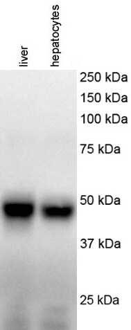
Application: Western BlotSample Tested: human liver and primary hepatocytesSpecies: HumanVerified Customer | Posted 12/15/2017Review for anti-CHOP antibody (NB600-1335)Name: Anti-CHOP antibody (NB600-1335) Catalog #: Anti-CHOP antibody (NB600-1335) Lot Number: Anti-CHOP antibody (NB600-1335, Lot # A-5) PO/Order Number: Click here to enter text.. WB Image Description (Please provide labels for all lanes): lane 1: human liver; lane 2: human primary hepatocytes Sample Information: Cell Line or Tissue: human liver and primary hepatocytes Species: human Treatment: No treatment Lysate Preparation: Date of lysate preparation: December 9, 2017 Lysis buffer used: 1X lysis buffer from Cell Signaling by adding PMSF Reducing agent: beta-mercaptoethanol, DTT If boiled (temperature/time): Yes Controls: Positive Control: No Negative Control: No Loading Control (please attach additional images if applicable): No Protein Amount Loaded per lane: 20 ug Antibody Storage Conditions: -20℃ Electrophoresis: Gel Percentage: 10% Electrophoresis Conditions: Tris-Glycine-SDS at room temperature Voltage: 120V Time: 2 hours Membrane Transfer: Method (Submersion/Semi-dry): wet transfer Membrane Type (PVDF/Nitrocellulose): Nitrocellulose Time: 2 hours Voltage: 100V Blocking: Blocking Solution: 5% milk in 1X TBST Time: 1 hour at room temperature Primary Antibody: Dilution: 1/1000 Diluent Buffer: 2.5% BSA Incubation Time: overnight Incubation Temperature: 4℃ Washing Conditions: Wash Solution: 1X TBST Time and Repetitions: 5 min each for 3 times Secondary Antibody Manufacturer and Catalog #: Promega, W402B, Lot # 0000191071 Secondary description: goat anti-mouse secondary antibody Dilution: 1/2000 Diluent Buffer: 3% milk Incubation Time: 1 hour Incubation Temperature: room temperature Detection Method: Detection: ECL (GE, cat # RPN2209, lot # 9838243) Procedure: Add equal volume of A and B, mix and apply on the membranes for 3-5 min before exposure Development Time: 40 seconds Molecular weight of band(s): ~ 48 kDa Experimental Concerns and Observations: A specific band around 48 kDa was observed

-
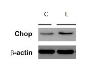
Application: Western BlotSample Tested: LiverSpecies: RatVerified Customer | Posted 09/20/2017Ethanol feeding increases CHOP expression.
-
Application: Western BlotSample Tested: Rat brainSpecies: RatVerified Customer | Posted 11/02/2014
There are no reviews that match your criteria.
Protocols
Find general support by application which include: protocols, troubleshooting, illustrated assays, videos and webinars.
- 7-Amino Actinomycin D (7-AAD) Cell Viability Flow Cytometry Protocol
- Antigen Retrieval Protocol (PIER)
- Antigen Retrieval for Frozen Sections Protocol
- Appropriate Fixation of IHC/ICC Samples
- Cellular Response to Hypoxia Protocols
- ChIP Protocol Video
- Chromatin Immunoprecipitation (ChIP) Protocol
- Chromatin Immunoprecipitation Protocol
- Chromogenic IHC Staining of Formalin-Fixed Paraffin-Embedded (FFPE) Tissue Protocol
- Chromogenic Immunohistochemistry Staining of Frozen Tissue
- ClariTSA™ Fluorophore Kits
- Detection & Visualization of Antibody Binding
- ELISA Sample Preparation & Collection Guide
- ELISA Troubleshooting Guide
- Extracellular Membrane Flow Cytometry Protocol
- Flow Cytometry Protocol for Cell Surface Markers
- Flow Cytometry Protocol for Staining Membrane Associated Proteins
- Flow Cytometry Staining Protocols
- Flow Cytometry Troubleshooting Guide
- Fluorescent IHC Staining of Frozen Tissue Protocol
- Graphic Protocol for Heat-induced Epitope Retrieval
- Graphic Protocol for the Preparation and Fluorescent IHC Staining of Frozen Tissue Sections
- Graphic Protocol for the Preparation and Fluorescent IHC Staining of Paraffin-embedded Tissue Sections
- Graphic Protocol for the Preparation of Gelatin-coated Slides for Histological Tissue Sections
- How to Run an R&D Systems DuoSet ELISA
- How to Run an R&D Systems Quantikine ELISA
- How to Run an R&D Systems Quantikine™ QuicKit™ ELISA
- ICC Cell Smear Protocol for Suspension Cells
- ICC Immunocytochemistry Protocol Videos
- ICC for Adherent Cells
- IHC Sample Preparation (Frozen sections vs Paraffin)
- Immunocytochemistry (ICC) Protocol
- Immunocytochemistry Troubleshooting
- Immunofluorescence of Organoids Embedded in Cultrex Basement Membrane Extract
- Immunofluorescent IHC Staining of Formalin-Fixed Paraffin-Embedded (FFPE) Tissue Protocol
- Immunohistochemistry (IHC) and Immunocytochemistry (ICC) Protocols
- Immunohistochemistry Frozen Troubleshooting
- Immunohistochemistry Paraffin Troubleshooting
- Immunoprecipitation Protocol
- Intracellular Flow Cytometry Protocol Using Alcohol (Methanol)
- Intracellular Flow Cytometry Protocol Using Detergents
- Intracellular Nuclear Staining Flow Cytometry Protocol Using Detergents
- Intracellular Staining Flow Cytometry Protocol Using Alcohol Permeabilization
- Intracellular Staining Flow Cytometry Protocol Using Detergents to Permeabilize Cells
- Preparing Samples for IHC/ICC Experiments
- Preventing Non-Specific Staining (Non-Specific Binding)
- Primary Antibody Selection & Optimization
- Propidium Iodide Cell Viability Flow Cytometry Protocol
- Protocol for Heat-Induced Epitope Retrieval (HIER)
- Protocol for Liperfluo
- Protocol for Making a 4% Formaldehyde Solution in PBS
- Protocol for VisUCyte™ HRP Polymer Detection Reagent
- Protocol for the Characterization of Human Th22 Cells
- Protocol for the Characterization of Human Th9 Cells
- Protocol for the Fluorescent ICC Staining of Cell Smears - Graphic
- Protocol for the Fluorescent ICC Staining of Cultured Cells on Coverslips - Graphic
- Protocol for the Preparation & Fixation of Cells on Coverslips
- Protocol for the Preparation and Chromogenic IHC Staining of Frozen Tissue Sections
- Protocol for the Preparation and Chromogenic IHC Staining of Frozen Tissue Sections - Graphic
- Protocol for the Preparation and Chromogenic IHC Staining of Paraffin-embedded Tissue Sections
- Protocol for the Preparation and Chromogenic IHC Staining of Paraffin-embedded Tissue Sections - Graphic
- Protocol for the Preparation and Fluorescent ICC Staining of Cells on Coverslips
- Protocol for the Preparation and Fluorescent ICC Staining of Non-adherent Cells
- Protocol for the Preparation and Fluorescent ICC Staining of Stem Cells on Coverslips
- Protocol for the Preparation and Fluorescent IHC Staining of Frozen Tissue Sections
- Protocol for the Preparation and Fluorescent IHC Staining of Paraffin-embedded Tissue Sections
- Protocol for the Preparation of Gelatin-coated Slides for Histological Tissue Sections
- Protocol for the Preparation of a Cell Smear for Non-adherent Cell ICC - Graphic
- Protocol: Annexin V and PI Staining by Flow Cytometry
- Protocol: Annexin V and PI Staining for Apoptosis by Flow Cytometry
- Quantikine HS ELISA Kit Assay Principle, Alkaline Phosphatase
- Quantikine HS ELISA Kit Principle, Streptavidin-HRP Polymer
- R&D Systems Quality Control Western Blot Protocol
- Sandwich ELISA (Colorimetric) – Biotin/Streptavidin Detection Protocol
- Sandwich ELISA (Colorimetric) – Direct Detection Protocol
- TUNEL and Active Caspase-3 Detection by IHC/ICC Protocol
- The Importance of IHC/ICC Controls
- Troubleshooting Guide: ELISA
- Troubleshooting Guide: Fluorokine Flow Cytometry Kits
- Troubleshooting Guide: Immunohistochemistry
- Troubleshooting Guide: Western Blot Figures
- Western Blot Conditions
- Western Blot Protocol
- Western Blot Protocol for Cell Lysates
- Western Blot Troubleshooting
- Western Blot Troubleshooting Guide
- View all Protocols, Troubleshooting, Illustrated assays and Webinars
Loading...