![Western Blot: Glut1 Antibody [NB300-666]](https://resources.rndsystems.com/images/products/Glut1-Antibody-Western-Blot-NB300-666-img0007.jpg "Western Blot: Glut1 Antibody [NB300-666]")
Loading...
Key Product Details
Species Reactivity
Validated:
Human, Mouse, Rat, Zebrafish
Cited:
Human, Mouse, Rat, Fish, Fish - Danio rerio (Zebrafish)
Applications
Validated:
Immunohistochemistry, Immunohistochemistry-Paraffin, Western Blot, ELISA, Immunocytochemistry/ Immunofluorescence, Immunoprecipitation
Cited:
Immunohistochemistry, Immunohistochemistry-Paraffin, Western Blot, Immunocytochemistry/ Immunofluorescence, IF/IHC
Label
Unconjugated
Antibody Source
Polyclonal Rabbit IgG
Loading...
Product Specifications
Immunogen
This Glut1 antibody is made against a synthetic peptide conjugated to KLH, corresponding to amino acids 478-492 of Human Glucose Transporter GLUT1. Peptide was covalently modified. Sequence: PEELFHPLGADSQV
Reactivity Notes
Zebrafish reactivity reported in scientific literature (PMID: 26657775).
Localization
Integral membrane protein. Localizes primarily at the cell surface (by similarity).
Marker
Plasma Membrane Marker
Specificity
Glucose Transporter GLUT1
Clonality
Polyclonal
Host
Rabbit
Isotype
IgG
Theoretical MW
54.1 kDa.
Disclaimer note: The observed molecular weight of the protein may vary from the listed predicted molecular weight due to post translational modifications, post translation cleavages, relative charges, and other experimental factors.
Disclaimer note: The observed molecular weight of the protein may vary from the listed predicted molecular weight due to post translational modifications, post translation cleavages, relative charges, and other experimental factors.
Scientific Data Images for Glut1 Antibody
Western Blot: Glut1 Antibody [NB300-666]
Western Blot: Glut1 Antibody [NB300-666] - Western blot analysis of 786-mock cells stably expressing shRNA constructs. For HIF2A (NB100-480) and HIF1B (NB100-124) analysis, nuclear extracts were generated and analyzed. Image collected and cropped by CiteAb from the following publication (//dx.plos.org/10.1371/journal.pone.0023936) licensed under a CC-BY license. Glut-1 was detected using NB300-666 in whole cell extracts.![Western Blot: Glut1 Antibody [NB300-666]](https://resources.rndsystems.com/images/products/Glut1-Antibody-Western-Blot-NB300-666-img0001.jpg "Western Blot: Glut1 Antibody [NB300-666]")
Western Blot: Glut1 Antibody [NB300-666]
Western Blot: Glut1 Antibody [NB300-666] - WB analysis of purified Glucose Transporter (GLUT1) with GLUT1 antibody at a dilution of 1:2000.![Western Blot: Glut1 Antibody [NB300-666]](https://resources.rndsystems.com/images/products/Glut1-Antibody-Western-Blot-NB300-666-img0008.jpg "Western Blot: Glut1 Antibody [NB300-666]")
Western Blot: Glut1 Antibody [NB300-666]
Glut1-Antibody-Western-Blot-NB300-666-img0008.jpg![Western Blot: Glut1 Antibody [NB300-666]](https://resources.rndsystems.com/images/products/Glut1-Antibody-Western-Blot-NB300-666-img0006.jpg "Western Blot: Glut1 Antibody [NB300-666]")
Western Blot: Glut1 Antibody [NB300-666]
Western Blot: Glut1 Antibody [NB300-666] - Rat brain cortical extracts (10 ug total protein per lane) on 10% SDS-PAGE gel. Antibody diluted 1:2000.Applications for Glut1 Antibody
Application
Recommended Usage
ELISA
1:12000
Immunocytochemistry/ Immunofluorescence
1:50 - 1:200
Immunohistochemistry
1:50 - 1:200
Immunohistochemistry-Paraffin
1:50 - 1:200
Immunoprecipitation
1:200
Western Blot
1:5000
Application Notes
Immunoprecipitation: 2 uL will immunoprecipitate 80-85% Glut1 from rat hippocampal membranes. By Western Blot, a 50 kDa band is seen (predicted MW is 54.1 kDa). Optimal dilutions should be determined by the end user.
Reviewed Applications
Read 2 reviews rated 3.5 using NB300-666 in the following applications:
Formulation, Preparation, and Storage
Purification
Immunogen affinity purified
Formulation
Tris/Glycine buffer (pH 7.4 - 7.8), HEPES, 0.5% BSA, 30% glycerol
Preservative
0.02% Sodium Azide
Concentration
Please see the vial label for concentration. If unlisted please contact technical services.
Shipping
The product is shipped with polar packs. Upon receipt, store it immediately at the temperature recommended below.
Stability & Storage
Store at -20C. Avoid freeze-thaw cycles.
Background: Glut1
GLUT1 (Human glycosylated form theoretical molecular weight 55kDa) functions primarily as a glucose transporter but can transport other substrates including mannose, galactose and glucosamine across the membrane (3). Like other GLUT family members, GLUT1 is broadly expressed, nevertheless it is the predominant glucose transporter expressed in red blood cells and brain endothelial cells (1). SLC2A1 mutations underscore the autosomal dominant disorder GLUT1 deficiency syndrome (GLUTI-DS) which is characterized by low glucose levels in the brain or hypoglycorrhachia due to insufficient glucose transport across the blood brain barrier (2, 4, 5). Phenotypically, GLUT1-DS is characterized by early onset seizures, neurologic developmental delay, microcephaly, and ataxia (4). GLUT1 is highly expressed in the endothelium of cutaneous vascular lesions and serves as a marker for the diagnosis of juvenile or infantile hemangiomas (6).
References
1. Augustin, R. (2010). The protein family of glucose transport facilitators: It's not only about glucose after all. IUBMB Life. https://doi.org/10.1002/iub.315
2. Mueckler, M., & Thorens, B. (2013). The SLC2 (GLUT) family of membrane transporters. Molecular Aspects of Medicine. https://doi.org/10.1016/j.mam.2012.07.001
3. Stein, W. D., & Litman, T. (2015). Carrier-Mediated Transport. In Channels, Carriers, and Pumps. https://doi.org/10.1016/b978-0-12-416579-3.00004-6
4. Pearson, T. S., Akman, C., Hinton, V. J., Engelstad, K., & De Vivo, D. C. (2013). Phenotypic spectrum of glucose transporter type 1 deficiency syndrome (Glut1 DS). Current Neurology and Neuroscience Reports. https://doi.org/10.1007/s11910-013-0342-7
5. Messana, T., Russo, A., Vergaro, R., Boni, A., Santucci, M., & Pini, A. (2018). Glucose transporter type 1 deficiency syndrome: Developmental delay and early-onset ataxia in a novel mutation of the SLC2A1 gene. Journal of Pediatric Neurosciences. https://doi.org/10.4103/JPN.JPN_169_17
6. van Vugt, L. J., van der Vleuten, C. J. M., Flucke, U., & Blokx, W. A. M. (2017). The utility of GLUT1 as a diagnostic marker in cutaneous vascular anomalies: A review of literature and recommendations for daily practice. Pathology Research and Practice. https://doi.org/10.1016/j.prp.2017.04.023
Long Name
Glucose Transporter Type 1
Alternate Names
DYT17, DYT18, DYT9, EIG12, GLUT1DS, SLC2A1
Gene Symbol
SLC2A1
UniProt
Additional Glut1 Products
Product Documents for Glut1 Antibody
Certificate of Analysis
To download a Certificate of Analysis, please enter a lot or batch number in the search box below.
Product Specific Notices for Glut1 Antibody
This product is for research use only and is not approved for use in humans or in clinical diagnosis. Primary Antibodies are guaranteed for 1 year from date of receipt.
Related Research Areas
Citations for Glut1 Antibody
 Powered by Bioz
Customer Reviews for Glut1 Antibody (2)
3.5 out of 5
2 Customer Ratings
Have you used Glut1 Antibody?
Submit a review and receive an Amazon gift card!
$25/€18/£15/$25CAN/¥2500 Yen for a review with an image
$10/€7/£6/$10CAN/¥1110 Yen for a review without an image
Submit a review
Customer Images
Showing
1
-
2 of
2 reviews
Showing All
Filter By:
-
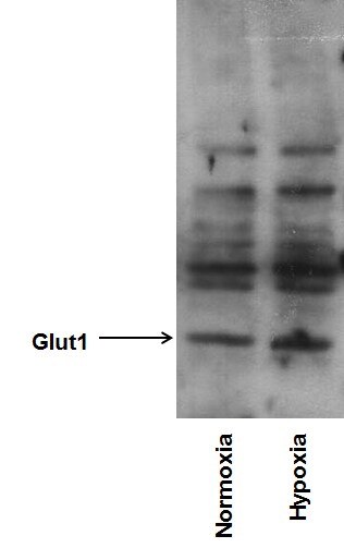
Application: Western BlotSample Tested: Human cancer cell whole cell lysateSpecies: HumanVerified Customer | Posted 10/04/2015Glut1 expression in MDA-MB-231 cells under normoxia and hypoxia.

-
Application: Western BlotSample Tested: See PMID 24260413Species: HumanVerified Customer | Posted 12/23/2014
There are no reviews that match your criteria.
Protocols
Find general support by application which include: protocols, troubleshooting, illustrated assays, videos and webinars.
- Antigen Retrieval Protocol (PIER)
- Antigen Retrieval for Frozen Sections Protocol
- Appropriate Fixation of IHC/ICC Samples
- Cellular Response to Hypoxia Protocols
- Chromogenic IHC Staining of Formalin-Fixed Paraffin-Embedded (FFPE) Tissue Protocol
- Chromogenic Immunohistochemistry Staining of Frozen Tissue
- ClariTSA™ Fluorophore Kits
- Detection & Visualization of Antibody Binding
- ELISA Sample Preparation & Collection Guide
- ELISA Troubleshooting Guide
- Fluorescent IHC Staining of Frozen Tissue Protocol
- Graphic Protocol for Heat-induced Epitope Retrieval
- Graphic Protocol for the Preparation and Fluorescent IHC Staining of Frozen Tissue Sections
- Graphic Protocol for the Preparation and Fluorescent IHC Staining of Paraffin-embedded Tissue Sections
- Graphic Protocol for the Preparation of Gelatin-coated Slides for Histological Tissue Sections
- How to Run an R&D Systems DuoSet ELISA
- How to Run an R&D Systems Quantikine ELISA
- How to Run an R&D Systems Quantikine™ QuicKit™ ELISA
- ICC Cell Smear Protocol for Suspension Cells
- ICC Immunocytochemistry Protocol Videos
- ICC for Adherent Cells
- IHC Sample Preparation (Frozen sections vs Paraffin)
- Immunocytochemistry (ICC) Protocol
- Immunocytochemistry Troubleshooting
- Immunofluorescence of Organoids Embedded in Cultrex Basement Membrane Extract
- Immunofluorescent IHC Staining of Formalin-Fixed Paraffin-Embedded (FFPE) Tissue Protocol
- Immunohistochemistry (IHC) and Immunocytochemistry (ICC) Protocols
- Immunohistochemistry Frozen Troubleshooting
- Immunohistochemistry Paraffin Troubleshooting
- Immunoprecipitation Protocol
- Preparing Samples for IHC/ICC Experiments
- Preventing Non-Specific Staining (Non-Specific Binding)
- Primary Antibody Selection & Optimization
- Protocol for Heat-Induced Epitope Retrieval (HIER)
- Protocol for Making a 4% Formaldehyde Solution in PBS
- Protocol for VisUCyte™ HRP Polymer Detection Reagent
- Protocol for the Fluorescent ICC Staining of Cell Smears - Graphic
- Protocol for the Fluorescent ICC Staining of Cultured Cells on Coverslips - Graphic
- Protocol for the Preparation & Fixation of Cells on Coverslips
- Protocol for the Preparation and Chromogenic IHC Staining of Frozen Tissue Sections
- Protocol for the Preparation and Chromogenic IHC Staining of Frozen Tissue Sections - Graphic
- Protocol for the Preparation and Chromogenic IHC Staining of Paraffin-embedded Tissue Sections
- Protocol for the Preparation and Chromogenic IHC Staining of Paraffin-embedded Tissue Sections - Graphic
- Protocol for the Preparation and Fluorescent ICC Staining of Cells on Coverslips
- Protocol for the Preparation and Fluorescent ICC Staining of Non-adherent Cells
- Protocol for the Preparation and Fluorescent ICC Staining of Stem Cells on Coverslips
- Protocol for the Preparation and Fluorescent IHC Staining of Frozen Tissue Sections
- Protocol for the Preparation and Fluorescent IHC Staining of Paraffin-embedded Tissue Sections
- Protocol for the Preparation of Gelatin-coated Slides for Histological Tissue Sections
- Protocol for the Preparation of a Cell Smear for Non-adherent Cell ICC - Graphic
- Quantikine HS ELISA Kit Assay Principle, Alkaline Phosphatase
- Quantikine HS ELISA Kit Principle, Streptavidin-HRP Polymer
- R&D Systems Quality Control Western Blot Protocol
- Sandwich ELISA (Colorimetric) – Biotin/Streptavidin Detection Protocol
- Sandwich ELISA (Colorimetric) – Direct Detection Protocol
- TUNEL and Active Caspase-3 Detection by IHC/ICC Protocol
- The Importance of IHC/ICC Controls
- Troubleshooting Guide: ELISA
- Troubleshooting Guide: Immunohistochemistry
- Troubleshooting Guide: Western Blot Figures
- Western Blot Conditions
- Western Blot Protocol
- Western Blot Protocol for Cell Lysates
- Western Blot Troubleshooting
- Western Blot Troubleshooting Guide
- View all Protocols, Troubleshooting, Illustrated assays and Webinars


