GPER/GPR30 Antibody - BSA Free
Novus Biologicals | Catalog # NLS4271
![Western Blot: GPER/GPR30 AntibodyBSA Free [NLS4271]](https://resources.rndsystems.com/images/products/GPER-GPR30-Antibody-Western-Blot-NLS4271-img0004.jpg "Western Blot: GPER/GPR30 AntibodyBSA Free [NLS4271]")
Key Product Details
Validated by
Species Reactivity
Validated:
Cited:
Predicted:
Applications
Validated:
Cited:
Label
Antibody Source
Format
Product Specifications
Immunogen
Epitope
Reactivity Notes
Localization
Specificity
Clonality
Host
Isotype
Description
Scientific Data Images for GPER/GPR30 Antibody - BSA Free
![Immunohistochemistry-Paraffin: GPER/GPR30 Antibody - BSA Free [NLS4271]](https://resources.rndsystems.com/images/products/GPER-GPR30-Antibody-Immunohistochemistry-Paraffin-NLS4271-img0003.jpg "Immunohistochemistry-Paraffin: GPER/GPR30 Antibody - BSA Free [NLS4271]")
Immunohistochemistry-Paraffin: GPER/GPR30 Antibody - BSA Free [NLS4271]
Immunohistochemistry-Paraffin: GPER/GPR30 Antibody [NLS4271] - Analysis of anti-GPR30 antibody with human brain, hippocampus.![Western Blot: GPER/GPR30 AntibodyBSA Free [NLS4271]](https://resources.rndsystems.com/images/products/GPER-GPR30-Antibody-Western-Blot-NLS4271-img0002.jpg "Western Blot: GPER/GPR30 AntibodyBSA Free [NLS4271]")
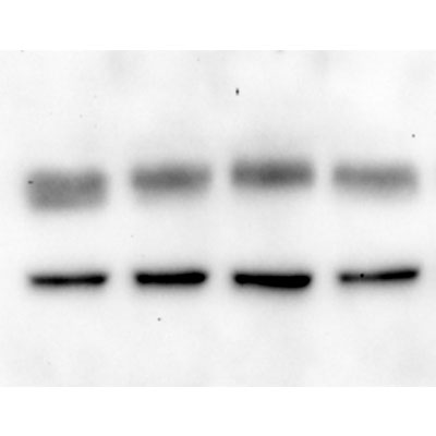
Western Blot: GPER/GPR30 AntibodyBSA Free [NLS4271]
Western Blot: GPER/GPR30 Antibody [NLS4271] - Analysis of anti-GPR30 antibody with rat hypothalamus 5-10ug protein loaded per lane. Image courtesy provided by Carrie McAllister.
Western Blot: GPER/GPR30 Antibody - BSA Free [NLS4271] -
Direct association of dopamine transporter and estrogen receptors on the plasma membrane vs. in total protein. Co-immunoprecipitation of dopamine transporter after 9 min 10-9 M E2 treatment and subsequent probing with antibodies for DAT, ER alpha, ER beta, and GPR30. This is a representative immunoblot of 3 experiments
Western Blot: GPER/GPR30 Antibody - BSA Free [NLS4271] -
Western Blot: GPER/GPR30 Antibody - BSA Free [NLS4271] - GPER/GPR30 expression in normal lung cell lines & lung adenocarcinoma cell lines. A) The expression of GPER was determined by realtime quantitative PCR in each of the normal lung cell lines (open bars), lung adenocarcinoma cell lines (grey bars), & MCF-7 human breast cancer cells (black bar). Values are the average of 4–6 biological replicates ± SEM. B-D) Representative western blot analysis of GPER/GPR30 expression in the indicted cell lines. 30 μg of whole cell extract (WCE) were separated on 10% SDS PAGE gels with MW marker migration indicated at the left of each blot. The diagrams at the top of B-C, & D indicate the region of GPER recognized by the 3 different polyclonal antibodies used: B) Novus NBP-1-31239, C) Novus NLS4271; D) Santa Cruz sc-4854-R. The MW of GPR30 is estimated to be 42 kDa, but higher MW sizes have been reported due to glycosylation & interaction with other proteins. Bands are identified as glycosylated & nonglycosylated based on reports cited in the text, but could include interaction with other proteins. For each GPER western, the membrane was stripped & reprobed for beta -actin as a loading control. Quantitation of GPER was evaluated by summing all immunoreactive bands & dividing by beta -actin, then normalizing to HBEC2-KT in each blot. Panel E is a summary of all westerns (not all westerns are shown) with each antibody. For Westerns with NBP1-31239 & sc-48524, values are the mean ± SEM from 3 separate blots using different WCE. Image collected & cropped by CiteAb from the following publication (https://pubmed.ncbi.nlm.nih.gov/23273253), licensed under a CC-BY license. Not internally tested by Novus Biologicals.Applications for GPER/GPR30 Antibody - BSA Free
Immunohistochemistry-Paraffin
Western Blot
Reviewed Applications
Read 1 review rated 4 using NLS4271 in the following applications:
Formulation, Preparation, and Storage
Purification
Formulation
Format
Preservative
Concentration
Shipping
Stability & Storage
Background: GPER/GPR30
Long Name
Alternate Names
Entrez Gene IDs
Gene Symbol
UniProt
Additional GPER/GPR30 Products
Product Documents for GPER/GPR30 Antibody - BSA Free
Certificate of Analysis
To download a Certificate of Analysis, please enter a lot or batch number in the search box below.
Product Specific Notices for GPER/GPR30 Antibody - BSA Free
This product is for research use only and is not approved for use in humans or in clinical diagnosis. Primary Antibodies are guaranteed for 1 year from date of receipt.
Related Research Areas
Citations for GPER/GPR30 Antibody - BSA Free
 Powered by Bioz
Customer Reviews for GPER/GPR30 Antibody - BSA Free (1)
Have you used GPER/GPR30 Antibody - BSA Free?
Submit a review and receive an Amazon gift card!
$25/€18/£15/$25CAN/¥2500 Yen for a review with an image
$10/€7/£6/$10CAN/¥1110 Yen for a review without an image
Submit a review
Customer Images
-
Application: Western BlotSample Tested: rat hypothalamusSpecies: RatVerified Customer | Posted 01/24/2013

There are no reviews that match your criteria.
Protocols
Find general support by application which include: protocols, troubleshooting, illustrated assays, videos and webinars.
- Antigen Retrieval Protocol (PIER)
- Antigen Retrieval for Frozen Sections Protocol
- Appropriate Fixation of IHC/ICC Samples
- Cellular Response to Hypoxia Protocols
- Chromogenic IHC Staining of Formalin-Fixed Paraffin-Embedded (FFPE) Tissue Protocol
- Chromogenic Immunohistochemistry Staining of Frozen Tissue
- ClariTSA™ Fluorophore Kits
- Detection & Visualization of Antibody Binding
- ELISA Sample Preparation & Collection Guide
- ELISA Troubleshooting Guide
- Fluorescent IHC Staining of Frozen Tissue Protocol
- Graphic Protocol for Heat-induced Epitope Retrieval
- Graphic Protocol for the Preparation and Fluorescent IHC Staining of Frozen Tissue Sections
- Graphic Protocol for the Preparation and Fluorescent IHC Staining of Paraffin-embedded Tissue Sections
- Graphic Protocol for the Preparation of Gelatin-coated Slides for Histological Tissue Sections
- How to Run an R&D Systems DuoSet ELISA
- How to Run an R&D Systems Quantikine ELISA
- How to Run an R&D Systems Quantikine™ QuicKit™ ELISA
- ICC Cell Smear Protocol for Suspension Cells
- ICC Immunocytochemistry Protocol Videos
- ICC for Adherent Cells
- IHC Sample Preparation (Frozen sections vs Paraffin)
- Immunocytochemistry (ICC) Protocol
- Immunocytochemistry Troubleshooting
- Immunofluorescence of Organoids Embedded in Cultrex Basement Membrane Extract
- Immunofluorescent IHC Staining of Formalin-Fixed Paraffin-Embedded (FFPE) Tissue Protocol
- Immunohistochemistry (IHC) and Immunocytochemistry (ICC) Protocols
- Immunohistochemistry Frozen Troubleshooting
- Immunohistochemistry Paraffin Troubleshooting
- Immunoprecipitation Protocol
- Preparing Samples for IHC/ICC Experiments
- Preventing Non-Specific Staining (Non-Specific Binding)
- Primary Antibody Selection & Optimization
- Protocol for Heat-Induced Epitope Retrieval (HIER)
- Protocol for Making a 4% Formaldehyde Solution in PBS
- Protocol for VisUCyte™ HRP Polymer Detection Reagent
- Protocol for the Fluorescent ICC Staining of Cell Smears - Graphic
- Protocol for the Fluorescent ICC Staining of Cultured Cells on Coverslips - Graphic
- Protocol for the Preparation & Fixation of Cells on Coverslips
- Protocol for the Preparation and Chromogenic IHC Staining of Frozen Tissue Sections
- Protocol for the Preparation and Chromogenic IHC Staining of Frozen Tissue Sections - Graphic
- Protocol for the Preparation and Chromogenic IHC Staining of Paraffin-embedded Tissue Sections
- Protocol for the Preparation and Chromogenic IHC Staining of Paraffin-embedded Tissue Sections - Graphic
- Protocol for the Preparation and Fluorescent ICC Staining of Cells on Coverslips
- Protocol for the Preparation and Fluorescent ICC Staining of Non-adherent Cells
- Protocol for the Preparation and Fluorescent ICC Staining of Stem Cells on Coverslips
- Protocol for the Preparation and Fluorescent IHC Staining of Frozen Tissue Sections
- Protocol for the Preparation and Fluorescent IHC Staining of Paraffin-embedded Tissue Sections
- Protocol for the Preparation of Gelatin-coated Slides for Histological Tissue Sections
- Protocol for the Preparation of a Cell Smear for Non-adherent Cell ICC - Graphic
- Quantikine HS ELISA Kit Assay Principle, Alkaline Phosphatase
- Quantikine HS ELISA Kit Principle, Streptavidin-HRP Polymer
- R&D Systems Quality Control Western Blot Protocol
- Sandwich ELISA (Colorimetric) – Biotin/Streptavidin Detection Protocol
- Sandwich ELISA (Colorimetric) – Direct Detection Protocol
- TUNEL and Active Caspase-3 Detection by IHC/ICC Protocol
- The Importance of IHC/ICC Controls
- Troubleshooting Guide: ELISA
- Troubleshooting Guide: Immunohistochemistry
- Troubleshooting Guide: Western Blot Figures
- Western Blot Conditions
- Western Blot Protocol
- Western Blot Protocol for Cell Lysates
- Western Blot Troubleshooting
- Western Blot Troubleshooting Guide
- View all Protocols, Troubleshooting, Illustrated assays and Webinars
FAQs for GPER/GPR30 Antibody - BSA Free
-
Q: What is the MW band size for GPER with your GPR30 Antibody (NLS4271)?
A: The expected molecular weight of GPR30 is 42.3, however, because this protein has at least 3 different glycosylation sites, you should not be surprised if you see a band at higher position in your samples.