Apoptosis signal-regulating kinase 1 (ASK1) is a MAP kinase kinase kinase also known as MEKK5 and MAP3K5. ASK1 is induced by inflammatory cytokines, UV light, and other stress-related stimuli to phosphorylate MAP kinase kinases that in turn activate the p38 and JNK families of MAP kinases. Mice lacking ASK1 are resistant to stress-induced p38 and JNK activation and cell death.

Key Product Details
Species Reactivity
Validated:
Human
Cited:
Human, Mouse
Applications
Validated:
Western Blot, Immunocytochemistry
Cited:
Immunohistochemistry, Western Blot
Label
Unconjugated
Antibody Source
Polyclonal Sheep IgG
Loading...
Product Specifications
Immunogen
E. coli-derived recombinant human ASK1
Lys1011-Asp1196
Accession # Q99683
Lys1011-Asp1196
Accession # Q99683
Specificity
Detects human ASK1 in direct ELISAs and Western blots.
Clonality
Polyclonal
Host
Sheep
Isotype
IgG
Scientific Data Images for Human ASK1 Antibody
Detection of Human ASK1 by Western Blot.
Western blot shows lysates of Raji human Burkitt's lymphoma cell line. PVDF membrane was probed with 1 µg/mL of Sheep Anti-Human ASK1 Antigen Affinity-purified Polyclonal Antibody (Catalog # AF3575) followed by HRP-conjugated Anti-Sheep IgG Secondary Antibody (Catalog # HAF016). A specific band was detected for ASK1 at approximately 154 kDa (as indicated). This experiment was conducted under reducing conditions and using Immunoblot Buffer Group 2.
Detection of Human ASK1 by Western Blot
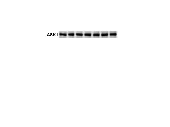
M. tb virulence induces ASK1 expression. Human MDMs were obtained after seven days in culture. 2 × 10⁶ MDMs were infected at MOI 1 and MOI 10 with an avirulent (H37Ra) and MOI 1 and MOI 5 with a virulent (H37Rv) strain of M. tb; 2 × 10⁶ MDMs were not infected as a control (Uninf). At 24 h postinfection, cells were recovered and prepared for Western Blot. Representative Western blot of ASK1, JNK1, JNK2, p-p38, and GAPDH (a). Band densities of ASK1 (b), JNK1 (c), JNK2 (d), and p-p38 (e) were normalized against GAPDH and quantified by densitometry analysis with the ImageJ software. Results are shown in relative units (RU) of concentration. The bar graphs show the mean ± SD from two independent experiments (n = 2 donors and two technical replicates). Statistical analysis was performed using Kruskal–Wallis analysis, followed by Dunn’s post hoc test. * p < 0.05. Image collected and cropped by CiteAb from the following open publication (https://pubmed.ncbi.nlm.nih.gov/35631013), licensed under a CC-BY license. Not internally tested by R&D Systems.
Detection of Human ASK1 by Western Blot
M. tb virulence induces ASK1 expression. Human MDMs were obtained after seven days in culture. 2 × 10⁶ MDMs were infected at MOI 1 and MOI 10 with an avirulent (H37Ra) and MOI 1 and MOI 5 with a virulent (H37Rv) strain of M. tb; 2 × 10⁶ MDMs were not infected as a control (Uninf). At 24 h postinfection, cells were recovered and prepared for Western Blot. Representative Western blot of ASK1, JNK1, JNK2, p-p38, and GAPDH (a). Band densities of ASK1 (b), JNK1 (c), JNK2 (d), and p-p38 (e) were normalized against GAPDH and quantified by densitometry analysis with the ImageJ software. Results are shown in relative units (RU) of concentration. The bar graphs show the mean ± SD from two independent experiments (n = 2 donors and two technical replicates). Statistical analysis was performed using Kruskal–Wallis analysis, followed by Dunn’s post hoc test. * p < 0.05. Image collected and cropped by CiteAb from the following open publication (https://pubmed.ncbi.nlm.nih.gov/35631013), licensed under a CC-BY license. Not internally tested by R&D Systems.Applications for Human ASK1 Antibody
Application
Recommended Usage
Immunocytochemistry
5-15 µg/mL
Sample: Immersion fixed human peripheral blood mononuclear cells
Sample: Immersion fixed human peripheral blood mononuclear cells
Western Blot
1 µg/mL
Sample: Raji human Burkitt's lymphoma cell line
Sample: Raji human Burkitt's lymphoma cell line
Reviewed Applications
Read 1 review rated 4 using AF3575 in the following applications:
Formulation, Preparation, and Storage
Purification
Antigen Affinity-purified
Reconstitution
Reconstitute at 0.2 mg/mL in sterile PBS. For liquid material, refer to CoA for concentration.
Loading...
Formulation
Lyophilized from a 0.2 μm filtered solution in PBS with Trehalose. *Small pack size (SP) is supplied either lyophilized or as a 0.2 µm filtered solution in PBS.
Shipping
Lyophilized product is shipped at ambient temperature. Liquid small pack size (-SP) is shipped with polar packs. Upon receipt, store immediately at the temperature recommended below.
Stability & Storage
Use a manual defrost freezer and avoid repeated freeze-thaw cycles.
- 12 months from date of receipt, -20 to -70 °C as supplied.
- 1 month, 2 to 8 °C under sterile conditions after reconstitution.
- 6 months, -20 to -70 °C under sterile conditions after reconstitution.
Calculators
Background: ASK1
Long Name
Apoptosis Signal-regulated Kinase 1
Alternate Names
MAP3K5, MEKK5
Gene Symbol
MAP3K5
UniProt
Additional ASK1 Products
Product Documents for Human ASK1 Antibody
Certificate of Analysis
To download a Certificate of Analysis, please enter a lot or batch number in the search box below.
Note: Certificate of Analysis not available for kit components.
Product Specific Notices for Human ASK1 Antibody
For research use only
Related Research Areas
Citations for Human ASK1 Antibody
 Powered by Bioz
Customer Reviews for Human ASK1 Antibody (1)
4 out of 5
1 Customer Rating
Have you used Human ASK1 Antibody?
Submit a review and receive an Amazon gift card!
$25/€18/£15/$25CAN/¥2500 Yen for a review with an image
$10/€7/£6/$10CAN/¥1110 Yen for a review without an image
Submit a review
Customer Images
Showing
1
-
1 of
1 review
Showing All
Filter By:
-
Application: Western BlotSample Tested: MDA-MB-231 human breast cancer cell lineSpecies: HumanVerified Customer | Posted 01/25/2018

There are no reviews that match your criteria.
Protocols
Find general support by application which include: protocols, troubleshooting, illustrated assays, videos and webinars.
- Appropriate Fixation of IHC/ICC Samples
- Cellular Response to Hypoxia Protocols
- ClariTSA™ Fluorophore Kits
- Detection & Visualization of Antibody Binding
- ICC Cell Smear Protocol for Suspension Cells
- ICC Immunocytochemistry Protocol Videos
- ICC for Adherent Cells
- Immunocytochemistry (ICC) Protocol
- Immunocytochemistry Troubleshooting
- Immunofluorescence of Organoids Embedded in Cultrex Basement Membrane Extract
- Immunohistochemistry (IHC) and Immunocytochemistry (ICC) Protocols
- Preparing Samples for IHC/ICC Experiments
- Preventing Non-Specific Staining (Non-Specific Binding)
- Primary Antibody Selection & Optimization
- Protocol for VisUCyte™ HRP Polymer Detection Reagent
- Protocol for the Fluorescent ICC Staining of Cell Smears - Graphic
- Protocol for the Fluorescent ICC Staining of Cultured Cells on Coverslips - Graphic
- Protocol for the Preparation and Fluorescent ICC Staining of Cells on Coverslips
- Protocol for the Preparation and Fluorescent ICC Staining of Non-adherent Cells
- Protocol for the Preparation and Fluorescent ICC Staining of Stem Cells on Coverslips
- Protocol for the Preparation of a Cell Smear for Non-adherent Cell ICC - Graphic
- R&D Systems Quality Control Western Blot Protocol
- TUNEL and Active Caspase-3 Detection by IHC/ICC Protocol
- The Importance of IHC/ICC Controls
- Troubleshooting Guide: Western Blot Figures
- Western Blot Conditions
- Western Blot Protocol
- Western Blot Protocol for Cell Lysates
- Western Blot Troubleshooting
- Western Blot Troubleshooting Guide
- View all Protocols, Troubleshooting, Illustrated assays and Webinars
Loading...
Associated Pathways
