Bone morphogenetic protein 15 (BMP-15), also known as GDF-9B, is a TGF-beta superfamily ligand that is expressed by oocytes throughout folliculogenesis, and plays an important role in oocyte development (1). BMP-15 promotes the FSH-independent proliferation of ovarian granulosa cells (GC) and induces GC glycolysis and cholesterol synthesis (2‑4). It also induces GC production of stem cell factor which, in turn, negatively regulates BMP-15 expression in oocytes (5). BMP-15 blocks the FSH-induced GC expression of FSH R and multiple steroidogenic molecules (6). BMP-15 is synthesized with a 249 amino acid (aa) N-terminal propeptide (7). The propeptide is cleaved intracellularly from the 50 kDa proBMP-15 but remains associated with mature BMP-15 (8). Mature BMP-15 exists in 16 kDa and 17 kDa forms which are distinguishable by the presence of O-linked glycosylation on the 17 kDa form (8). Mature BMP-15 is phosphorylated, a modification which is required for the stimulation of GC proliferation (9). BMP-15 exerts its effects through interactions with BMPR-IB/ALK6 and BMPR-II (9‑11). Mature BMP-15 forms 34 kDa noncovalently-linked homodimers and 37 kDa heterodimers with mature GDF-9 (12). Both of these oocyte-expressed factors lack the cysteine that mediates disulfide-linked dimerization in most TGF-beta superfamily ligands (1). Although heterodimerization with GDF-9 may limit the secretion of active BMP-15, these two factors synergize in promoting oocyte survival and folliculogenesis (12, 13). Mature human BMP-15 shares 70%, 68%, and 78% aa sequence identity with mouse, rat, and sheep BMP-15, respectively. It shares 27%‑38% aa sequence with other BMPs.

Key Product Details
Species Reactivity
Applications
Label
Antibody Source
Product Specifications
Immunogen
Gln268-Arg392
Accession # O95972
Specificity
Clonality
Host
Isotype
Scientific Data Images for Human BMP-15/GDF-9B Antibody (317809)
BMP‑15/GDF‑9B in Mouse Ovary.
BMP‑15/GDF‑9B was detected in immersion fixed frozen sections of mouse ovary using 10 µg/mL Rat Anti-Human BMP‑15/GDF‑9B Monoclonal Antibody (Catalog # MAB2925) overnight at 4 °C. Tissue was stained with the NorthernLights™ 557-conjugated Anti-Rat IgG Secondary Antibody (red; Catalog # NL013) and counterstained with DAPI (blue). View our protocol for Fluorescent IHC Staining of Frozen Tissue Sections.Applications for Human BMP-15/GDF-9B Antibody (317809)
Immunohistochemistry
Sample: Immersion fixed frozen sections of mouse ovary
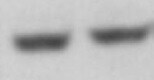
Western Blot
Sample: Recombinant Human BMP‑15/GDF‑9B (Catalog # 5096-BM)
Reviewed Applications
Read 1 review rated 5 using MAB2925 in the following applications:
Formulation, Preparation, and Storage
Purification
Reconstitution
Reconstitute at 0.5 mg/mL in sterile PBS. For liquid material, refer to CoA for concentration.
Formulation
Shipping
Stability & Storage
- 12 months from date of receipt, -20 to -70 °C as supplied.
- 1 month, 2 to 8 °C under sterile conditions after reconstitution.
- 6 months, -20 to -70 °C under sterile conditions after reconstitution.
Calculators
Background: BMP-15/GDF-9B
References
- Moore, R.K. and S. Shimasaki (2005) Mol. Cell. Endocrinol. 234:67.
- Otsuka, F. et al. (2000) J. Biol. Chem. 275:39523.
- Sugiura, K. et al. (2007) Development 134:2593.
- Su, Y.-Q. et al. (2008) Development 135:111.
- Otsuka, F. and S. Shimasaki (2002) Proc. Natl. Acad. Sci. 99:8060.
- Otsuka, F. et al. (2001) J. Biol. Chem. 276:11387.
- Dube, J.L. et al. (1998) Mol. Endocrinol. 12:1809.
- Saito, S. et al. (2008) Prot. Sci. 17:362.
- McMahon, H.E. et al. (2008) Endocrinology 149:812.
- Moore, R.K. et al. (2003) J. Biol. Chem. 278:304.
- Edwards, S.J. et al. (2008) Endocrinology 149:1026.
- Liao, W.X. et al. (2003) J. Biol. Chem. 278:3713.
- Yan, C. et al. (2001) Mol. Endocrinol. 15:854.
Long Name
Alternate Names
Gene Symbol
UniProt
Additional BMP-15/GDF-9B Products
Product Documents for Human BMP-15/GDF-9B Antibody (317809)
Certificate of Analysis
To download a Certificate of Analysis, please enter a lot or batch number in the search box below.
Note: Certificate of Analysis not available for kit components.
Product Specific Notices for Human BMP-15/GDF-9B Antibody (317809)
This product is sold under license from Vanderbilt University for research and laboratory use only. Licenses for commercial manufacture, commercial use, diagnostics or therapeutics may be obtained by contacting Vanderbilt University, Nashville, Tennessee.
For research use only
Related Research Areas
Customer Reviews for Human BMP-15/GDF-9B Antibody (317809) (1)
Have you used Human BMP-15/GDF-9B Antibody (317809)?
Submit a review and receive an Amazon gift card!
$25/€18/£15/$25CAN/¥2500 Yen for a review with an image
$10/€7/£6/$10CAN/¥1110 Yen for a review without an image
Submit a review
Customer Images
-
Application: Western BlotSample Tested: Ovary tissueSpecies: HumanVerified Customer | Posted 01/21/2022

There are no reviews that match your criteria.
Protocols
Find general support by application which include: protocols, troubleshooting, illustrated assays, videos and webinars.
- Antigen Retrieval Protocol (PIER)
- Antigen Retrieval for Frozen Sections Protocol
- Appropriate Fixation of IHC/ICC Samples
- Cellular Response to Hypoxia Protocols
- Chromogenic IHC Staining of Formalin-Fixed Paraffin-Embedded (FFPE) Tissue Protocol
- Chromogenic Immunohistochemistry Staining of Frozen Tissue
- ClariTSA™ Fluorophore Kits
- Detection & Visualization of Antibody Binding
- Fluorescent IHC Staining of Frozen Tissue Protocol
- Graphic Protocol for Heat-induced Epitope Retrieval
- Graphic Protocol for the Preparation and Fluorescent IHC Staining of Frozen Tissue Sections
- Graphic Protocol for the Preparation and Fluorescent IHC Staining of Paraffin-embedded Tissue Sections
- Graphic Protocol for the Preparation of Gelatin-coated Slides for Histological Tissue Sections
- IHC Sample Preparation (Frozen sections vs Paraffin)
- Immunofluorescent IHC Staining of Formalin-Fixed Paraffin-Embedded (FFPE) Tissue Protocol
- Immunohistochemistry (IHC) and Immunocytochemistry (ICC) Protocols
- Immunohistochemistry Frozen Troubleshooting
- Immunohistochemistry Paraffin Troubleshooting
- Preparing Samples for IHC/ICC Experiments
- Preventing Non-Specific Staining (Non-Specific Binding)
- Primary Antibody Selection & Optimization
- Protocol for Heat-Induced Epitope Retrieval (HIER)
- Protocol for Making a 4% Formaldehyde Solution in PBS
- Protocol for VisUCyte™ HRP Polymer Detection Reagent
- Protocol for the Preparation & Fixation of Cells on Coverslips
- Protocol for the Preparation and Chromogenic IHC Staining of Frozen Tissue Sections
- Protocol for the Preparation and Chromogenic IHC Staining of Frozen Tissue Sections - Graphic
- Protocol for the Preparation and Chromogenic IHC Staining of Paraffin-embedded Tissue Sections
- Protocol for the Preparation and Chromogenic IHC Staining of Paraffin-embedded Tissue Sections - Graphic
- Protocol for the Preparation and Fluorescent IHC Staining of Frozen Tissue Sections
- Protocol for the Preparation and Fluorescent IHC Staining of Paraffin-embedded Tissue Sections
- Protocol for the Preparation of Gelatin-coated Slides for Histological Tissue Sections
- R&D Systems Quality Control Western Blot Protocol
- TUNEL and Active Caspase-3 Detection by IHC/ICC Protocol
- The Importance of IHC/ICC Controls
- Troubleshooting Guide: Immunohistochemistry
- Troubleshooting Guide: Western Blot Figures
- Western Blot Conditions
- Western Blot Protocol
- Western Blot Protocol for Cell Lysates
- Western Blot Troubleshooting
- Western Blot Troubleshooting Guide
- View all Protocols, Troubleshooting, Illustrated assays and Webinars