Dickkopf related protein 1 (Dkk-1) is a member of the Dkk protein family that includes Dkk-1, -2, -3, and -4 (1). All four members are secreted proteins that are synthesized as precursor proteins with an N-terminal signal peptide and 2 conserved cysteine-rich domains, which are separated by a linker region. Dkk proteins have potential furin type proteolytic cleavage sites, and short forms of Dkk-2 and Dkk-4 containing only the second cysteine-rich domain can be generated by proteolytic processing (1). Dkk proteins have distinct patterns of expression in adult and embryonic tissues, suggesting that they may play diverse roles in these tissues. The Dkk proteins have distinct effects on Wnt signaling. Dkk-1 and Dkk-4 are Wnt antagonists. Dkk-3 has not been demonstrated to affect Wnt signaling, and Dkk-2 acts as an agonist or antagonist, depending on the cellular context. Wnt signaling regulates many important developmental processes including neural crest differentiation, brain development, kidney morphogenesis, and sex determination. In addition, Wnt signaling has also been implicated in tumor formation. Canonical Wnt signaling via the beta-catenin pathway is transduced by a receptor complex composed of the Frizzled proteins (Fz) and low-density lipoprotein (LDL) receptor-related proteins (LRP5/6) (2, 3). Unlike many soluble Wnt antagonists that function by binding extracellular Wnt ligands to prevent interaction of Wnt with the Fz-LRP5/6 receptor complex, Dkk-1 and Dkk-4 antagonize Wnt/ beta -catenin signaling by direct high-affinity binding to the Wnt coreceptor LRP5/6 and inhibiting interaction of LRP5/6 with the Wnt-Frizzled complex (4). Dkk-1 and Dkk-4 also bind the transmembrane proteins Kremen1 (Krm1) and Krm2 with high-affinity (5). Krm2 has been shown to form a ternary complex with Dkk-1 or -4 and LRP5/6 to trigger internalization of the complex and removal LRP6 from the cell surface. Thus, Dkk-1/4 and Kremens function synergistically to antagonize LRP5/6-mediated Wnt activity. Dkk-2 also binds to LRP5/6 and the Kremens, but Dkk-2 acts as antagonist of the Wnt signaling pathway only in the presence of Krm2 (5, 6). Dkk 2 binding to LRP5/6 in the absence of Krm2 activates rather than inhibits Wnt signaling (6).

Key Product Details
Validated by
Knockout/Knockdown
Species Reactivity
Validated:
Human, Mouse
Cited:
Human, Mouse, Avian - Chicken, Transgenic Mouse
Applications
Validated:
Knockout Validated, Western Blot, Blockade of Receptor-ligand Interaction
Cited:
Immunohistochemistry, Immunohistochemistry-Paraffin, Western Blot, Neutralization, Immunocytochemistry, Bioassay, ELISA Development, Immunodepletion, Neutralizing
Label
Unconjugated
Antibody Source
Polyclonal Goat IgG
Loading...
Product Specifications
Immunogen
S. frugiperda insect ovarian cell line Sf 21-derived recombinant human Dkk-1
Thr32-His266
Accession # O94907
Thr32-His266
Accession # O94907
Specificity
Detects human and mouse Dkk-1 in direct ELISAs and Western blots. In direct ELISAs, less than 1% cross-reactivity with recombinant human (rh) Dkk-2, and rhDkk-4 is observed.
Clonality
Polyclonal
Host
Goat
Isotype
IgG
Endotoxin Level
<0.10 EU per 1 μg of the antibody by the LAL method.
Scientific Data Images for Dkk-1 Antibody
Detection of Human Dkk‑1 by Western Blot.
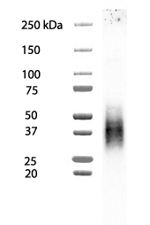
Western blot shows lysates of MCF-7 human breast cancer cell line, HepG2 human hepatocellular carcinoma cell line, A431 human epithelial carcinoma cell line, and A549 human lung carcinoma cell line. PVDF membrane was probed with 1 µg/mL of Goat Anti-Human/Mouse Dkk-1 Antigen Affinity-purified Polyclonal Antibody (Catalog # AF1096) followed by HRP-conjugated Anti-Goat IgG Secondary Antibody (Catalog # HAF017). Specific bands were detected for Dkk-1 at approximately 28-40 kDa (as indicated). This experiment was conducted under reducing conditions and using Immunoblot Buffer Group 1.
Detection of Mouse Dkk‑1 by Western Blot.
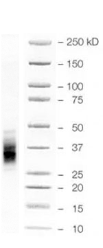
Western blot shows lysates of Neuro-2A mouse neuroblastoma cell line. PVDF membrane was probed with 1 µg/mL of Goat Anti-Human/Mouse Dkk-1 Antigen Affinity-purified Polyclonal Antibody (Catalog # AF1096) followed by HRP-conjugated Anti-Goat IgG Secondary Antibody (Catalog # HAF017). Specific bands were detected for Dkk-1 at approximately 28-40 kDa (as indicated). This experiment was conducted under reducing conditions and using Immunoblot Buffer Group 1.
Western Blot Shows Human Dkk‑1 Specificity by Using Knockout Cell Line.
Western blot shows lysates of HeLa human cervical epithelial carcinoma parental cell line and Dkk-1 knockout HeLa cell line (KO). PVDF membrane was probed with 0.2 µg/mL of Goat Anti-Human/Mouse Dkk-1 Antigen Affinity-purified Polyclonal Antibody (Catalog # AF1096) followed by HRP-conjugated Anti-Goat IgG Secondary Antibody (Catalog # HAF017). A specific band was detected for Dkk-1 at approximately 35 kDa (as indicated) in the parental HeLa cell line, but is not detectable in knockout HeLa cell line. GAPDH (Catalog # AF5718) is shown as a loading control. This experiment was conducted under reducing conditions and using Immunoblot Buffer Group 1.
Detection of Mouse Dkk-1 by Immunohistochemistry
Thrombocyte is a major source of DKK1. (A-C) Upon BLM challenge, C57Bl/6J mice (n = 4 per group) were intraperitoneally injected with an isotype control antibody (Iso Ab) or anti-CD41a antibody ( alpha -CD41 Ab) on day 0. The lungs were harvested on day 2 (A). DKK1 protein levels were analyzed by IHC and Image J (B). Representative IHC images are shown (C). The boxed areas are shown in higher magnification below. DKK1-positive cells are indicated by red arrows. Bar = 20 μm (top and middle panels); 5 μm (bottom panels). (D) Peripheral blood was collected from Dkk1PKO mice (n = 5) and their Dkk1fl/fl littermate controls (n = 5). Circulating DKK1 protein levels were analyzed by DKK1 ELISA. (E) Hemavet analysis of inflammatory cells in the peripheral blood was performed from Dkk1PKO mice (n = 6) and their Dkk1fl/fl littermate controls (n = 6). Shown are white blood cells (WBC), lymphocytes, monocytes, neutrophils, eosinophils, basophils, red blood cells (RBC), platelets, and mean platelet volume (MPV). A one-way ANOVA analysis with Bonferroni’s post hoc test was performed for (B). Student’s t-test was performed for (D, E). ****p < 0.0001, ns, not significant. See also Supplementary Figure 5. Image collected and cropped by CiteAb from the following open publication (https://pubmed.ncbi.nlm.nih.gov/38162655), licensed under a CC-BY license. Not internally tested by R&D Systems.
Human Dkk-1 ELISA Standard Curve
Recombinant Human Dkk‑1 (Catalog # 5439-DK) was serially diluted and captured by Mouse Anti-Human Dkk‑1 Monoclonal Antibody (Catalog # MAB10962) coated on a Clear Polystyrene Microplate (Catalog # DY990). Goat Anti-Human/Mouse Dkk‑1 Antigen Affinity-purified Polyclonal Antibody (Catalog # AF1096) was biotinylated and incubated with the protein captured on the plate. Detection of the standard curve was achieved by incubating Streptavidin-HRP (Catalog # DY998)Applications for Dkk-1 Antibody
Application
Recommended Usage
Blockade of Receptor-ligand Interaction
In a functional ELISA, 0.1-0.4 μg/mL of this antibody will block 50% of the binding of 50 ng/mL of recombinant human biotinylated Dkk-1 to immobilized recombinant human LRP-6 Fc Chimera (Catalog # 1505-LR) coated at 1 µg/mL (100 µL/well). At 10 μg/mL, this antibody will block >90% of the binding.
Knockout Validated
Dkk‑1
is specifically detected in HeLa human cervical epithelial carcinoma parental cell line but is not detectable in
Dkk‑1 knockout HeLa cell line.
Western Blot
1 µg/mL
Sample: MCF‑7 human breast cancer cell line, HepG2 human hepatocellular carcinoma cell line, A431 human epithelial carcinoma cell line, A549 human lung carcinoma cell line, and Neuro‑2A mouse neuroblastoma cell line
Sample: MCF‑7 human breast cancer cell line, HepG2 human hepatocellular carcinoma cell line, A431 human epithelial carcinoma cell line, A549 human lung carcinoma cell line, and Neuro‑2A mouse neuroblastoma cell line
Reviewed Applications
Read 3 reviews rated 4.7 using AF1096 in the following applications:
Formulation, Preparation, and Storage
Purification
Antigen Affinity-purified
Reconstitution
Reconstitute at 0.2 mg/mL in sterile PBS. For liquid material, refer to CoA for concentration.
Loading...
Formulation
Lyophilized from a 0.2 μm filtered solution in PBS with Trehalose. *Small pack size (SP) is supplied either lyophilized or as a 0.2 µm filtered solution in PBS.
Shipping
Lyophilized product is shipped at ambient temperature. Liquid small pack size (-SP) is shipped with polar packs. Upon receipt, store immediately at the temperature recommended below.
Stability & Storage
Use a manual defrost freezer and avoid repeated freeze-thaw cycles.
- 12 months from date of receipt, -20 to -70 °C as supplied.
- 1 month, 2 to 8 °C under sterile conditions after reconstitution.
- 6 months, -20 to -70 °C under sterile conditions after reconstitution.
Calculators
Background: Dkk-1
References
- Krupnik, V.E. et al. (1999) Gene 238:301.
- Zorn, A.M (2001) Current Biology R592.
- Mao, J. et al. (2001) Mol. Cell 7:801.
- Nusse, R. et al. (2001) Nature 411:255.
- Mao, J. et al. (2002) Nature 417:664.
- Mao, B. and C. Niehrs (2003) Gene 302:179.
Long Name
Dickkopf-1
Alternate Names
Dkk1
Gene Symbol
DKK1
UniProt
Additional Dkk-1 Products
Product Documents for Dkk-1 Antibody
Certificate of Analysis
To download a Certificate of Analysis, please enter a lot or batch number in the search box below.
Note: Certificate of Analysis not available for kit components.
Product Specific Notices for Dkk-1 Antibody
For research use only
Related Research Areas
Citations for Dkk-1 Antibody
 Powered by Bioz
Customer Reviews for Dkk-1 Antibody (3)
4.7 out of 5
3 Customer Ratings
Have you used Dkk-1 Antibody?
Submit a review and receive an Amazon gift card!
$25/€18/£15/$25CAN/¥2500 Yen for a review with an image
$10/€7/£6/$10CAN/¥1110 Yen for a review without an image
Submit a review
Customer Images
Showing
1
-
3 of
3 reviews
Showing All
Filter By:
-
Application: Western BlotSample Tested: MCF-7 human breast cancer cell lineSpecies: HumanVerified Customer | Posted 01/01/2026Whole cell lysate of MCF7

-
Application: Western BlotSample Tested: HCT-116 human colorectal carcinoma cell lineSpecies: HumanVerified Customer | Posted 01/01/2026Used at 0.7 ug/mL
-
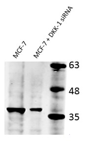
Application: Western BlotSample Tested: Cell LysatesSpecies: HumanVerified Customer | Posted 12/04/2016Silencing of DKK-1 in MCF-7 cells. Buffer: 5% BSA. Dilution: 1/1,000.
There are no reviews that match your criteria.
Protocols
Find general support by application which include: protocols, troubleshooting, illustrated assays, videos and webinars.
- Cellular Response to Hypoxia Protocols
- R&D Systems Quality Control Western Blot Protocol
- Troubleshooting Guide: Western Blot Figures
- Western Blot Conditions
- Western Blot Protocol
- Western Blot Protocol for Cell Lysates
- Western Blot Troubleshooting
- Western Blot Troubleshooting Guide
- View all Protocols, Troubleshooting, Illustrated assays and Webinars
Loading...
Associated Pathways
