ErbB2IP (ErbB2 Interacting Protein/Erbin; also Densin-180-like protein and LAP2) is both a nuclear and cytoplasmic 175-180 kDa member of the LAP (LRR And PDZ) family of proteins. It is widely expressed, and found in cell types as diverse as intestinal columnar epithelium, neurons, keratinocytes and skeletal plus cardiac muscle cells. Functionally, ErbB2IP interacts with multiple proteins through a PZD-mediated interaction. Partners include ErbB2, delta -catenin, PSD-95 and ARVCF. The ErbB2 interaction localizes this receptor to the basolateral membrane of epithelium, while the delta -catenin interaction impacts dendritic arborization, possibly through involvement with type 1 or 2 catenin(s) and the F-actin cytoskeleton. Finally, ErbB2IP/Erbin is suggested to play a role in maintaining the G1 phase of the cell cycle. Human ErbB2IP is 1412 amino acids (aa) in length. It contains 17 LRRs (Leu-rich repeats) (aa 23-413) followed by one PDZ domain (aa 1321-1410). There are more than 10 utilized Tyr, and 15 utilized Ser/Thr phosphorylation sites. A series of deletions over aa 1211-1377 generate at least six known splice variants. All show a deletion of aa 1212-1252, and at least one additional 10-41 aa deletion C-terminal to the common deletion. Over aa 496-687, human and mouse ErbB2IP share 88% aa sequence identity.

Key Product Details
Species Reactivity
Validated:
Human
Cited:
Human
Applications
Validated:
Western Blot, Immunocytochemistry
Cited:
Immunocytochemistry
Label
Unconjugated
Antibody Source
Polyclonal Sheep IgG
Loading...
Product Specifications
Immunogen
E. coli-derived recombinant human Erbin
Lys496-Thr687
Accession # Q96RT1
Lys496-Thr687
Accession # Q96RT1
Specificity
Detects human Erbin in direct ELISAs and Western blots. In direct ELISAs, less than 1% cross-reactivity with recombinant mouse ErbB2 is observed.
Clonality
Polyclonal
Host
Sheep
Isotype
IgG
Scientific Data Images for Human Erbin Antibody
Detection of Human Erbin by Western Blot.
Western blot shows lysates of A172 human glioblastoma cell line and HeLa human cervical epithelial carcinoma cell line. PVDF membrane was probed with 0.2 µg/mL of Sheep Anti-Human Erbin Antigen Affinity-purified Polyclonal Antibody (Catalog # AF7866) followed by HRP-conjugated Anti-Sheep IgG Secondary Antibody (Catalog # HAF016). A specific band was detected for Erbin at approximately 180 kDa (as indicated). This experiment was conducted under reducing conditions and using Immunoblot Buffer Group 1.
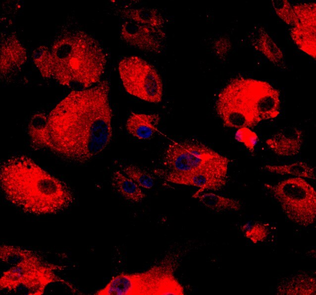
Erbin in DU145 Human Cell Line.
Erbin was detected in immersion fixed DU145 human prostate carcinoma cell line using Sheep Anti-Human Erbin Antigen Affinity-purified Polyclonal Antibody (Catalog # AF7866) at 10 µg/mL for 3 hours at room temperature. Cells were stained using the NorthernLights™ 557-conjugated Anti-Sheep IgG Secondary Antibody (red; Catalog # NL010) and counterstained with DAPI (blue). Specific staining was localized to cell surfaces. View our protocol for Fluorescent ICC Staining of Cells on Coverslips.Applications for Human Erbin Antibody
Application
Recommended Usage
Immunocytochemistry
5-15 µg/mL
Sample: Immersion fixed DU145 human prostate carcinoma cell line
Sample: Immersion fixed DU145 human prostate carcinoma cell line
Western Blot
0.2 µg/mL
Sample: A172 human glioblastoma cell line and HeLa human cervical epithelial carcinoma cell line
Sample: A172 human glioblastoma cell line and HeLa human cervical epithelial carcinoma cell line
Reviewed Applications
Read 2 reviews rated 4.5 using AF7866 in the following applications:
Formulation, Preparation, and Storage
Purification
Antigen Affinity-purified
Reconstitution
Sterile PBS to a final concentration of 0.2 mg/mL. For liquid material, refer to CoA for concentration.
Loading...
Formulation
Lyophilized from a 0.2 μm filtered solution in PBS with Trehalose. *Small pack size (SP) is supplied either lyophilized or as a 0.2 µm filtered solution in PBS.
Shipping
Lyophilized product is shipped at ambient temperature. Liquid small pack size (-SP) is shipped with polar packs. Upon receipt, store immediately at the temperature recommended below.
Stability & Storage
Use a manual defrost freezer and avoid repeated freeze-thaw cycles.
- 12 months from date of receipt, -20 to -70 °C as supplied.
- 1 month, 2 to 8 °C under sterile conditions after reconstitution.
- 6 months, -20 to -70 °C under sterile conditions after reconstitution.
Calculators
Background: Erbin
Long Name
ErbB2-interacting Protein
Alternate Names
Densin-180-like protein, ERBB2IP
Gene Symbol
ERBIN
UniProt
Additional Erbin Products
Product Documents for Human Erbin Antibody
Certificate of Analysis
To download a Certificate of Analysis, please enter a lot or batch number in the search box below.
Note: Certificate of Analysis not available for kit components.
Product Specific Notices for Human Erbin Antibody
For research use only
Related Research Areas
Citations for Human Erbin Antibody
 Powered by Bioz
Customer Reviews for Human Erbin Antibody (2)
4.5 out of 5
2 Customer Ratings
Have you used Human Erbin Antibody?
Submit a review and receive an Amazon gift card!
$25/€18/£15/$25CAN/¥2500 Yen for a review with an image
$10/€7/£6/$10CAN/¥1110 Yen for a review without an image
Submit a review
Customer Images
Showing
1
-
2 of
2 reviews
Showing All
Filter By:
-
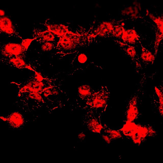
Application: ImmunocytochemistrySample Tested: pig SC cellsSpecies: PigVerified Customer | Posted 06/07/2017Human Erbin Antibody AF7866 labeled pig SC cells with Alexa 594

-
Application: ImmunocytochemistrySample Tested: pig trabecular meshwork cellsSpecies: PigVerified Customer | Posted 06/07/2017AF7866, ERbin antibody labeling pig trabecular meshwork cells with Alexa 594
There are no reviews that match your criteria.
Protocols
Find general support by application which include: protocols, troubleshooting, illustrated assays, videos and webinars.
- Appropriate Fixation of IHC/ICC Samples
- Cellular Response to Hypoxia Protocols
- ClariTSA™ Fluorophore Kits
- Detection & Visualization of Antibody Binding
- ICC Cell Smear Protocol for Suspension Cells
- ICC Immunocytochemistry Protocol Videos
- ICC for Adherent Cells
- Immunocytochemistry (ICC) Protocol
- Immunocytochemistry Troubleshooting
- Immunofluorescence of Organoids Embedded in Cultrex Basement Membrane Extract
- Immunohistochemistry (IHC) and Immunocytochemistry (ICC) Protocols
- Preparing Samples for IHC/ICC Experiments
- Preventing Non-Specific Staining (Non-Specific Binding)
- Primary Antibody Selection & Optimization
- Protocol for VisUCyte™ HRP Polymer Detection Reagent
- Protocol for the Fluorescent ICC Staining of Cell Smears - Graphic
- Protocol for the Fluorescent ICC Staining of Cultured Cells on Coverslips - Graphic
- Protocol for the Preparation and Fluorescent ICC Staining of Cells on Coverslips
- Protocol for the Preparation and Fluorescent ICC Staining of Non-adherent Cells
- Protocol for the Preparation and Fluorescent ICC Staining of Stem Cells on Coverslips
- Protocol for the Preparation of a Cell Smear for Non-adherent Cell ICC - Graphic
- R&D Systems Quality Control Western Blot Protocol
- TUNEL and Active Caspase-3 Detection by IHC/ICC Protocol
- The Importance of IHC/ICC Controls
- Troubleshooting Guide: Western Blot Figures
- Western Blot Conditions
- Western Blot Protocol
- Western Blot Protocol for Cell Lysates
- Western Blot Troubleshooting
- Western Blot Troubleshooting Guide
- View all Protocols, Troubleshooting, Illustrated assays and Webinars
Loading...