Fibroblast growth factor 20 (FGF-20) is a member of the FGF gene family, which currently contains 22 members. Based on its structure, it is further classified as an FGF-9 subfamily member. All FGF family members are heparin-binding growth factors with a 120 amino acid (aa) core FGF domain that exhibits a beta -trefoil structure (1). The cDNA of FGF-20 predicts a 211 aa polypeptide without a canonical signal peptide sequence, a feature shared with other members of this subfamily (2‑4). Nevertheless, it is secreted with a molecular weight of 27 kDa (2‑4). FGF-20 is known to bind to heparin (4). No alternate splice forms have been reported. However, three amino acid polymorphisms are known, and single nucleotide polymorphisms in noncoding regions that may effect expression show a strong correlation with a risk of developing Parkinson’s disease (5, 6). Human FGF-20 shows 98% aa identity to bovine FGF-20 and 95% aa identity to both rat and mouse FGF-20. Within the FGF-9 subfamily, FGF-20 is 69% and 63% aa identical to human FGF-9 and FGF-16, respectively. Human FGF-20 is reported to be promiscuous in its selection of receptors which include FGF R1c, FGF R2c, FGF R3b, FGF R3c and FGF R4 (4, 7, 8). FGF-20 is expressed a variety of cells, including dopaminergic neurons (2), fibroblasts, keratinocytes and breast epithelium (4), and multiple sites in the fetus (2, 7). Finally, the expression of FGF-20 and DKK-1 is regulated by beta -catenin during development and tumorigenesis, implying that FGF-20 may play a role in the oncogenesis induced by the Wnt signaling pathway (9).

Key Product Details
Species Reactivity
Validated:
Human
Cited:
Human
Applications
Validated:
Western Blot, Immunocytochemistry
Cited:
Western Blot
Label
Unconjugated
Antibody Source
Monoclonal Rat IgG2A Clone # 272317
Loading...
Product Specifications
Immunogen
E. coli-derived recombinant human FGF-20
Met1-Thr211
Accession # Q9NP95
Met1-Thr211
Accession # Q9NP95
Specificity
Detects human FGF-20 in direct ELISAs and Western blots. In direct ELISAs and Western blots, no cross‑reactivity with recombinant human (rh) FGF-3, -4, -5, -6, -7, -9, -10, -11, -12, -16, -17, -18, -19, -23, recombinant mouse FGF-8b, -8c, -15, -23, rhFGF acidic, or rhFGF basic is observed.
Clonality
Monoclonal
Host
Rat
Isotype
IgG2A
Scientific Data Images for Human FGF-20 Antibody (272317)
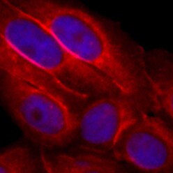
FGF‑20 in SW480 Human Cell Line.
FGF-20 was detected in immersion fixed SW480 human colorectal adenocarcinoma cell line using Rat Anti-Human FGF-20 Monoclonal Antibody (Catalog # MAB2547) at 10 µg/mL for 3 hours at room temperature. Cells were stained using the NorthernLights™ 557-conjugated Anti-Rat IgG Secondary Antibody (red; Catalog # NL013) and counterstained with DAPI (blue). Specific staining was localized to cytoplasm. View our protocol for Fluorescent ICC Staining of Cells on Coverslips.Applications for Human FGF-20 Antibody (272317)
Application
Recommended Usage
Immunocytochemistry
8-25 µg/mL
Sample: Immersion fixed SW480 human colorectal adenocarcinoma cell line
Sample: Immersion fixed SW480 human colorectal adenocarcinoma cell line
Western Blot
1 µg/mL
Sample: Recombinant Human FGF‑20 (Catalog # 2547-FG)
Sample: Recombinant Human FGF‑20 (Catalog # 2547-FG)
Reviewed Applications
Read 2 reviews rated 5 using MAB2547 in the following applications:
Formulation, Preparation, and Storage
Purification
Protein A or G purified from hybridoma culture supernatant
Reconstitution
Reconstitute at 0.5 mg/mL in sterile PBS. For liquid material, refer to CoA for concentration.
Loading...
Formulation
Lyophilized from a 0.2 μm filtered solution in PBS with Trehalose. *Small pack size (SP) is supplied either lyophilized or as a 0.2 µm filtered solution in PBS.
Shipping
Lyophilized product is shipped at ambient temperature. Liquid small pack size (-SP) is shipped with polar packs. Upon receipt, store immediately at the temperature recommended below.
Stability & Storage
Use a manual defrost freezer and avoid repeated freeze-thaw cycles.
- 12 months from date of receipt, -20 to -70 °C as supplied.
- 1 month, 2 to 8 °C under sterile conditions after reconstitution.
- 6 months, -20 to -70 °C under sterile conditions after reconstitution.
Calculators
Background: FGF-20
References
- Itoh, N. and D.M. Ornitz (2004) Trends Genet. 20:563.
- Ohmachi, S. et al. (2000) Biochem. Biophys. Res. Commun. 277:355.
- Kirikoshi, H. et al. (2000) Biochem. Biophys. Res. Commun. 274:337.
- Jeffers, M. et al. (2001) Cancer Res. 61:3131.
- van der Walt, J. et al. (2004) Am. J. Hum. Genet. 74:1121.
- Genbank Accession # Q9NP95.
- Lavine, K.J. et al. (2005) Dev. Cell 8:85.
- Ohmachi, S. et al. (2003) J. Neurosci. Res. 72:436.
- Chamorro, M.N. et al. (2005) EMBO J. 24:73.
Long Name
Fibroblast Growth Factor 20
Alternate Names
FGF20
Entrez Gene IDs
26281 (Human)
Gene Symbol
FGF20
UniProt
Additional FGF-20 Products
Product Documents for Human FGF-20 Antibody (272317)
Certificate of Analysis
To download a Certificate of Analysis, please enter a lot or batch number in the search box below.
Note: Certificate of Analysis not available for kit components.
Product Specific Notices for Human FGF-20 Antibody (272317)
For research use only
Related Research Areas
Citations for Human FGF-20 Antibody (272317)
 Powered by Bioz
Customer Reviews for Human FGF-20 Antibody (272317) (2)
5 out of 5
2 Customer Ratings
Have you used Human FGF-20 Antibody (272317)?
Submit a review and receive an Amazon gift card!
$25/€18/£15/$25CAN/¥2500 Yen for a review with an image
$10/€7/£6/$10CAN/¥1110 Yen for a review without an image
Submit a review
Customer Images
Showing
1
-
2 of
2 reviews
Showing All
Filter By:
-
Application: Immunocytochemistry/ImmunofluorescenceSample Tested: SW480 human colorectal adenocarcinoma cell lineSpecies: HumanVerified Customer | Posted 03/25/2022

-
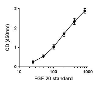
Application: ELISASample Tested: Purified StandardSpecies: HumanVerified Customer | Posted 07/17/2017Sandwich ELISA. Tested against hFGF20 standards (800 pg/ml-25 pg/ml), run in duplicate.
There are no reviews that match your criteria.
Protocols
Find general support by application which include: protocols, troubleshooting, illustrated assays, videos and webinars.
- Appropriate Fixation of IHC/ICC Samples
- Cellular Response to Hypoxia Protocols
- ClariTSA™ Fluorophore Kits
- Detection & Visualization of Antibody Binding
- ICC Cell Smear Protocol for Suspension Cells
- ICC Immunocytochemistry Protocol Videos
- ICC for Adherent Cells
- Immunocytochemistry (ICC) Protocol
- Immunocytochemistry Troubleshooting
- Immunofluorescence of Organoids Embedded in Cultrex Basement Membrane Extract
- Immunohistochemistry (IHC) and Immunocytochemistry (ICC) Protocols
- Preparing Samples for IHC/ICC Experiments
- Preventing Non-Specific Staining (Non-Specific Binding)
- Primary Antibody Selection & Optimization
- Protocol for VisUCyte™ HRP Polymer Detection Reagent
- Protocol for the Fluorescent ICC Staining of Cell Smears - Graphic
- Protocol for the Fluorescent ICC Staining of Cultured Cells on Coverslips - Graphic
- Protocol for the Preparation and Fluorescent ICC Staining of Cells on Coverslips
- Protocol for the Preparation and Fluorescent ICC Staining of Non-adherent Cells
- Protocol for the Preparation and Fluorescent ICC Staining of Stem Cells on Coverslips
- Protocol for the Preparation of a Cell Smear for Non-adherent Cell ICC - Graphic
- R&D Systems Quality Control Western Blot Protocol
- TUNEL and Active Caspase-3 Detection by IHC/ICC Protocol
- The Importance of IHC/ICC Controls
- Troubleshooting Guide: Western Blot Figures
- Western Blot Conditions
- Western Blot Protocol
- Western Blot Protocol for Cell Lysates
- Western Blot Troubleshooting
- Western Blot Troubleshooting Guide
- View all Protocols, Troubleshooting, Illustrated assays and Webinars
Loading...