Human Fibroblast Activation Protein alpha /FAP Antibody
Human Fibroblast Activation Protein alpha /FAP Antibody Summary
Leu26-Asp760
Accession # Q12884
*Small pack size (-SP) is supplied either lyophilized or as a 0.2 µm filtered solution in PBS.
Applications
Please Note: Optimal dilutions should be determined by each laboratory for each application. General Protocols are available in the Technical Information section on our website.
Scientific Data
 View Larger
Detection of Human Fibroblast Activation Protein alpha /FAP by Western Blot. Western blot shows lysates of WI-38 human lung fibroblast cell line. PVDF membrane was probed with 0.5 µg/mL of Sheep Anti-Human Fibroblast Activation Protein a/FAP Antigen Affinity-purified Polyclonal Antibody (Catalog # AF3715) followed by HRP-conjugated Anti-Sheep IgG Secondary Antibody (Catalog # HAF016). A specific band was detected for Fibroblast Activation Protein a/FAP at approximately 97 kDa (as indicated). This experiment was conducted under reducing conditions and using Immunoblot Buffer Group 1.
 View Larger
Fibroblast Activation Protein alpha /FAP in Human Squamous Cell Carcinoma. Fibroblast Activation Protein a/FAP was detected in immersion fixed paraffin-embedded sections of human squamous cell carcinoma using Sheep Anti-Human Fibroblast Activation Protein a/FAP Antigen Affinity-purified Polyclonal Antibody (Catalog # AF3715) at 15 µg/mL overnight at 4 °C. Tissue was stained using the Anti-Sheep HRP-DAB Cell & Tissue Staining Kit (brown; Catalog # CTS019) and counterstained with hematoxylin (blue). Specific staining was localized to connective tissue. View our protocol for Chromogenic IHC Staining of Paraffin-embedded Tissue Sections.
 View Larger
Fibroblast Activation Protein alpha /FAP in Human Basal Cell Carcinoma. Fibroblast Activation Protein alpha /FAP was detected in immersion fixed paraffin-embedded sections of human basal cell carcinoma using Sheep Anti-Human Fibroblast Activation Protein alpha /FAP Antigen Affinity-purified Polyclonal Antibody (Catalog # AF3715) at 10 µg/mL overnight at 4 °C. Tissue was stained using the Anti-Sheep HRP-DAB Cell & Tissue Staining Kit (brown; Catalog # CTS019) and counterstained with hematoxylin (blue). Specific staining was localized to cytoplasm. View our protocol for Chromogenic IHC Staining of Paraffin-embedded Tissue Sections.
 View Larger
Detection of Human Fibroblast Activation Protein alpha /FAP by Simple WesternTM. Simple Western lane view shows lysates of IMR‑90 human lung fibroblast cell line and WI‑38 human lung fibroblast cell line, loaded at 0.2 mg/mL. A specific band was detected for Fibroblast Activation Protein alpha /FAP at approximately 130 kDa (as indicated) using 10 µg/mL of Sheep Anti-Human Fibroblast Activation Protein alpha /FAP Antigen Affinity-purified Polyclonal Antibody (Catalog # AF3715) followed by 1:50 dilution of HRP-conjugated Anti-Sheep IgG Secondary Antibody (HAF016). This experiment was conducted under reducing conditions and using the 12-230 kDa separation system.
 View Larger
Western Blot Shows Human Fibroblast Activation Protein alpha /FAP Specificity by Using Knockout Cell Line. Western blot shows lysates of WI‑38 human lung fibroblast cell line and human FAP knockout WI‑38 human lung fibroblast cell line. PVDF membrane was probed with 0.5 µg/mL of Sheep Anti-Human Fibroblast Activation Protein alpha /FAP Antigen Affinity-purified Polyclonal Antibody (Catalog # AF3715) followed by HRP-conjugated Anti-Sheep IgG Secondary Antibody (HAF016). A specific band was detected for Fibroblast Activation Protein alpha /FAP at approximately 97 kDa (as indicated) in the parental WI‑38 human lung fibroblast cell line, but is not detectable in knockout WI‑38 human lung fibroblast cell line. GAPDH (AF5718) is shown as a loading control. This experiment was conducted under reducing conditions and using Western Blot Buffer Group 1.
 View Larger
Detection of Fibroblast Activation Protein alpha /FAP in Human Esophagus. Formalin-fixed paraffin-embedded tissue sections of human squamous cell carcinoma in the esophagus were probed for FAP mRNA (ACD RNAScope Probe, catalog # 411971; Fast Red chromogen, ACD catalog # 322360). Adjacent tissue section was processed for immunohistochemistry using sheep anti-human FAP polyclonal antibody (R&D Systems catalog # AF3715) at 5ug/mL with overnight incubation at 4 degrees Celsius followed by incubation with anti-sheep IgG VisUCyte HRP Polymer Antibody (Catalog # VC006) and DAB chromogen (yellow-brown). Tissue was counterstained with hematoxylin (blue). Specific staining was localized to connective tissue.
 View Larger
Detection of Fibroblast Activation Protein alpha /FAP by Western Blot FGFR2 drives the process of liver fibrosis. (A) The expression of liver fibrosis markers (ACTA2, fibroblast activation protein alpha (FAP), alpha-1 type I collagen (COL1A1)) was measured by qPCR. The samples were divided into four groups according to whether cells were induced with or without TGF-beta and whether cells were overexpressed by FGFR2. (B) The expression of markers in wild-type and FGFR2-OE cell lines under TGF-beta induction was analyzed by Western blot analysis. (C) The expression levels of alpha -SMA and (D) collagen secretion in LX-2 and Huh-7 were evaluated, when FGFR2 was overexpressed or knocked down upon equal TGF-beta induction. (E) Cells were collected after 48 h of co-culture, and the expression of alpha -SMA and (F) type I collagen in the lower layer cells was determined by ELISA. The results are marked as significant “*” when p < 0.05, “**” when p < 0.01, and not significant (ns) if p ≥ 0.05. Image collected and cropped by CiteAb from the following open publication (https://pubmed.ncbi.nlm.nih.gov/37111305), licensed under a CC-BY license. Not internally tested by R&D Systems.
 View Larger
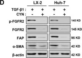
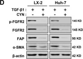
Detection of Fibroblast Activation Protein alpha /FAP by Western Blot CYN blocks the activation and development of liver fibrosis in vitro. (A) Evaluation of the inhibitory effect of CYN on the fibrosis-promoting effect of FGFR2. The fibrotic transformation of wild-type and FGFR2-OE LX-2 cells and Huh-7 cells was induced through TGF-beta activation, followed by an intervention with CYN for the relevant groups. The expression changes of the fibrosis markers ACTA2 and COL1A1 were analyzed by qPCR. (B) Wild-type cell lines were employed in the aforementioned experiments, and the activation of FGFR2 was triggered by supplementation with the exogenous basic fibroblast growth factor (bFGF) factor. (C) Analysis of the extent of antagonism of CYN towards TGF-beta Signaling. Activation induction models were established by adding or not adding TGF-beta to the cell culture environment with or without the CYN intervention, and the expression of liver fibrosis markers was determined by qPCR, (D) Western blot, and (E,F) ELISA analyses. (G) A co-culture model was used to evaluate the blocking effect of CYN on liver fibrosis activation of signaling transmission. The activation intensity of lower-layer wild-type cells was measured and compared using alpha -SMA expression and collagen secretion. The results are marked as significant “*” when p < 0.05, “**” when p < 0.01, and not significant (ns) if p ≥ 0.05. Image collected and cropped by CiteAb from the following open publication (https://pubmed.ncbi.nlm.nih.gov/37111305), licensed under a CC-BY license. Not internally tested by R&D Systems.
 View Larger
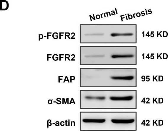
Detection of Fibroblast Activation Protein alpha /FAP by Western Blot High expression of fibroblast growth factor receptor 2 (FGFR2) coincides with liver fibrosis. (A) The expression of FGFR2 in fibrotic liver and normal liver tissues was evaluated through data mining of human samples affected by Hepatitis B infection (GSE38941), alcohol abuse (GSE28619), and nonalcoholic steatohepatitis (GSE48452), as well as mouse samples induced with carbon tetrachloride (CCl4) (GSE152329, GSE98577). The differential fold expression was determined by comparing the expression of FGFR2 in the fibrotic liver and normal liver tissues. (B) A comparison of changes in FGFR2 expression in liver fibrosis patients in remission (improved) or not in remission (not improved) with mining data of GSE175448. (C) The correlation between FGFR2 expression and the liver fibrosis score (Ishak score) was analyzed by comparing FGFR2 expression in three pairs of liver fibrotic tissues and normal liver tissues through immunohistochemistry. (D) FGFR2 expression in liver fibrosis tissues and normal liver tissues was verified by Western blot and (E) q-PCR analyses. (E) Liver tissues from mice treated with CCl4 for different durations were used to determine the expression of the liver fibrosis markers Actin Alpha 2 (ACTA2) and FGFR2 by q-PCR and the trends of their expression with increasing days of induction. (F) The expression of ACTA2 and (G) FGFR2 was determined by q-PCR in liver tissues from mice treated with CCl4 for varying durations. The statistical significance was determined based on the p-value. The results are marked as significant “*” when p < 0.05, “**” when p < 0.01, “***” when p < 0.001, and not significant (ns) if p ≥ 0.05. Image collected and cropped by CiteAb from the following open publication (https://pubmed.ncbi.nlm.nih.gov/37111305), licensed under a CC-BY license. Not internally tested by R&D Systems.
 View Larger
Detection of Fibroblast Activation Protein alpha /FAP by Western Blot Expression analysis of Fap in OA synovium. a Immunostaining of human FAP in the synovium of control and OA patients. DAPI staining indicates the nucleus. Scale bars: 100 μm. b qPCR analysis of human FAP mRNA levels in the synovium of control and OA patients (n = 8 samples per group). c Western blot analysis of human FAP protein levels in the synovium of control and OA patients (n = 3 samples per group). d Immunostaining of mouse Fap in the knee joint of sham and DMM-treated mice. Sham or DMM surgery was performed in 8-week-old FapLacZ/+ mice, which were sacrificed 8 weeks later, and LacZ immunostaining was used to detect Fap expression at the posterior horn of the medial meniscus (F: femur; T: tibia; M: meniscus; S: synovium). DAPI staining indicates the nucleus. Scale bars: 100 μm. The statistical significance was assessed using two-tailed Student’s unpaired t tests. Data are presented as the mean ± SD (**P < 0.01) Image collected and cropped by CiteAb from the following open publication (https://pubmed.ncbi.nlm.nih.gov/36588124), licensed under a CC-BY license. Not internally tested by R&D Systems.
 View Larger
Detection of Fibroblast Activation Protein alpha /FAP by Western Blot FGFR2 drives the process of liver fibrosis. (A) The expression of liver fibrosis markers (ACTA2, fibroblast activation protein alpha (FAP), alpha-1 type I collagen (COL1A1)) was measured by qPCR. The samples were divided into four groups according to whether cells were induced with or without TGF-beta and whether cells were overexpressed by FGFR2. (B) The expression of markers in wild-type and FGFR2-OE cell lines under TGF-beta induction was analyzed by Western blot analysis. (C) The expression levels of alpha -SMA and (D) collagen secretion in LX-2 and Huh-7 were evaluated, when FGFR2 was overexpressed or knocked down upon equal TGF-beta induction. (E) Cells were collected after 48 h of co-culture, and the expression of alpha -SMA and (F) type I collagen in the lower layer cells was determined by ELISA. The results are marked as significant “*” when p < 0.05, “**” when p < 0.01, and not significant (ns) if p ≥ 0.05. Image collected and cropped by CiteAb from the following open publication (https://pubmed.ncbi.nlm.nih.gov/37111305), licensed under a CC-BY license. Not internally tested by R&D Systems.
 View Larger
Detection of Fibroblast Activation Protein alpha /FAP by Western Blot Expression analysis of Fap in OA synovium. a Immunostaining of human FAP in the synovium of control and OA patients. DAPI staining indicates the nucleus. Scale bars: 100 μm. b qPCR analysis of human FAP mRNA levels in the synovium of control and OA patients (n = 8 samples per group). c Western blot analysis of human FAP protein levels in the synovium of control and OA patients (n = 3 samples per group). d Immunostaining of mouse Fap in the knee joint of sham and DMM-treated mice. Sham or DMM surgery was performed in 8-week-old FapLacZ/+ mice, which were sacrificed 8 weeks later, and LacZ immunostaining was used to detect Fap expression at the posterior horn of the medial meniscus (F: femur; T: tibia; M: meniscus; S: synovium). DAPI staining indicates the nucleus. Scale bars: 100 μm. The statistical significance was assessed using two-tailed Student’s unpaired t tests. Data are presented as the mean ± SD (**P < 0.01) Image collected and cropped by CiteAb from the following open publication (https://pubmed.ncbi.nlm.nih.gov/36588124), licensed under a CC-BY license. Not internally tested by R&D Systems.
 View Larger
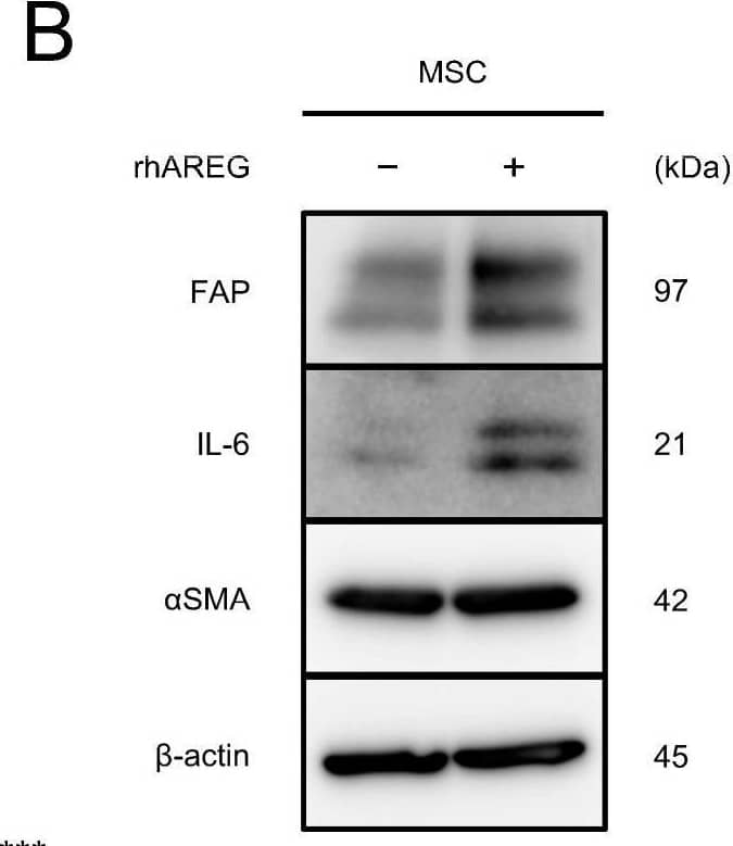
Detection of Fibroblast Activation Protein alpha /FAP by Western Blot AREG promotes migration and CAF-like differentiation of MSCs: (A,B) To investigate the differentiation of MSCs into CAFs upon treatment with rhAREG (100 ng/mL), the mRNA and protein expression levels of FAP, IL-6, and alpha SMA were compared using qRT-PCR (A) and Western blotting (B). beta -actin was used as a loading control (B). (C,D) The effects of rhAREG (100 ng/mL) on the survival (C) and growth (D) of MSCs were evaluated using the MTS assay. (E) The effect of rhAREG (100 ng/mL) on the migration of MSCs was evaluated using the transwell migration assay. MSCs were seeded in the upper chamber, and migrated cells were counted in five fields of view after 48 h. Representative images for each condition are presented below the graph (E). The data are presented as the mean ± SEM of three independent experiments (A,C–E). N.S., not significant; ** p < 0.01, *** p < 0.001. Scale bars: 100 μm (E). Image collected and cropped by CiteAb from the following open publication (https://pubmed.ncbi.nlm.nih.gov/39451251), licensed under a CC-BY license. Not internally tested by R&D Systems.
Reconstitution Calculator
Preparation and Storage
- 12 months from date of receipt, -20 to -70 °C as supplied.
- 1 month, 2 to 8 °C under sterile conditions after reconstitution.
- 6 months, -20 to -70 °C under sterile conditions after reconstitution.
Background: Fibroblast Activation Protein alpha/FAP
FAP (also known as seprase) is a Type II transmembrane serine protease that is structurally related to dipeptidyl peptidase IV (1). FAP has substrate specificity similar to dipeptidyl peptidase IV, which is specific for N-terminal Xaa-Pro sequences, but FAP is also an endopeptidase able to degrade gelatin and Type I collagen (2). The enzymatically active form of FAP is a dimer (3). FAP has a restricted tissue distribution. It is not detectable in normal tissues or resting fibroblasts, but is highly expressed on reactive stromal fibroblasts in epithelial cancers (4), in granulation tissue during wound healing, and in bone and soft tissue sarcomas (5). Because of its expression patterns and enzymatic activities, FAP is believed to play roles in tumor invasion, tissue remodeling, and wound repair.
- Scanlan, M.J. et al. (1994) Proc. Natl. Acad. Sci. USA 91:5657.
- Park, J.E. et al. (1999) J. Biol. Chem. 274:36505.
- Pineiro-Sanchez, M.L. et al. (1997) J. Biol. Chem. 272:7595.
- Garin-Chesa, P. et al. (1990) Proc. Natl. Acad. Sci. USA 87:7235.
- Rettig, W.J. et al. (1988) Proc. Natl. Acad. Sci. USA 85:3110.
Product Datasheets
Citations for Human Fibroblast Activation Protein alpha /FAP Antibody
R&D Systems personnel manually curate a database that contains references using R&D Systems products. The data collected includes not only links to publications in PubMed, but also provides information about sample types, species, and experimental conditions.
48
Citations: Showing 1 - 10
Filter your results:
Filter by:
-
Macrophages are exploited from an innate wound healing response to facilitate cancer metastasis
Authors: T Muliaditan, J Caron, M Okesola, JW Opzoomer, P Kosti, M Georgouli, P Gordon, S Lall, DM Kuzeva, L Pedro, JD Shields, CE Gillett, SS Diebold, V Sanz-Moren, T Ng, E Hoste, JN Arnold
Nat Commun, 2018-07-27;9(1):2951.
-
Preclinical platforms to study therapeutic efficacy of human gamma δ T cells
Authors: Lingling Ou, Huaishan Wang, Hui Huang, Zhiyan Zhou, Qiang Lin, Yeye Guo et al.
Clinical and Translational Medicine
-
Increased expression of cancer-associated fibroblast markers at the invasive front and its association with tumor-stroma ratio in colorectal cancer
Authors: TP Sandberg, MPME Stuart, J Oosting, RAEM Tollenaar, CFM Sier, WE Mesker
BMC Cancer, 2019-03-29;19(1):284.
-
Collagen Family and Other Matrix Remodeling Proteins Identified by Bioinformatics Analysis as Hub Genes Involved in Gastric Cancer Progression and Prognosis
Authors: Chivu-Economescu M, Necula LG, Matei L et al.
International journal of molecular sciences
-
Fibroblast Activation Protein (FAP)+ cancer-associated fibroblasts induce macrophage M2-like polarization via the Fibronectin 1-Integrin ?5?1 axis in breast cancer
Authors: Chen, W;Jiang, M;Zou, X;Chen, Z;Shen, L;Hu, J;Kong, M;Huang, J;Ni, C;Xia, W;
Oncogene
Species: Human
Sample Types: Whole Tissue
Applications: Immunohistochemistry -
Injured Cardiac Tissue-Targeted Delivery of TGF?1 siRNA by FAP Aptamer-Functionalized Extracellular Vesicles Promotes Cardiac Repair
Authors: Kang, JY;Mun, D;Park, M;Yoo, G;Kim, H;Yun, N;Joung, B;
International journal of nanomedicine
Species: Human, Mouse
Sample Types: Cell Lysates, Whole Tissue
Applications: Immunohistochemistry, Western Blot -
The multifaceted role of the stroma in the healthy prostate and prostate cancer
Authors: Di Carlo, E;Sorrentino, C;
Journal of translational medicine
Species: Human
Sample Types: Whole Tissue
Applications: Immunohistochemistry -
Senescent fibroblasts in the tumor stroma rewire lung cancer metabolism and plasticity
Authors: Lee, JY;Reyes, N;Woo, SH;Goel, S;Stratton, F;Kuang, C;Mansfield, AS;LaFave, LM;Peng, T;
bioRxiv : the preprint server for biology
Species: Transgenic Mouse
Sample Types: Whole Tissue
Applications: Immunohistochemistry -
Expression of fibroblast activation protein-? in human deep vein thrombosis
Authors: Oguri, N;Gi, T;Nakamura, E;Furukoji, E;Goto, H;Maekawa, K;Tsuji, AB;Nishii, R;Aman, M;Moriguchi-Goto, S;Sakae, T;Azuma, M;Yamashita, A;
Thrombosis research
Species: Human
Sample Types: Whole Cells
Applications: Immunocytochemistry -
Obesity defined molecular endotypes in the synovium of patients with osteoarthritis provides a rationale for therapeutic targeting of fibroblast subsets
Authors: Susanne N. Wijesinghe, Amel Badoume, Dominika E. Nanus, Archana Sharma‐Oates, Hussein Farah, Michelangelo Certo et al.
Clinical and Translational Medicine
-
Functional roles of FAP-alpha in metabolism, migration and invasion of human cancer cells
Authors: Noriko Mori, Jiefu Jin, Balaji Krishnamachary, Yelena Mironchik, Flonné Wildes, Farhad Vesuna et al.
Frontiers in Oncology
-
Engineering edgeless human skin with enhanced biomechanical properties
Authors: A Pappalardo, D Alvarez Ce, S Fang, AR Herschman, EY Jeon, KM Myers, JW Kysar, HE Abaci
Science Advances, 2023-01-27;9(4):eade2514.
Species: Human
Sample Types: Whole Tissue
Applications: IHC -
Inhibition of fibroblast activation protein ameliorates cartilage matrix degradation and osteoarthritis progression
Authors: Aoyuan Fan, Genbin Wu, Jianfang Wang, Laiya Lu, Jingyi Wang, Hanjing Wei et al.
Bone Research
-
3D bioprinting of an implantable xeno‐free vascularized human skin graft
Authors: Tania Baltazar, Bo Jiang, Alejandra Moncayo, Jonathan Merola, Mohammad Z. Albanna, W. Mark Saltzman et al.
Bioengineering & Translational Medicine
-
Evaluating near‐infrared photoimmunotherapy for targeting fibroblast activation protein‐ alpha expressing cells in vitro and in vivo
Authors: Jiefu Jin, James D. Barnett, Balaji Krishnamachary, Yelena Mironchik, Catherine K. Luo, Hisataka Kobayashi et al.
Cancer Science
-
FAP is critical for ovarian cancer cell survival by sustaining NF-kappaB activation through recruitment of PRKDC in lipid rafts
Authors: B Li, Z Ding, O Calbay, Y Li, T Li, L Jin, S Huang
Cancer Gene Therapy, 2022-12-09;0(0):.
Species: Mouse
Sample Types: Whole Cell
Applications: IP -
CCL18 signaling from tumor-associated macrophages activates fibroblasts to adopt a chemoresistance-inducing phenotype
Authors: W Zeng, L Xiong, W Wu, S Li, J Liu, L Yang, L Lao, P Huang, M Zhang, H Chen, N Miao, Z Lin, Z Liu, X Yang, J Wang, P Wang, E Song, Y Yao, Y Nie, J Chen, D Huang
Oncogene, 2022-11-22;0(0):.
Species: Human
Sample Types: Cell Lysates, Whole Cells, Whole Tissue
Applications: Flow Cytometry, IHC, Western Blot -
Thy-1 plays a pathogenic role and is a potential biomarker for skin fibrosis in scleroderma
Authors: RG Marangoni, P Datta, A Paine, S Duemmel, MA Nuzzo, L Sherwood, J Varga, C Ritchlin, BD Korman
JCI Insight, 2022-10-10;0(0):.
Species: Mouse
Sample Types: Whole Tissue
Applications: IHC -
Type IV Collagen in Human Colorectal Liver Metastases-Cellular Origin and a Circulating Biomarker
Authors: M Lindgren, G Rask, J Jonsson, A Berglund, C Lundin, P Jonsson, I Ljuslinder, H Nyström
Cancers, 2022-07-13;14(14):.
Species: Human
Sample Types: Whole Tissue
Applications: IHC -
A stromal Integrated Stress Response activates perivascular cancer-associated fibroblasts to drive angiogenesis and tumour progression
Authors: II Verginadis, H Avgousti, J Monslow, G Skoufos, F Chinga, K Kim, NM Leli, IV Karagounis, BI Bell, A Velalopoul, CS Salinas, VS Wu, Y Li, J Ye, DA Scott, AL Osterman, A Sengupta, A Weljie, M Huang, D Zhang, Y Fan, E Radaelli, JW Tobias, F Rambow, P Karras, JC Marine, X Xu, AG Hatzigeorg, S Ryeom, JA Diehl, SY Fuchs, E Puré, C Koumenis
Nature Cell Biology, 2022-06-02;0(0):.
Species: Mouse
Sample Types: Whole Tissue
Applications: IHC -
Single-cell proteomics defines the cellular heterogeneity of localized prostate cancer
Authors: Laura De Vargas Roditi, Andrea Jacobs, Jan H. Rueschoff, Pete Bankhead, Stéphane Chevrier, Hartland W. Jackson et al.
Cell Reports Medicine
-
Fibroblast Activation Protein Specific Optical Imaging in Non-Small Cell Lung Cancer
Authors: Layla Mathieson, Richard A. O’Connor, Hazel Stewart, Paige Shaw, Kevin Dhaliwal, Gareth O. S. Williams et al.
Frontiers in Oncology
Species: Human
Sample Types: Whole Cells
Applications: Flow Cytometry -
The FAP alpha -activated prodrug Z-GP-DAVLBH inhibits the growth and pulmonary metastasis of osteosarcoma cells by suppressing the AXL pathway
Authors: Geni Ye, Maohua Huang, Yong Li, Jie Ouyang, Minfeng Chen, Qing Wen et al.
Acta Pharmaceutica Sinica B
-
IL-1-driven stromal-neutrophil interactions define a subset of patients with inflammatory bowel disease that does not respond to therapies
Authors: M Friedrich, M Pohin, MA Jackson, I Korsunsky, SJ Bullers, K Rue-Albrec, Z Christofor, D Sathananth, T Thomas, R Ravindran, R Tandon, RS Peres, H Sharpe, K Wei, GFM Watts, EH Mann, A Geremia, M Attar, Oxford IBD, Roche Fibr, S McCuaig, L Thomas, E Collantes, HH Uhlig, SN Sansom, A Easton, S Raychaudhu, SP Travis, FM Powrie
Nature Medicine, 2021-10-21;0(0):.
Species: Human
Sample Types: Whole Tissue
Applications: IHC -
Metallothionein 2A Expression in Cancer-Associated Fibroblasts and Cancer Cells Promotes Esophageal Squamous Cell Carcinoma Progression
Authors: M Shimizu, YI Koma, H Sakamoto, S Tsukamoto, Y Kitamura, S Urakami, K Tanigawa, T Kodama, N Higashino, M Nishio, M Shigeoka, Y Kakeji, H Yokozaki
Cancers, 2021-09-10;13(18):.
Species: Human
Sample Types: Cell Lysates
Applications: Western Blot -
T cells drive negative feedback mechanisms in cancer associated fibroblasts, promoting expression of co-inhibitory ligands, CD73 and IL-27 in non-small cell lung cancer
Authors: Richard A O’Connor, Vishwani Chauhan, Layla Mathieson, Helen Titmarsh, Lilian Koppensteiner, Irene Young et al.
OncoImmunology
-
Proteomic Analyses of Fibroblast- and Serum-Derived Exosomes Identify QSOX1 as a Marker for Non-invasive Detection of Colorectal Cancer
Authors: N Ganig, F Baenke, ML Thepkayson, K Lin, VS Rao, FC Wong, H Polster, M Schneider, D Helm, M Pecqueux, AM Seifert, L Seifert, J Weitz, NN Rahbari, C Kahlert
Cancers, 2021-03-17;13(6):.
Species: Human
Sample Types: Cell Lysates, Whole Tissue
Applications: IHC, Western Blot -
Extreme Diversity of the Human Vascular Mesenchymal Cell Landscape
Authors: LE Bruijn, BEWM van den Ak, CM van Rhijn, JF Hamming, JHN Lindeman
J Am Heart Assoc, 2020-11-16;9(23):e017094.
Species: Human
Sample Types: Whole Cells, Whole Tissue
Applications: ICC, IHC -
Cancer-associated fibroblasts downregulate type I interferon receptor to stimulate intratumoral stromagenesis
Authors: C Cho, R Mukherjee, AR Peck, Y Sun, N McBrearty, KV Katlinski, J Gui, PK Govindaraj, E Puré, H Rui, SY Fuchs
Oncogene, 2020-08-17;0(0):.
Species: Human
Sample Types: Whole Cells, Whole Tissue
Applications: ICC, IHC -
Overexpression of B7-H3 in alpha -SMA-Positive Fibroblasts Is Associated With Cancer Progression and Survival in Gastric Adenocarcinomas
Authors: Shenghua Zhan, Zhiju Liu, Min Zhang, Tianwei Guo, Qiuying Quan, Lili Huang et al.
Frontiers in Oncology
-
Patch repair of deep wounds by mobilized fascia
Authors: D Correa-Gal, D Jiang, S Christ, P Ramesh, H Ye, J Wannemache, S Kalgudde G, Q Yu, M Aichler, A Walch, U Mirastschi, T Volz, Y Rinkevich
Nature, 2019-11-27;0(0):.
Species: Human
Sample Types: Whole Tissue
Applications: IHC -
Highly versatile cancer photoimmunotherapy using photosensitizer-conjugated avidin and biotin-conjugated targeting antibodies
Authors: N Shirasu, H Shibaguchi, H Yamada, M Kuroki, S Yasunaga
Cancer Cell Int., 2019-11-15;19(0):299.
Species: Human
Sample Types: Whole Cells
Applications: Photoimmunotherapy -
A Single-Cell Atlas of the Tumor and Immune Ecosystem of Human Breast Cancer
Authors: Johanna Wagner, Maria Anna Rapsomaniki, Stéphane Chevrier, Tobias Anzeneder, Claus Langwieder, August Dykgers et al.
Cell
Species: Mouse
Sample Types: Whole Cells
Applications: Flow Cytometry -
Fibroblast activation protein-positive fibroblasts promote tumor progression through secretion of CCL2 and interleukin-6 in esophageal squamous cell carcinoma
Authors: N Higashino, YI Koma, M Hosono, N Takase, M Okamoto, H Kodaira, M Nishio, M Shigeoka, Y Kakeji, H Yokozaki
Lab. Invest., 2019-01-25;0(0):.
Species: Human
Sample Types: Whole Cells, Whole Tissue
Applications: IHC -
Pro-tumorigenic roles of fibroblast activation protein in cancer: back to the basics
Authors: Ellen Puré, Rachel Blomberg
Oncogene
-
Cancer-associated fibroblasts induce antigen-specific deletion of CD8+T Cells to protect tumour cells
Authors: MA Lakins, E Ghorani, H Munir, CP Martins, JD Shields
Nat Commun, 2018-03-05;9(1):948.
Species: Mouse
Sample Types: Whole Cells
Applications: Flow Cytometry -
Tumor-targeting efficacy of a BF211 prodrug through hydrolysis by fibroblast activation protein-?
Authors: XP Chai, GL Sun, YF Fang, LH Hu, X Liu, XW Zhang
Acta Pharmacol. Sin., 2017-11-09;0(0):.
Species: Mouse
Sample Types: Tissue Homogenates
Applications: Western Blot -
Fibroblast activation protein-alpha promotes the growth and migration of lung cancer cells via the PI3K and sonic hedgehog pathways
Authors: Jun Jia, Tracey A. Martin, Lin Ye, Lin Meng, Nan Xia, Wen G. Jiang et al.
International Journal of Molecular Medicine
-
Fibroblast activation protein augments progression and metastasis of pancreatic ductal adenocarcinoma
Authors: A Lo, CP Li, EL Buza, R Blomberg, P Govindaraj, D Avery, J Monslow, M Hsiao, E Puré
JCI Insight, 2017-10-05;2(19):.
Species: Human
Sample Types: Whole Tissue
Applications: IHC-P -
Adrenergic-mediated increases in INHBA drive CAF phenotype and collagens
Authors: AS Nagaraja, RL Dood, G Armaiz-Pen, Y Kang, SY Wu, JK Allen, NB Jennings, LS Mangala, S Pradeep, Y Lyons, M Haemmerle, KM Gharpure, NC Sadaoui, C Rodriguez-, C Ivan, Y Wang, K Baggerly, P Ram, G Lopez-Bere, J Liu, SC Mok, L Cohen, SK Lutgendorf, SW Cole, AK Sood
JCI Insight, 2017-08-17;2(16):.
Species: Mouse
Sample Types: Whole Tissue
Applications: IHC -
A synthetic urinary probe-coated nanoparticles sensitive to fibroblast activation protein ? for solid tumor diagnosis
Authors: X Feng, Q Wang, Y Liao, X Zhou, Y Wang, W Liu, G Zhang
Int J Nanomedicine, 2017-07-27;12(0):5359-5372.
Species: Human, Mouse
Sample Types: Cell Lysates
Applications: Western Blot -
GM-CSF Mediates Mesenchymal–Epithelial Cross-talk in Pancreatic Cancer
Authors: Meghna Waghray, Malica Yalamanchili, Michele Dziubinski, Mina Zeinali, Marguerite Erkkinen, Huibin Yang et al.
Cancer Discovery
-
Fibroblast Activation Protein (FAP) Accelerates Collagen Degradation and Clearance from Lungs in Mice
Authors: MH Fan, Q Zhu, HH Li, HJ Ra, S Majumdar, DL Gulick, JA Jerome, DH Madsen, M Christofid, DW Speicher, WW Bachovchin, C Feghali-Bo, E Puré
J. Biol. Chem., 2015-12-09;291(15):8070-89.
Species: Mouse
Sample Types: Tissue Homogenates, Whole Tissue
Applications: IHC, Immunoprecipitation, Western Blot -
miR-142-5p and miR-130a-3p are regulated by IL-4 and IL-13 and control profibrogenic macrophage program
Authors: Shicheng Su, Qiyi Zhao, Chonghua He, Di Huang, Jiang Liu, Fei Chen et al.
Nature Communications
Species: Human
Sample Types: Whole Cells
Applications: Immunocytochemistry -
Antitumor effects of chimeric receptor engineered human T cells directed to tumor stroma.
Authors: Kakarla S, Chow K, Mata M, Shaffer D, Song X, Wu M, Liu H, Wang L, Rowley D, Pfizenmaier K, Gottschalk S
Mol Ther, 2013-06-04;21(8):1611-20.
Species: Human
Sample Types: Whole Cells
Applications: CAR-T (Flow Cytometry), CAR-T (Flow Cytomety) -
Depletion of stromal cells expressing fibroblast activation protein-alpha from skeletal muscle and bone marrow results in cachexia and anemia
Authors: Edward W. Roberts, Andrew Deonarine, James O. Jones, Alice E. Denton, Christine Feig, Scott K. Lyons et al.
Journal of Experimental Medicine
-
Suppression of antitumor immunity by stromal cells expressing fibroblast activation protein-alpha.
Authors: Kraman M, Bambrough P, Arnold J, Roberts E, Magiera L, Jones J, Gopinathan A, Tuveson D, Fearon D
Science, 2010-11-05;330(6005):827-30.
Species: Mouse
Sample Types: Whole Cells, Whole Tissue
Applications: Flow Cytometry, IHC -
Macrophages orchestrate the expansion of a proangiogenic perivascular niche during cancer progression
Authors: JW Opzoomer, JE Anstee, I Dean, EJ Hill, I Bouybayoun, J Caron, T Muliaditan, P Gordon, D Sosnowska, R Nuamah, SE Pinder, T Ng, F Dazzi, S Kordasti, DR Withers, T Lawrence, JN Arnold
Science Advances, 2021-11-03;7(45):eabg9518.
FAQs
No product specific FAQs exist for this product, however you may
View all Antibody FAQsReviews for Human Fibroblast Activation Protein alpha /FAP Antibody
Average Rating: 4.5 (Based on 2 Reviews)
Have you used Human Fibroblast Activation Protein alpha /FAP Antibody?
Submit a review and receive an Amazon gift card.
$25/€18/£15/$25CAN/¥75 Yuan/¥2500 Yen for a review with an image
$10/€7/£6/$10 CAD/¥70 Yuan/¥1110 Yen for a review without an image
Filter by:
Human Fibroblast Activation Protein alpha
FAP Antibody ref AF3715 RetD Systems
Polyclonal Sheep . 95kd mais peut présenter
Des glycosylations d’où la taille aux alentours de
120kd!
La prochaine fois:
- Utiliser des concentrations de protéines >0,6mg/ml
- Diluer l’anticorps secondaire sheep 1/25è au lieu du
1/10è pour éviter un peu plus le bruit de fond.
- Reconstituer l’anticorps à 0,5mg/ml
Au lieu de 0,2mg/ml recommandé par R et D.
Efficacité validée dans conditions suivantes:
Dilution anticorps FAP= 1/10è (0,2mg/ml solution stock)
[CMLh]= 0,7mg/ml
Dilution anticorps 2aire sheep = 1/10è


