Human Fibronectin Antibody (P1H11)
R&D Systems | Catalog # MAB1918

Key Product Details
Species Reactivity
Validated:
Human
Cited:
Human, Mouse, Canine
Applications
Validated:
Immunohistochemistry, Western Blot, Flow Cytometry, Immunocytochemistry, Simple Western, Immunoprecipitation
Cited:
Immunohistochemistry, Immunohistochemistry-Paraffin, Western Blot, Neutralization, Immunocytochemistry
Label
Unconjugated
Antibody Source
Monoclonal Mouse IgG1 Clone # P1H11
Loading...
Product Specifications
Immunogen
Human plasma-derived Fibronectin
Specificity
Detects human Fibronectin in Western blots.
Clonality
Monoclonal
Host
Mouse
Isotype
IgG1
Scientific Data Images for Human Fibronectin Antibody (P1H11)
Detection of Fibronectin in HepG2 cells by Flow Cytometry
HepG2 (filled histogram) and Jurkat (open histogram) cells were stained with Mouse Anti-Human Fibronectin Monoclonal Antibody (Catalog # MAB1918, filled histogram) followed by Allophycocyanin-conjugated Anti-Mouse IgG Secondary Antibody (Catalog # F0101B). To facilitate intracellular staining, cells were fixed with Flow Cytometry Fixation Buffer (Catalog # FC004) and permeabilized with Flow Cytometry Permeabilization/Wash Buffer I (Catalog # FC005). View our protocol for Staining Intracellular Molecules.
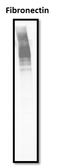
Detection of Human Fibronectin by Western Blot.
Western blot shows lysates of HepG2 human hepatocellular carcinoma cell line and C6 rat glioma cell line. PVDF membrane was probed with 1 µg/mL of Mouse Anti-Human Fibronectin Monoclonal Antibody (Catalog # MAB1918) followed by HRP-conjugated Anti-Mouse IgG Secondary Antibody (HAF018). A specific band was detected for Fibronectin at approximately 300 kDa (as indicated). This experiment was conducted under reducing conditions and using Western Blot Buffer Group 1.
Detection of Human Fibronectin by Simple WesternTM.
Simple Western shows lysates of Exosome Standards (PC‑3) (NBP2-49856), Exosome Standards (Human Urine) (NBP2-49840) and HepG2 human hepatocellular carcinoma cell line, loaded at 0.5 mg/ml. A specific band was detected for Fibronectin at approximately 290 kDa (as indicated) using 10 µg/mL of Mouse Anti-Human Fibronectin Monoclonal Antibody (Catalog # MAB1918). This experiment was conducted under reducing conditions and using the 66-440kDa separation system.
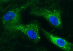
Fibronectin in HepG2 and Daudi Human Cell Lines.
Fibronectin was detected in immersion fixed HepG2 human hepatocellular carcinoma cell line (positive staining; left panel) and Daudi human Burkitt's lymphoma cell line (negative staining; right panel) using Mouse Anti-Human Fibronectin Monoclonal Antibody (Catalog # MAB1918) at 3 µg/mL for 3 hours at room temperature. Cells were stained using the NorthernLights™ 557-conjugated Anti-Mouse IgG Secondary Antibody (red; Catalog # NL007) and counterstained with DAPI (blue). Specific staining was localized to cytoplasm. View our protocol for Fluorescent ICC Staining of Cells on Coverslips.
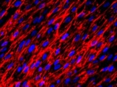
Fibronectin in Human Breast Cancer Tissue.
Fibronectin was detected in immersion fixed paraffin-embedded sections of human breast cancer tissue using Mouse Anti-Human Fibronectin Monoclonal Antibody (Catalog # MAB1918) at 15 µg/mL overnight at 4 °C. Tissue was stained using the Anti-Mouse HRP-DAB Cell & Tissue Staining Kit (brown; Catalog # CTS002) and counterstained with hematoxylin (blue). Specific staining was localized to connective tissue and fibroblasts. View our protocol for Chromogenic IHC Staining of Paraffin-embedded Tissue Sections.
Detection of Fibronectin by Western Blot
DCN inhibits E-cadherin expression and EGFR pathway activation in IBC.a DCN suppresses E-cadherin expression and EGFR signaling in IBC cells. Expression levels of E-cadherin and EGFR are decreased in DCN-overexpressing MDA-IBC3, SUM190, SUM149, and BCX010 cells; also, the phosphorylation of EGFR (pEGFR) and ERK1/2 (pERK1/2) was suppressed in DCN-overexpressing IBC cell lines. Total ERK1/2 (tERK1/2) remains unchanged. GAPDH served as a loading control. b Treatment of IBC cells with DCN protein (4 or 8 μg/mL) for 2 h suppresses E-cadherin expression and EGFR pathway activation. Tubulin served as a loading control. c and d Western blot validation of E-cadherin and EGFR downregulation in tumor samples obtained from mammary fatpad transplantation of control or DCN-overexpressing MDA-IBC3 (c) or SUM149 (d) cells. e and f Immunohistochemical staining validation of E-cadherin and EGFR downregulation in tumor samples obtained from mammary fatpad transplantation of control or DCN-overexpressing MDA-IBC3 (e) or SUM149 (f) cells. Scale bar: 100 μm. g DCN inhibits EGFR signaling in IBC cells independently of EGF stimulation. DCN-overexpressing and control IBC cell lines were stimulated with 50 ng/mL EGF for the indicated number of hours, and total cell lysates were analyzed by western blotting. Both the total levels and the phosphorylation levels of EGFR and ERK1/2 were detected by western blotting. Tubulin served as a loading control. h DCN-mediated inhibition of E-cadherin does not affect expression of epithelial–mesenchymal transition markers. Cell lysates containing 40 μg of total protein were analyzed by western blotting with anti-E-cadherin, fibronectin, vimentin, and DCN antibodies. GAPDH served as a loading control. Image collected and cropped by CiteAb from the following open publication (https://pubmed.ncbi.nlm.nih.gov/33452400), licensed under a CC-BY license. Not internally tested by R&D Systems.Applications for Human Fibronectin Antibody (P1H11)
Application
Recommended Usage
Flow Cytometry
0.25 µg/106 cells
Sample: HepG2 human hepatocellular carcinoma cell line
Sample: HepG2 human hepatocellular carcinoma cell line
Immunocytochemistry
3-25 µg/mL
Sample: Immersion fixed HepG2 human hepatocellular carcinoma cell line
Sample: Immersion fixed HepG2 human hepatocellular carcinoma cell line
Immunohistochemistry
8-25 µg/mL
Sample: Immersion fixed paraffin-embedded sections of human intestine, kidney, and liver, and human breast cancer tissue
Sample: Immersion fixed paraffin-embedded sections of human intestine, kidney, and liver, and human breast cancer tissue
Immunoprecipitation
Yu, H. et al. (2004) Mol. Carcinog. 41:85.
Simple Western
10 µg/mL
Sample: Exosome Standards (PC-3) (Catalog # NBP2-49856), Exosome Standards (Human Urine) (Catalog # NBP2-49840) and HepG2 human hepatocellular carcinoma cell line
Sample: Exosome Standards (PC-3) (Catalog # NBP2-49856), Exosome Standards (Human Urine) (Catalog # NBP2-49840) and HepG2 human hepatocellular carcinoma cell line
Western Blot
1 µg/mL
Sample: HepG2 human hepatocellular carcinoma cell line and C6 rat glioma cell line
Sample: HepG2 human hepatocellular carcinoma cell line and C6 rat glioma cell line
Reviewed Applications
Read 7 reviews rated 4.4 using MAB1918 in the following applications:
Flow Cytometry Panel Builder
Bio-Techne Knows Flow Cytometry
Save time and reduce costly mistakes by quickly finding compatible reagents using the Panel Builder Tool.
Advanced Features
- Spectra Viewer - Custom analysis of spectra from multiple fluorochromes
- Spillover Popups - Visualize the spectra of individual fluorochromes
- Antigen Density Selector - Match fluorochrome brightness with antigen density

Formulation, Preparation, and Storage
Purification
Protein A or G purified from hybridoma culture supernatant
Reconstitution
Reconstitute at 0.5 mg/mL in sterile PBS. For liquid material, refer to CoA for concentration.
Loading...
Formulation
Lyophilized from a 0.2 μm filtered solution in PBS with Trehalose. See Certificate of Analysis for details.
*Small pack size (-SP) is supplied either lyophilized or as a 0.2 µm filtered solution in PBS.
*Small pack size (-SP) is supplied either lyophilized or as a 0.2 µm filtered solution in PBS.
Shipping
Lyophilized product is shipped at ambient temperature. Liquid small pack size (-SP) is shipped with polar packs. Upon receipt, store immediately at the temperature recommended below.
Stability & Storage
Use a manual defrost freezer and avoid repeated freeze-thaw cycles.
- 12 months from date of receipt, -20 to -70 °C as supplied.
- 1 month, 2 to 8 °C under sterile conditions after reconstitution.
- 6 months, -20 to -70 °C under sterile conditions after reconstitution.
Calculators
Background: Fibronectin
Alternate Names
CIG, ED-B, FINC, FN1, FNZ, GFND2, LETS, SMDCF
Gene Symbol
FN1
Additional Fibronectin Products
Product Documents for Human Fibronectin Antibody (P1H11)
Certificate of Analysis
To download a Certificate of Analysis, please enter a lot or batch number in the search box below.
Note: Certificate of Analysis not available for kit components.
Product Specific Notices for Human Fibronectin Antibody (P1H11)
For research use only
Citations for Human Fibronectin Antibody (P1H11)
 Powered by Bioz
Customer Reviews for Human Fibronectin Antibody (P1H11) (7)
4.4 out of 5
7 Customer Ratings
Have you used Human Fibronectin Antibody (P1H11)?
Submit a review and receive an Amazon gift card!
$25/€18/£15/$25CAN/¥2500 Yen for a review with an image
$10/€7/£6/$10CAN/¥1110 Yen for a review without an image
Submit a review
Customer Images
Showing
1
-
5 of
7 reviews
Showing All
Filter By:
-
Application: Immunocytochemistry/ImmunofluorescenceSample Tested: Cardiac fibroblastsSpecies: HumanVerified Customer | Posted 07/08/2022

-
Application: Immunocytochemistry/ImmunofluorescenceSample Tested: fibroblastsSpecies: HumanVerified Customer | Posted 09/28/2021
-
Application: ELISASample Tested: 293T human embryonic kidney cell lineSpecies: HumanVerified Customer | Posted 05/15/2020tested the antibody in whole cell ELISA using fibronectin overexpression cells. It works well.
-
Application: ELISASample Tested: Recombinant proteinSpecies: HumanVerified Customer | Posted 10/01/2019Paired with AF1918
-
Application: Western BlotSample Tested: Purified proteinSpecies: HumanVerified Customer | Posted 08/12/2019
-
Application: Western BlotSample Tested: See PMID 22321604Species: HumanVerified Customer | Posted 02/12/2015
-
Application: ELISASample Tested: See PMID 22225866Species: HumanVerified Customer | Posted 02/12/2015
There are no reviews that match your criteria.
Protocols
Find general support by application which include: protocols, troubleshooting, illustrated assays, videos and webinars.
- 7-Amino Actinomycin D (7-AAD) Cell Viability Flow Cytometry Protocol
- Antigen Retrieval Protocol (PIER)
- Antigen Retrieval for Frozen Sections Protocol
- Appropriate Fixation of IHC/ICC Samples
- Cellular Response to Hypoxia Protocols
- Chromogenic IHC Staining of Formalin-Fixed Paraffin-Embedded (FFPE) Tissue Protocol
- Chromogenic Immunohistochemistry Staining of Frozen Tissue
- ClariTSA™ Fluorophore Kits
- Detection & Visualization of Antibody Binding
- Extracellular Membrane Flow Cytometry Protocol
- Flow Cytometry Protocol for Cell Surface Markers
- Flow Cytometry Protocol for Staining Membrane Associated Proteins
- Flow Cytometry Staining Protocols
- Flow Cytometry Troubleshooting Guide
- Fluorescent IHC Staining of Frozen Tissue Protocol
- Graphic Protocol for Heat-induced Epitope Retrieval
- Graphic Protocol for the Preparation and Fluorescent IHC Staining of Frozen Tissue Sections
- Graphic Protocol for the Preparation and Fluorescent IHC Staining of Paraffin-embedded Tissue Sections
- Graphic Protocol for the Preparation of Gelatin-coated Slides for Histological Tissue Sections
- ICC Cell Smear Protocol for Suspension Cells
- ICC Immunocytochemistry Protocol Videos
- ICC for Adherent Cells
- IHC Sample Preparation (Frozen sections vs Paraffin)
- Immunocytochemistry (ICC) Protocol
- Immunocytochemistry Troubleshooting
- Immunofluorescence of Organoids Embedded in Cultrex Basement Membrane Extract
- Immunofluorescent IHC Staining of Formalin-Fixed Paraffin-Embedded (FFPE) Tissue Protocol
- Immunohistochemistry (IHC) and Immunocytochemistry (ICC) Protocols
- Immunohistochemistry Frozen Troubleshooting
- Immunohistochemistry Paraffin Troubleshooting
- Immunoprecipitation Protocol
- Intracellular Flow Cytometry Protocol Using Alcohol (Methanol)
- Intracellular Flow Cytometry Protocol Using Detergents
- Intracellular Nuclear Staining Flow Cytometry Protocol Using Detergents
- Intracellular Staining Flow Cytometry Protocol Using Alcohol Permeabilization
- Intracellular Staining Flow Cytometry Protocol Using Detergents to Permeabilize Cells
- Preparing Samples for IHC/ICC Experiments
- Preventing Non-Specific Staining (Non-Specific Binding)
- Primary Antibody Selection & Optimization
- Propidium Iodide Cell Viability Flow Cytometry Protocol
- Protocol for Heat-Induced Epitope Retrieval (HIER)
- Protocol for Liperfluo
- Protocol for Making a 4% Formaldehyde Solution in PBS
- Protocol for VisUCyte™ HRP Polymer Detection Reagent
- Protocol for the Characterization of Human Th22 Cells
- Protocol for the Characterization of Human Th9 Cells
- Protocol for the Fluorescent ICC Staining of Cell Smears - Graphic
- Protocol for the Fluorescent ICC Staining of Cultured Cells on Coverslips - Graphic
- Protocol for the Preparation & Fixation of Cells on Coverslips
- Protocol for the Preparation and Chromogenic IHC Staining of Frozen Tissue Sections
- Protocol for the Preparation and Chromogenic IHC Staining of Frozen Tissue Sections - Graphic
- Protocol for the Preparation and Chromogenic IHC Staining of Paraffin-embedded Tissue Sections
- Protocol for the Preparation and Chromogenic IHC Staining of Paraffin-embedded Tissue Sections - Graphic
- Protocol for the Preparation and Fluorescent ICC Staining of Cells on Coverslips
- Protocol for the Preparation and Fluorescent ICC Staining of Non-adherent Cells
- Protocol for the Preparation and Fluorescent ICC Staining of Stem Cells on Coverslips
- Protocol for the Preparation and Fluorescent IHC Staining of Frozen Tissue Sections
- Protocol for the Preparation and Fluorescent IHC Staining of Paraffin-embedded Tissue Sections
- Protocol for the Preparation of Gelatin-coated Slides for Histological Tissue Sections
- Protocol for the Preparation of a Cell Smear for Non-adherent Cell ICC - Graphic
- Protocol: Annexin V and PI Staining by Flow Cytometry
- Protocol: Annexin V and PI Staining for Apoptosis by Flow Cytometry
- R&D Systems Quality Control Western Blot Protocol
- TUNEL and Active Caspase-3 Detection by IHC/ICC Protocol
- The Importance of IHC/ICC Controls
- Troubleshooting Guide: Fluorokine Flow Cytometry Kits
- Troubleshooting Guide: Immunohistochemistry
- Troubleshooting Guide: Western Blot Figures
- Western Blot Conditions
- Western Blot Protocol
- Western Blot Protocol for Cell Lysates
- Western Blot Troubleshooting
- Western Blot Troubleshooting Guide
- View all Protocols, Troubleshooting, Illustrated assays and Webinars


