
Key Product Details
Species Reactivity
Validated:
Human
Cited:
Human, Mouse
Applications
Validated:
Western Blot, Immunocytochemistry, Simple Western
Cited:
Immunohistochemistry, Western Blot, Immunocytochemistry
Label
Unconjugated
Antibody Source
Polyclonal Sheep IgG
Loading...
Product Specifications
Immunogen
E. coli-derived recombinant human GM130/GOLGA2
Met528-Gln606
Accession # Q08379
Met528-Gln606
Accession # Q08379
Specificity
Detects human GM130/GOLGA2 in direct ELISAs and Western blots.
Clonality
Polyclonal
Host
Sheep
Isotype
IgG
Scientific Data Images for Human GM130/GOLGA2 Antibody
Detection of Human GM130/GOLGA2 by Western Blot.
Western blot shows lysates of MCF-7 human breast cancer cell line and ZR-75 human breast cancer cell line. PVDF membrane was probed with 0.25 µg/mL of Sheep Anti-Human GM130/GOLGA2 Antigen Affinity-purified Polyclonal Antibody (Catalog # AF8199) followed by HRP-conjugated Anti-Sheep IgG Secondary Antibody (Catalog # HAF016). A specific band was detected for GM130/GOLGA2 at approximately 130 kDa (as indicated). This experiment was conducted under reducing conditions and using Immunoblot Buffer Group 1.
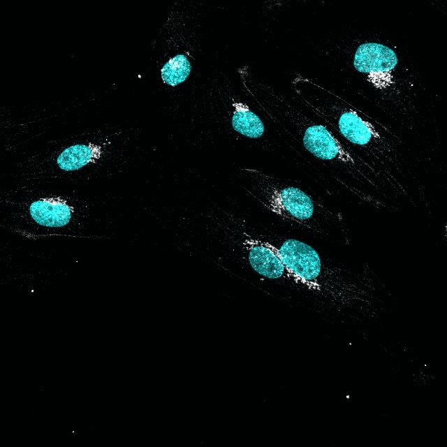
GM130/GOLGA2 in HeLa Human Cell Line.
GM130/GOLGA2 was detected in immersion fixed HeLa human cervical epithelial carcinoma cell line using Sheep Anti-Human GM130/GOLGA2 Antigen Affinity-purified Polyclonal Antibody (Catalog # AF8199) at 1.7 µg/mL for 3 hours at room temperature. Cells were stained using the NorthernLights™ 557-conjugated Anti-Sheep IgG Secondary Antibody (red; Catalog # NL010) and counterstained with DAPI (blue). Specific staining was localized to Golgi apparatus vesicles. View our protocol for Fluorescent ICC Staining of Cells on Coverslips.
Detection of Human GM130/GOLGA2 by Simple WesternTM.
Simple Western lane view shows lysates of HeLa human cervical epithelial carcinoma cell line, MCF-7 human breast cancer cell line, and ZR-75 human breast cancer cell line, loaded at 0.2 mg/mL. A specific band was detected for GM130/GOLGA2 at approximately 141 kDa (as indicated) using 50 µg/mL of Sheep Anti-Human GM130/GOLGA2 Antigen Affinity-purified Polyclonal Antibody (Catalog # AF8199) followed by 1:50 dilution of HRP-conjugated Anti-Sheep IgG Secondary Antibody (Catalog # HAF016). This experiment was conducted under reducing conditions and using the 12-230 kDa separation system.
Detection of Mouse GM130/GOLGA2 by Immunocytochemistry/Immunofluorescence
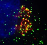
Kcnj13 orchestrates SM cell alignment and polarity. a Immunostaining for alpha SMA (red) and DAPI staining (blue) in dorsal views of E14.5 WT (n = 8) and Kcnj13T38C/T38C (n = 8) tracheas. b Quantification of E14.5 WT (n = 8) and Kcnj13T38C/T38C (n = 8) tracheal SM cell orientation. c Quantification of E14.5 WT (n = 8) and Kcnj13T38C/T38C (n = 8) tracheal SM cell nuclear aspect ratio (NAR). d Immunostaining for alpha SMA (red) and DAPI staining (blue) of transverse sections of E14.5 WT (n = 10) and Kcnj13T38C/T38C (n = 10) tracheas. e Quantification of WT (n = 10) and Kcnj13T38C/T38C (n = 10) tracheal SM cell layers. f Immunostaining for alpha SMA (red) and GM130 (green) and DAPI staining (blue) in dorsal views of E14.5 WT (n = 8) and Kcnj13T38C/T38C (n = 8) tracheas. g Quantification of WT (n = 8) and Kcnj13T38C/T38C (n = 8) Golgi apparatus (green) position relative to the nucleus (blue). h Timeline for tamoxifen administration. i Immunostaining for KCNJ13 (red), alpha SMA (green), and DAPI staining (blue) of transverse sections of E14.5 control (n = 6) and Myh11-CreERT2;Kcnj13flox/flox (n = 6) tracheas. j Representative images of ventral views of E14.5 control (n = 9) and Myh11-CreERT2;Kcnj13flox/flox (n = 9) tracheas. Double-sided arrows indicate tracheal tube length. k Quantification of E14.5 control (n = 9) and Myh11-CreERT2;Kcnj13flox/flox (n = 9) tracheal tube length. l Immunostaining for alpha SMA (red) and DAPI staining (blue) in dorsal views of E14.5 control (n = 9) and Myh11-CreERT2;Kcnj13flox/flox (n = 9) tracheas. m Quantification of E14.5 control (n = 9) and Myh11-CreERT2;Kcnj13flox/flox (n = 9) tracheal SM cell orientation. n Immunostaining for SOX9 (green) in ventral views of E15.5 control (n = 6) and Myh11-CreERT2;Kcnj13flox/flox (n = 6) tracheas. Arrows point to tracheal mesenchymal condensations. Scale bars: 1000 μm (j), 100 μm (n), 50 μm (d), 20 μm (a, f, i, l). *P < 0.05; **P < 0.01; unpaired Student’s t-test, mean ± s.d Image collected and cropped by CiteAb from the folApplications for Human GM130/GOLGA2 Antibody
Application
Recommended Usage
Immunocytochemistry
5-15 µg/mL
Sample: Immersion fixed HeLa human cervical epithelial carcinoma cell line
Sample: Immersion fixed HeLa human cervical epithelial carcinoma cell line
Simple Western
50 µg/mL
Sample: HeLa human cervical epithelial carcinoma cell line, MCF‑7 human breast cancer cell line, and ZR‑75 human breast cancer cell line
Sample: HeLa human cervical epithelial carcinoma cell line, MCF‑7 human breast cancer cell line, and ZR‑75 human breast cancer cell line
Western Blot
0.25 µg/mL
Sample: MCF‑7 human breast cancer cell line and ZR‑75 human breast cancer cell line
Sample: MCF‑7 human breast cancer cell line and ZR‑75 human breast cancer cell line
Reviewed Applications
Read 3 reviews rated 4 using AF8199 in the following applications:
Formulation, Preparation, and Storage
Purification
Antigen Affinity-purified
Reconstitution
Reconstitute at 0.2 mg/mL in sterile PBS. For liquid material, refer to CoA for concentration.
Loading...
Formulation
Lyophilized from a 0.2 μm filtered solution in PBS with Trehalose. *Small pack size (SP) is supplied either lyophilized or as a 0.2 µm filtered solution in PBS.
Shipping
Lyophilized product is shipped at ambient temperature. Liquid small pack size (-SP) is shipped with polar packs. Upon receipt, store immediately at the temperature recommended below.
Stability & Storage
Use a manual defrost freezer and avoid repeated freeze-thaw cycles.
- 12 months from date of receipt, -20 to -70 °C as supplied.
- 1 month, 2 to 8 °C under sterile conditions after reconstitution.
- 6 months, -20 to -70 °C under sterile conditions after reconstitution.
Calculators
Background: GM130/GOLGA2
Long Name
Golgin A2
Alternate Names
GOLGA2, Golgin-95, SY11 Protein
Gene Symbol
GOLGA2
UniProt
Additional GM130/GOLGA2 Products
Product Documents for Human GM130/GOLGA2 Antibody
Certificate of Analysis
To download a Certificate of Analysis, please enter a lot or batch number in the search box below.
Note: Certificate of Analysis not available for kit components.
Product Specific Notices for Human GM130/GOLGA2 Antibody
For research use only
Related Research Areas
Citations for Human GM130/GOLGA2 Antibody
 Powered by Bioz
Customer Reviews for Human GM130/GOLGA2 Antibody (3)
4 out of 5
3 Customer Ratings
Have you used Human GM130/GOLGA2 Antibody?
Submit a review and receive an Amazon gift card!
$25/€18/£15/$25CAN/¥2500 Yen for a review with an image
$10/€7/£6/$10CAN/¥1110 Yen for a review without an image
Submit a review
Customer Images
Showing
1
-
3 of
3 reviews
Showing All
Filter By:
-
Application: Immunocytochemistry/ImmunofluorescenceSample Tested: 293T human embryonic kidney cell lineSpecies: HumanVerified Customer | Posted 10/06/2021

-
Application: Immunocytochemistry/ImmunofluorescenceSample Tested: HUVEC human umbilical vein endothelial cellsSpecies: HumanVerified Customer | Posted 07/16/2021
-
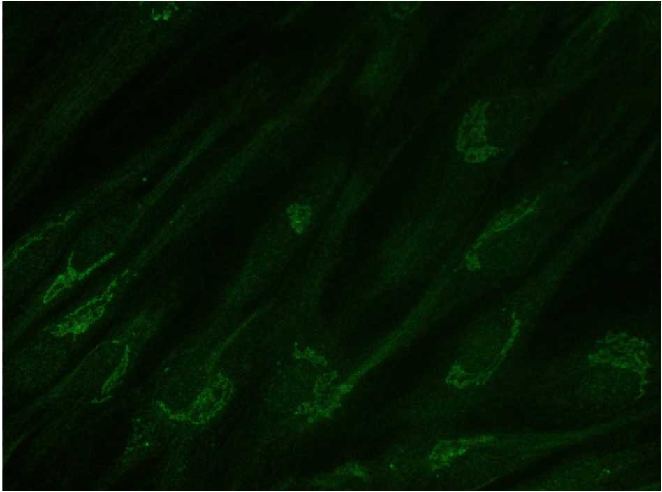
Application: ImmunocytochemistrySample Tested: Human primary fibroblastSpecies: HumanVerified Customer | Posted 11/08/2020GM130 in human primary fibroblasts (Golgi). ICC using recently reconstituted GM130. It precipitates becoming useless in 6 month at -20ºC. PFA 4% fixed cells. Primary ab at 5 ug/mL O/N 4ºC. Donkey anti-Sheep Alexafluor 488.Good antbody to label Golgi in multiple ICC because it is produced in Sheep. It comes lyophilized, and after reconstitution and saved at -20ºC it precipitates in 6 month, becoming useless. ICC conditions: PFA 4% 20 min fixed cells. Blocking 10% DS in PBST (0.1% Triton) 1h RT. Primary ab O/N incubation 4ºC 5 ug/mL in DS 10% in PBST (0.1% Triton). Secondary Donkey anti-Sheep Alexa fluor 488 1:500 1h RT.
There are no reviews that match your criteria.
Protocols
Find general support by application which include: protocols, troubleshooting, illustrated assays, videos and webinars.
- Appropriate Fixation of IHC/ICC Samples
- Cellular Response to Hypoxia Protocols
- ClariTSA™ Fluorophore Kits
- Detection & Visualization of Antibody Binding
- ICC Cell Smear Protocol for Suspension Cells
- ICC Immunocytochemistry Protocol Videos
- ICC for Adherent Cells
- Immunocytochemistry (ICC) Protocol
- Immunocytochemistry Troubleshooting
- Immunofluorescence of Organoids Embedded in Cultrex Basement Membrane Extract
- Immunohistochemistry (IHC) and Immunocytochemistry (ICC) Protocols
- Preparing Samples for IHC/ICC Experiments
- Preventing Non-Specific Staining (Non-Specific Binding)
- Primary Antibody Selection & Optimization
- Protocol for VisUCyte™ HRP Polymer Detection Reagent
- Protocol for the Fluorescent ICC Staining of Cell Smears - Graphic
- Protocol for the Fluorescent ICC Staining of Cultured Cells on Coverslips - Graphic
- Protocol for the Preparation and Fluorescent ICC Staining of Cells on Coverslips
- Protocol for the Preparation and Fluorescent ICC Staining of Non-adherent Cells
- Protocol for the Preparation and Fluorescent ICC Staining of Stem Cells on Coverslips
- Protocol for the Preparation of a Cell Smear for Non-adherent Cell ICC - Graphic
- R&D Systems Quality Control Western Blot Protocol
- TUNEL and Active Caspase-3 Detection by IHC/ICC Protocol
- The Importance of IHC/ICC Controls
- Troubleshooting Guide: Western Blot Figures
- Western Blot Conditions
- Western Blot Protocol
- Western Blot Protocol for Cell Lysates
- Western Blot Troubleshooting
- Western Blot Troubleshooting Guide
- View all Protocols, Troubleshooting, Illustrated assays and Webinars
Loading...