HOXB1 (Homeobox protein B1; also HOX-2I) is a 32 kDa (predicted) member of the Antp homeobox family, labial subfamily of transcription factors. It is expressed in rhombomere-4 (r-4) of the hindbrain, where it drives the development of r-4 derived structures. Among these are the motor neurons of the facial nerve, and neural crest cells that form Schwann cells plus cartilage and bone of the neck and ear. Human HOXB1 is 301 amino acids (aa) in length. It contains an Antp-type hexapeptide (aa 179-184) that mediates heterodimerization, and a DNA-binding homeobox domain (aa 203-262). There is one potential splice variant that shows a 40 aa substitution for aa 193-301. Over aa 1-192, human HOXB1 shares 82% aa identity with mouse HOXB1.

Key Product Details
Validated by
Biological Validation
Species Reactivity
Validated:
Human
Cited:
Human
Applications
Validated:
Immunocytochemistry
Cited:
Flow Cytometry
Label
Unconjugated
Antibody Source
Polyclonal Sheep IgG
Loading...
Product Specifications
Immunogen
E. coli-derived recombinant human HOXB1
Met1-Thr192
Accession # P14653
Met1-Thr192
Accession # P14653
Specificity
Detects human HOXB1 in direct ELISAs. In direct ELISAs, less than 2% cross-reactivity with recombinant human (rh) HOXA1, rhHOXB4, and rhHOXA9 is observed.
Clonality
Polyclonal
Host
Sheep
Isotype
IgG
Scientific Data Images for Human HOXB1 Antibody
HOXB1 in NTera‑2 Human Cell Line.
HOXB1 was detected in immersion fixed NTera-2 human testicular embryonic carcinoma cells, treated with (upper panel) and without (lower panel) retinoic acid, using Sheep Anti-Human HOXB1 Antigen Affinity-purified Polyclonal Antibody (Catalog # AF6318) at 10 µg/mL for 3 hours at room temperature. Cells were stained using the NorthernLights™ 557-conjugated Anti-Sheep IgG Secondary Antibody (red; Catalog # NL010) and counterstained with DAPI (blue). Specific staining was localized to nuclei. View our protocol for Fluorescent ICC Staining of Cells on Coverslips.Applications for Human HOXB1 Antibody
Application
Recommended Usage
Immunocytochemistry
5-15 µg/mL
Sample: Immersion fixed NTera‑2 human testicular embryonic carcinoma cells treated with retinoic acid
Sample: Immersion fixed NTera‑2 human testicular embryonic carcinoma cells treated with retinoic acid
Reviewed Applications
Read 1 review rated 4 using AF6318 in the following applications:
Formulation, Preparation, and Storage
Purification
Antigen Affinity-purified
Reconstitution
Sterile PBS to a final concentration of 0.2 mg/mL. For liquid material, refer to CoA for concentration.
Loading...
Formulation
Lyophilized from a 0.2 μm filtered solution in PBS with Trehalose. *Small pack size (SP) is supplied either lyophilized or as a 0.2 µm filtered solution in PBS.
Shipping
Lyophilized product is shipped at ambient temperature. Liquid small pack size (-SP) is shipped with polar packs. Upon receipt, store immediately at the temperature recommended below.
Stability & Storage
Use a manual defrost freezer and avoid repeated freeze-thaw cycles.
- 12 months from date of receipt, -20 to -70 °C as supplied.
- 1 month, 2 to 8 °C under sterile conditions after reconstitution.
- 6 months, -20 to -70 °C under sterile conditions after reconstitution.
Calculators
Background: HOXB1
Long Name
Homeobox B1
Alternate Names
HOX2, HOX2I
Gene Symbol
HOXB1
UniProt
Additional HOXB1 Products
Product Documents for Human HOXB1 Antibody
Certificate of Analysis
To download a Certificate of Analysis, please enter a lot or batch number in the search box below.
Note: Certificate of Analysis not available for kit components.
Product Specific Notices for Human HOXB1 Antibody
For research use only
Related Research Areas
Citations for Human HOXB1 Antibody
 Powered by Bioz
Customer Reviews for Human HOXB1 Antibody (1)
4 out of 5
1 Customer Rating
Have you used Human HOXB1 Antibody?
Submit a review and receive an Amazon gift card!
$25/€18/£15/$25CAN/¥2500 Yen for a review with an image
$10/€7/£6/$10CAN/¥1110 Yen for a review without an image
Submit a review
Customer Images
Showing
1
-
1 of
1 review
Showing All
Filter By:
-
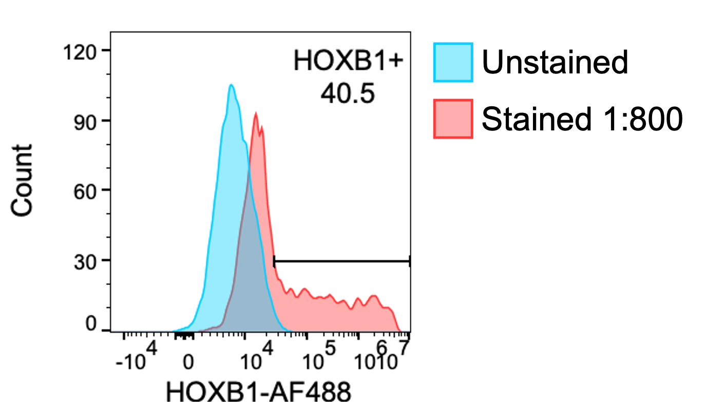
Application: Flow CytometrySample Tested: 293T human embryonic kidney cell lineSpecies: HumanVerified Customer | Posted 09/01/2021Before use conjugated 100ug of the HOXB1 antibody (AF6318) with Alexa Fluor 488 using the Alexa Fluor™ 488 Antibody Labeling Kit from ThermoFisher and eluted in 100 μL. 293T cells were transfected with a HOXB1 expressing construct, fixed in 4% paraformaldehyde for 10 minutes at room temperature, and permeabilized/stained using BioLegends True-Nuclear™ Transcription Factor Buffer Set according to the instructions. I stained at 1:800 and got robust staining. Data was acquired on a Cytek Aurora and analyzed in FlowJo.

There are no reviews that match your criteria.
Protocols
Find general support by application which include: protocols, troubleshooting, illustrated assays, videos and webinars.
- Appropriate Fixation of IHC/ICC Samples
- Cellular Response to Hypoxia Protocols
- ClariTSA™ Fluorophore Kits
- Detection & Visualization of Antibody Binding
- ICC Cell Smear Protocol for Suspension Cells
- ICC Immunocytochemistry Protocol Videos
- ICC for Adherent Cells
- Immunocytochemistry (ICC) Protocol
- Immunocytochemistry Troubleshooting
- Immunofluorescence of Organoids Embedded in Cultrex Basement Membrane Extract
- Immunohistochemistry (IHC) and Immunocytochemistry (ICC) Protocols
- Preparing Samples for IHC/ICC Experiments
- Preventing Non-Specific Staining (Non-Specific Binding)
- Primary Antibody Selection & Optimization
- Protocol for VisUCyte™ HRP Polymer Detection Reagent
- Protocol for the Fluorescent ICC Staining of Cell Smears - Graphic
- Protocol for the Fluorescent ICC Staining of Cultured Cells on Coverslips - Graphic
- Protocol for the Preparation and Fluorescent ICC Staining of Cells on Coverslips
- Protocol for the Preparation and Fluorescent ICC Staining of Non-adherent Cells
- Protocol for the Preparation and Fluorescent ICC Staining of Stem Cells on Coverslips
- Protocol for the Preparation of a Cell Smear for Non-adherent Cell ICC - Graphic
- TUNEL and Active Caspase-3 Detection by IHC/ICC Protocol
- The Importance of IHC/ICC Controls
- View all Protocols, Troubleshooting, Illustrated assays and Webinars
Loading...