TGF-beta 1 (transforming growth factor beta 1) and the closely related TGF-beta 2 and -beta 3 are members of the large TGF-beta superfamily. TGF‑ beta proteins are highly pleiotropic cytokines that regulate processes such as immune function, proliferation and epithelial-mesenchymal transition (1‑3). Human TGF-beta 1 cDNA encodes a 390 amino acid (aa) precursor that contains a 29 aa signal peptide and a 361 aa proprotein (4). A furin-like convertase processes the proprotein within the trans-Golgi to generate an N‑terminal 249 aa (aa 30-278) latency-associated peptide (LAP) and a C-terminal 112 aa mature TGF-beta 1 (aa 279-390) (4‑6). Disulfide-linked homodimers of LAP and TGF-beta 1 remain non‑covalently associated after secretion, forming the small latent TGF-beta 1 complex (4‑8). Purified LAP is also capable of associating with active TGF‑ beta with high affinity, and can neutralize TGF-beta activity (9). Covalent linkage of LAP to one of three latent TGF-beta binding proteins (LTBPs) creates a large latent complex that may interact with the extracellular matrix (5-7). TGF-beta activation from latency is controlled both spatially and temporally, by multiple pathways that include actions of proteases such as plasmin and MMP9, and/or by thrombospondin 1 or selected integrins (5, 8). The LAP portion of human TGF-beta 1 shares 91%, 92%, 85%, 86% and 88% aa identity with porcine, canine, mouse, rat and equine TGF-beta 1 LAP, respectively, while mature human TGF-beta 1 portion shares 100% aa identity with porcine, canine and bovine TGF-beta 1, and 99% aa identity with mouse, rat and equine TGF-beta 1. Although different isoforms of TGF-beta are naturally associated with their own distinct LAPs, the TGF-beta 1 LAP is capable of complexing with, and inactivating, all other human TGF-beta isoforms and those of most other species (9). Mutations within the LAP are associated with Camurati-Engelmann disease, a rare sclerosing bone dysplasia characterized by inappropriate presence of active TGF‑ beta 1 (10).
Human LAP (TGF-beta 1) Antibody
R&D Systems | Catalog # AF-246-NA
 by Western Blot.")
Key Product Details
Species Reactivity
Validated:
Human
Cited:
Human, Mouse, Rat, Porcine, Mink, Rabbit, Transgenic Mouse
Applications
Validated:
Western Blot, Neutralization, Immunocytochemistry, Simple Western
Cited:
Immunohistochemistry, Immunohistochemistry-Paraffin, Immunohistochemistry-Frozen, Western Blot, Neutralization, Immunocytochemistry, Bioassay, Dot Blot
Label
Unconjugated
Antibody Source
Polyclonal Goat IgG
Loading...
Product Specifications
Immunogen
S. frugiperda insect ovarian cell line Sf 21-derived and Chinese hamster ovary cell line CHO-derived recombinant human LAP TGF‑ beta 1
Leu30-Ser390
Accession # P01137
Leu30-Ser390
Accession # P01137
Specificity
Detects human LAP TGF‑ beta 1 in direct ELISAs and Western blots.
Clonality
Polyclonal
Host
Goat
Isotype
IgG
Endotoxin Level
<0.10 EU per 1 μg of the antibody by the LAL method.
Scientific Data Images for Human LAP (TGF-beta 1) Antibody
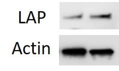
Detection of Human LAP (TGF-beta 1) by Western Blot.
Western blot shows lysates of Saos-2 human osteosarcoma cell line. PVDF membrane was probed with 2 µg/mL of Goat Anti-Human LAP (TGF-beta 1) Antigen Affinity-purified Polyclonal Antibody (Catalog # AF-246-NA) followed by HRP-conjugated Anti-Goat IgG Secondary Antibody (Catalog # HAF017). A specific band was detected for LAP (TGF-beta 1) at approximately 48 kDa (as indicated). This experiment was conducted under reducing conditions and using Immunoblot Buffer Group 1. in Human PBMCs.")
LAP (TGF-beta 1) in Human PBMCs.
LAP (TGF-beta 1) was detected in immersion fixed Human peripheral blood mononuclear cells using Goat Anti-Human LAP (TGF-beta 1) Antigen Affinity-purified Polyclonal Antibody (Catalog # AF-246-NA) at 15 µg/mL for 3 hours at room temperature. Cells were stained using the NorthernLights™ 557-conjugated Anti-Goat IgG Secondary Antibody (red; NL001) and counterstained with DAPI (blue). Specific staining was localized to cytoplasm. Staining was performed using our protocol for Fluorescent ICC Staining of Non-adherent Cells. by Simple Western<sup>TM</sup>.")
Detection of Human LAP (TGF-beta 1) by Simple WesternTM.
Simple Western lane view shows lysates of K562 human chronic myelogenous leukemia cell line and A549 human lung carcinoma cell line, loaded at 0.2 mg/mL. A specific band was detected for LAP (TGF-beta 1) at approximately 61 kDa (as indicated) using 20 µg/mL of Goat Anti-Human LAP (TGF-beta 1) Antigen Affinity-purified Polyclonal Antibody (Catalog # AF-246-NA) followed by 1:50 dilution of HRP-conjugated Anti-Goat IgG Secondary Antibody (Catalog # HAF109). This experiment was conducted under reducing conditions and using the 12-230 kDa separation system.
LAP TGF‑ beta 1 Inhibition of TGF‑ beta 1 Activity and Neutralization by Human LAP TGF‑ beta 1 Antibody.
Recombinant Human LAP TGF-beta 1 (Catalog # 246-LP) inhibits Recom-binant Human TGF-beta 1 (Catalog # 240-B) growth inhibition activity in the HT-2 mouse T cell line in a dose-dependent manner (orange line). Inhibition of Recom-binant Human TGF-beta 1 (1 ng/mL) activity elicited by Recombin-ant Human LAP TGF-beta 1 (500 ng/mL) is neutralized (green line) by increasing concentrations of Goat Anti-Human LAP TGF-beta 1 Antigen Affinity-purified Polyclonal Antibody (Catalog # AF-246-NA). The ND50 is typically 0.4-2 µg/mL.Applications for Human LAP (TGF-beta 1) Antibody
Application
Recommended Usage
Immunocytochemistry
5-15 µg/mL
Sample: Immersion fixed Human peripheral blood mononuclear cells
Sample: Immersion fixed Human peripheral blood mononuclear cells
Simple Western
20 µg/mL
Sample: K562 human chronic myelogenous leukemia cell line and A549 human lung carcinoma cell line
Sample: K562 human chronic myelogenous leukemia cell line and A549 human lung carcinoma cell line
Western Blot
2 µg/mL
Sample: Saos‑2 human osteosarcoma cell line
Sample: Saos‑2 human osteosarcoma cell line
Neutralization
Measured by its ability to neutralize LAP TGF‑ beta 1 inhibition of TGF‑ beta 1 growth inhibition in the HT‑2 mouse T cell line. Tsang, M. et al. (1995) Cytokine 7:389. The Neutralization Dose (ND50) is typically 0.4-2 µg/mL in the presence of 500 ng/mL Recombinant Human LAP TGF‑ beta 1 and 1 ng/mL TGF‑ beta 1.
Reviewed Applications
Read 4 reviews rated 4 using AF-246-NA in the following applications:
Formulation, Preparation, and Storage
Purification
Antigen Affinity-purified
Reconstitution
Reconstitute at 0.2 mg/mL in sterile PBS. For liquid material, refer to CoA for concentration.
Loading...
Formulation
Lyophilized from a 0.2 μm filtered solution in PBS with Trehalose. *Small pack size (SP) is supplied either lyophilized or as a 0.2 µm filtered solution in PBS.
Shipping
Lyophilized product is shipped at ambient temperature. Liquid small pack size (-SP) is shipped with polar packs. Upon receipt, store immediately at the temperature recommended below.
Stability & Storage
Use a manual defrost freezer and avoid repeated freeze-thaw cycles.
- 12 months from date of receipt, -20 to -70 °C as supplied.
- 1 month, 2 to 8 °C under sterile conditions after reconstitution.
- 6 months, -20 to -70 °C under sterile conditions after reconstitution.
Calculators
Background: LAP (TGF-beta 1)
References
- Dunker, N. & K. Krieglstein (2000) Eur. J. Biochem. 267:6982.
- Wahl, S.M. (2006) Immunol. Rev. 213:213.
- Chang, H. et al. (2002) Endocr. Rev. 23:787.
- Derynck, R. et al. (1985) Nature 316:701.
- Dabovic, B. and D.B. Rifkin (2008) “TGF-beta Bioavailability” in The TGF-beta Family. Derynck, R. and K. Miyazono (eds): Cold Spring Harbor Laboratory Press, p. 179.
- Brunner, A.M. et al. (1989) J. Biol. Chem. 264:13660.
- Miyazono, K. et al. (1991) EMBO J. 10:1091.
- Oklu, R. and R. Hesketh (2000) Biochem. J. 352:601.
- Miller, D.M. et al. (1992) Mol. Endocrinol. 6:694.
- Janssens, K. et al. (2003) J. Biol. Chem. 278:7718.
Long Name
Latency-associated Peptide
Alternate Names
LAP (TGFbeta 1)
Entrez Gene IDs
7040 (Human)
Gene Symbol
TGFB1
UniProt
Additional LAP (TGF-beta 1) Products
Product Documents for Human LAP (TGF-beta 1) Antibody
Certificate of Analysis
To download a Certificate of Analysis, please enter a lot or batch number in the search box below.
Note: Certificate of Analysis not available for kit components.
Product Specific Notices for Human LAP (TGF-beta 1) Antibody
For research use only
Citations for Human LAP (TGF-beta 1) Antibody
 Powered by Bioz
Customer Reviews for Human LAP (TGF-beta 1) Antibody (4)
4 out of 5
4 Customer Ratings
Have you used Human LAP (TGF-beta 1) Antibody?
Submit a review and receive an Amazon gift card!
$25/€18/£15/$25CAN/¥2500 Yen for a review with an image
$10/€7/£6/$10CAN/¥1110 Yen for a review without an image
Submit a review
Customer Images

Showing
1
-
4 of
4 reviews
Showing All
Filter By:
-
Application: Western BlotSample Tested: Pancreatic cancer cellsSpecies: HumanVerified Customer | Posted 01/27/2021

-
Application: Western BlotSample Tested: Aorta tissueSpecies: RatVerified Customer | Posted 02/03/2020
-
Application: Western BlotSample Tested: See PMID 21998117Species: OtherVerified Customer | Posted 01/07/2015
-
Application: Western BlotSample Tested: See PMID 22835852Species: OtherVerified Customer | Posted 01/07/2015
There are no reviews that match your criteria.
Protocols
Find general support by application which include: protocols, troubleshooting, illustrated assays, videos and webinars.
- Appropriate Fixation of IHC/ICC Samples
- Cellular Response to Hypoxia Protocols
- ClariTSA™ Fluorophore Kits
- Detection & Visualization of Antibody Binding
- ICC Cell Smear Protocol for Suspension Cells
- ICC Immunocytochemistry Protocol Videos
- ICC for Adherent Cells
- Immunocytochemistry (ICC) Protocol
- Immunocytochemistry Troubleshooting
- Immunofluorescence of Organoids Embedded in Cultrex Basement Membrane Extract
- Immunohistochemistry (IHC) and Immunocytochemistry (ICC) Protocols
- Preparing Samples for IHC/ICC Experiments
- Preventing Non-Specific Staining (Non-Specific Binding)
- Primary Antibody Selection & Optimization
- Protocol for VisUCyte™ HRP Polymer Detection Reagent
- Protocol for the Fluorescent ICC Staining of Cell Smears - Graphic
- Protocol for the Fluorescent ICC Staining of Cultured Cells on Coverslips - Graphic
- Protocol for the Preparation and Fluorescent ICC Staining of Cells on Coverslips
- Protocol for the Preparation and Fluorescent ICC Staining of Non-adherent Cells
- Protocol for the Preparation and Fluorescent ICC Staining of Stem Cells on Coverslips
- Protocol for the Preparation of a Cell Smear for Non-adherent Cell ICC - Graphic
- R&D Systems Quality Control Western Blot Protocol
- TUNEL and Active Caspase-3 Detection by IHC/ICC Protocol
- The Importance of IHC/ICC Controls
- Troubleshooting Guide: Western Blot Figures
- Western Blot Conditions
- Western Blot Protocol
- Western Blot Protocol for Cell Lysates
- Western Blot Troubleshooting
- Western Blot Troubleshooting Guide
- View all Protocols, Troubleshooting, Illustrated assays and Webinars
Loading...