LRIG1 (leucine-rich repeats and Ig-like domains-1; also LIG-1) is an approximately 134-145 kDa glycoprotein that belongs to the LRIG gene family. It is widely expressed and appears on the surface of prostatic epithelium, endothelial cells, vascular and visceral smooth muscle, mammary epithelium, cardiac muscle, keratinocytes and neurons. LRIG1 is believed to negatively regulate the ErbB family of receptors. In particular, and in a ligand-independent manner, LRIG1 complexes with all four ErbBs, promoting their ubiquitination and decreasing their number. Alternatively, LRIG1 is suggested to bind to the ErbBs, preventing their dimerization and signal transduction. Mature human LRIG1 is a 1059 amino acid (aa) type I transmembrane protein. It contains a large 760 amino acid (aa) extracellular domain (ECD) (aa 35-794) plus a 278 aa cytoplasmic region. The ECD contains 17 LRRs (aa 35‑491) and three C2-type Ig-like domains (aa 495-780). These two domain types are each sufficient for EGFR binding. There are two potential alternative splice forms. One contains a 27 aa insertion after Gly874, while another shows a 24 aa insertion after Lys387 coupled to a Gln substitution for aa 644-691. The LRIG1 ECD undergoes proteolysis, generating 100-110 and 55-60 kDa soluble fragments. Over aa 35‑779, human LRIG1 shares 90% aa sequence identity with mouse LRIG1.

Key Product Details
Species Reactivity
Validated:
Human
Cited:
Human
Applications
Validated:
Immunohistochemistry, Flow Cytometry, Immunocytochemistry, CyTOF-ready
Cited:
Immunohistochemistry, Immunocytochemistry
Label
Unconjugated
Antibody Source
Monoclonal Mouse IgG2B Clone # 789211
Loading...
Product Specifications
Immunogen
HEK293 human embryonic kidney cell line transfected with human LRIG1
Ala35-Ser779
Accession # Q96JA1
Ala35-Ser779
Accession # Q96JA1
Specificity
Detects human LRIG1 in ELISAs.
In direct ELISAs, less than 5%
cross-reactivity with recombinant mouse LRIG1 and recombinant human LRIG3 is
observed.
Clonality
Monoclonal
Host
Mouse
Isotype
IgG2B
Scientific Data Images for Human LRIG1 Antibody (789211)
Detection of LRIG1 in LNCaP Human Cell Line by Flow Cytometry.
LNCaP human prostate cancer cell line was stained with Mouse Anti-Human LRIG1 Monoclonal Antibody (Catalog # MAB7498, filled histogram) or isotype control antibody (Catalog # MAB0041, open histogram), followed by Allophycocyanin-conjugated Anti-Mouse IgG Secondary Antibody (Catalog # F0101B).
LRIG1 in SK‑BR‑3 Human Cell Line.
LRIG1 was detected in immersion fixed SK-BR-3 human breast cancer cell line using Mouse Anti-Human LRIG1 Monoclonal Antibody (Catalog # MAB7498) at 10 µg/mL for 3 hours at room temperature. Cells were stained using the Northern-Lights™ 557-conjugated Anti-Mouse IgG Secondary Antibody (red; Catalog # NL007) and counter-stained with DAPI (blue). Specific staining was localized to cytoplasm and cell surfaces. View our protocol for Fluorescent ICC Staining of Cells on Coverslips.
LRIG1 in Human Kidney.
LRIG1 was detected in immersion fixed paraffin-embedded sections of human kidney using Mouse Anti-Human LRIG1 Monoclonal Antibody (Catalog # MAB7498) at 15 µg/mL overnight at 4 °C. Before incubation with the primary antibody, tissue was subjected to heat-induced epitope retrieval using Antigen Retrieval Reagent-Basic (Catalog # CTS013). Tissue was stained using the Anti-Mouse HRP-DAB Cell & Tissue Staining Kit (brown; Catalog # CTS002) and counter-stained with hematoxylin (blue). Specific staining was localized to cytoplasm of epithelial cells in convoluted tubules. View our protocol for Chromogenic IHC Staining of Paraffin-embedded Tissue Sections.Applications for Human LRIG1 Antibody (789211)
Application
Recommended Usage
CyTOF-ready
Ready to be labeled using established conjugation methods. No BSA or other carrier proteins that could interfere with conjugation.
Flow Cytometry
2.5 µg/106 cells
Sample: LNCaP Human Cell Line
Sample: LNCaP Human Cell Line
Immunocytochemistry
8-25 µg/mL
Sample: Immersion fixed SK‑BR‑3 human breast cancer cell line
Sample: Immersion fixed SK‑BR‑3 human breast cancer cell line
Immunohistochemistry
8-25 µg/mL
Sample: Immersion fixed paraffin-embedded sections of human kidney
Sample: Immersion fixed paraffin-embedded sections of human kidney
Reviewed Applications
Read 3 reviews rated 3.7 using MAB7498 in the following applications:
Flow Cytometry Panel Builder
Bio-Techne Knows Flow Cytometry
Save time and reduce costly mistakes by quickly finding compatible reagents using the Panel Builder Tool.
Advanced Features
- Spectra Viewer - Custom analysis of spectra from multiple fluorochromes
- Spillover Popups - Visualize the spectra of individual fluorochromes
- Antigen Density Selector - Match fluorochrome brightness with antigen density

Formulation, Preparation, and Storage
Purification
Protein A or G purified from hybridoma culture supernatant
Reconstitution
Sterile PBS to a final concentration of 0.5 mg/mL. For liquid material, refer to CoA for concentration.
Loading...
Formulation
Lyophilized from a 0.2 μm filtered solution in PBS with Trehalose. *Small pack size (SP) is supplied either lyophilized or as a 0.2 µm filtered solution in PBS.
Shipping
Lyophilized product is shipped at ambient temperature. Liquid small pack size (-SP) is shipped with polar packs. Upon receipt, store immediately at the temperature recommended below.
Stability & Storage
Use a manual defrost freezer and avoid repeated freeze-thaw cycles.
- 12 months from date of receipt, -20 to -70 °C as supplied.
- 1 month, 2 to 8 °C under sterile conditions after reconstitution.
- 6 months, -20 to -70 °C under sterile conditions after reconstitution.
Calculators
Background: LRIG1
Long Name
Leucine-rich Repeats and Immunoglobulin-like Domains 1
Alternate Names
Img, LIG1
Gene Symbol
LRIG1
UniProt
Additional LRIG1 Products
Product Documents for Human LRIG1 Antibody (789211)
Certificate of Analysis
To download a Certificate of Analysis, please enter a lot or batch number in the search box below.
Note: Certificate of Analysis not available for kit components.
Product Specific Notices for Human LRIG1 Antibody (789211)
For research use only
Related Research Areas
Citations for Human LRIG1 Antibody (789211)
 Powered by Bioz
Customer Reviews for Human LRIG1 Antibody (789211) (3)
3.7 out of 5
3 Customer Ratings
Have you used Human LRIG1 Antibody (789211)?
Submit a review and receive an Amazon gift card!
$25/€18/£15/$25CAN/¥2500 Yen for a review with an image
$10/€7/£6/$10CAN/¥1110 Yen for a review without an image
Submit a review
Customer Images
Showing
1
-
3 of
3 reviews
Showing All
Filter By:
-
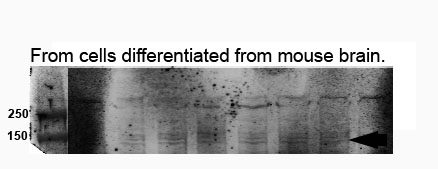
Application: Western BlotSample Tested: Brain tissueSpecies: HumanVerified Customer | Posted 09/18/2023A very fain band appeared at 150 kDa.

-
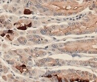
Application: ImmunohistochemistrySample Tested: Gastric epitheliumSpecies: HumanVerified Customer | Posted 10/30/2021
-
Application: Flow CytometrySample Tested: Peripheral blood lymphocytes (PBL)Species: HumanVerified Customer | Posted 06/28/2016Used with anti-mouse secondary in PE channel for detection. Could not see shift above background staining. One published report of expression on cell type, trying to replicate. May be antigen is not expressed rather than antibody poor as we have no real control.
There are no reviews that match your criteria.
Protocols
Find general support by application which include: protocols, troubleshooting, illustrated assays, videos and webinars.
- 7-Amino Actinomycin D (7-AAD) Cell Viability Flow Cytometry Protocol
- Antigen Retrieval Protocol (PIER)
- Antigen Retrieval for Frozen Sections Protocol
- Appropriate Fixation of IHC/ICC Samples
- Cellular Response to Hypoxia Protocols
- Chromogenic IHC Staining of Formalin-Fixed Paraffin-Embedded (FFPE) Tissue Protocol
- Chromogenic Immunohistochemistry Staining of Frozen Tissue
- ClariTSA™ Fluorophore Kits
- Detection & Visualization of Antibody Binding
- Extracellular Membrane Flow Cytometry Protocol
- Flow Cytometry Protocol for Cell Surface Markers
- Flow Cytometry Protocol for Staining Membrane Associated Proteins
- Flow Cytometry Staining Protocols
- Flow Cytometry Troubleshooting Guide
- Fluorescent IHC Staining of Frozen Tissue Protocol
- Graphic Protocol for Heat-induced Epitope Retrieval
- Graphic Protocol for the Preparation and Fluorescent IHC Staining of Frozen Tissue Sections
- Graphic Protocol for the Preparation and Fluorescent IHC Staining of Paraffin-embedded Tissue Sections
- Graphic Protocol for the Preparation of Gelatin-coated Slides for Histological Tissue Sections
- ICC Cell Smear Protocol for Suspension Cells
- ICC Immunocytochemistry Protocol Videos
- ICC for Adherent Cells
- IHC Sample Preparation (Frozen sections vs Paraffin)
- Immunocytochemistry (ICC) Protocol
- Immunocytochemistry Troubleshooting
- Immunofluorescence of Organoids Embedded in Cultrex Basement Membrane Extract
- Immunofluorescent IHC Staining of Formalin-Fixed Paraffin-Embedded (FFPE) Tissue Protocol
- Immunohistochemistry (IHC) and Immunocytochemistry (ICC) Protocols
- Immunohistochemistry Frozen Troubleshooting
- Immunohistochemistry Paraffin Troubleshooting
- Intracellular Flow Cytometry Protocol Using Alcohol (Methanol)
- Intracellular Flow Cytometry Protocol Using Detergents
- Intracellular Nuclear Staining Flow Cytometry Protocol Using Detergents
- Intracellular Staining Flow Cytometry Protocol Using Alcohol Permeabilization
- Intracellular Staining Flow Cytometry Protocol Using Detergents to Permeabilize Cells
- Preparing Samples for IHC/ICC Experiments
- Preventing Non-Specific Staining (Non-Specific Binding)
- Primary Antibody Selection & Optimization
- Propidium Iodide Cell Viability Flow Cytometry Protocol
- Protocol for Heat-Induced Epitope Retrieval (HIER)
- Protocol for Liperfluo
- Protocol for Making a 4% Formaldehyde Solution in PBS
- Protocol for VisUCyte™ HRP Polymer Detection Reagent
- Protocol for the Characterization of Human Th22 Cells
- Protocol for the Characterization of Human Th9 Cells
- Protocol for the Fluorescent ICC Staining of Cell Smears - Graphic
- Protocol for the Fluorescent ICC Staining of Cultured Cells on Coverslips - Graphic
- Protocol for the Preparation & Fixation of Cells on Coverslips
- Protocol for the Preparation and Chromogenic IHC Staining of Frozen Tissue Sections
- Protocol for the Preparation and Chromogenic IHC Staining of Frozen Tissue Sections - Graphic
- Protocol for the Preparation and Chromogenic IHC Staining of Paraffin-embedded Tissue Sections
- Protocol for the Preparation and Chromogenic IHC Staining of Paraffin-embedded Tissue Sections - Graphic
- Protocol for the Preparation and Fluorescent ICC Staining of Cells on Coverslips
- Protocol for the Preparation and Fluorescent ICC Staining of Non-adherent Cells
- Protocol for the Preparation and Fluorescent ICC Staining of Stem Cells on Coverslips
- Protocol for the Preparation and Fluorescent IHC Staining of Frozen Tissue Sections
- Protocol for the Preparation and Fluorescent IHC Staining of Paraffin-embedded Tissue Sections
- Protocol for the Preparation of Gelatin-coated Slides for Histological Tissue Sections
- Protocol for the Preparation of a Cell Smear for Non-adherent Cell ICC - Graphic
- Protocol: Annexin V and PI Staining by Flow Cytometry
- Protocol: Annexin V and PI Staining for Apoptosis by Flow Cytometry
- TUNEL and Active Caspase-3 Detection by IHC/ICC Protocol
- The Importance of IHC/ICC Controls
- Troubleshooting Guide: Fluorokine Flow Cytometry Kits
- Troubleshooting Guide: Immunohistochemistry
- View all Protocols, Troubleshooting, Illustrated assays and Webinars
Loading...
Associated Pathways
