MMP-19 (Matrix metalloprotease 19; also MMP-18 and MMP RASI) is a 55-59 kDa member of the peptidase M10A family of enzymes. It is widely expressed, being secreted by stratum basale keratinocytes, smooth muscle cells, epiphysial cartilage chondrocytes and monocytes/macrophages. MMP-19 has multiple substrates, including components of the basement membrane (type IV collagen; laminin; nidogen), fibronectin, aggrecan plus COMP, and IGFBP3, this latter cleavage resulting in the release of active IGF-I. Studies involving MMP-19 demonstrate an antiangiogenic function. This is attributable to the processing of plasminogen, generating angiostatin-like molecules, and the creation of an environment that promotes the ECM retention of soluble VEGF. Human proMMP-19 is 490 amino acids (aa) in length. It contains an autocleavable 9 kDa propeptide (aa 19-97) plus a 411 aa mature region (aa 98-508). The mature region contains a Zn catalytic region (aa 103-256) plus four hemopexin-like domains (aa 293-508). There are three additional potential isoforms. One shows an 88 aa substitution for aa 300-508, a second contains a 12 aa substitution for aa 1-298, while a third possesses an alternative start site at Met80. Over aa 229-508, human MMP-19 shares 76% aa identity with mouse MMP-19.

Key Product Details
Species Reactivity
Validated:
Human
Cited:
Human
Applications
Validated:
Immunohistochemistry, Western Blot
Cited:
Western Blot
Label
Unconjugated
Antibody Source
Polyclonal Goat IgG
Loading...
Product Specifications
Immunogen
Mouse myeloma cell line NS0-derived recombinant human MMP-19
Leu229-Tyr508
Accession # Q99542
Leu229-Tyr508
Accession # Q99542
Specificity
Detects human MMP-19 in direct ELISAs.
Clonality
Polyclonal
Host
Goat
Isotype
IgG
Scientific Data Images for Human MMP-19 Antibody
Detection of Human MMP-19 by Western Blot.
Western blot shows lysates of HeLa human cervical epithelial carcinoma cell line and human small intestine tissue. PVDF membrane was probed with 1 µg/mL of Goat Anti-Human MMP-19 Antigen Affinity-purified Polyclonal Antibody (Catalog # AF6790) followed by HRP-conjugated Anti-Goat IgG Secondary Antibody (Catalog # HAF019). Specific bands were detected for MMP-19 at approximately 57 and 50 kDa (as indicated). This experiment was conducted under reducing conditions and using Immunoblot Buffer Group 1.
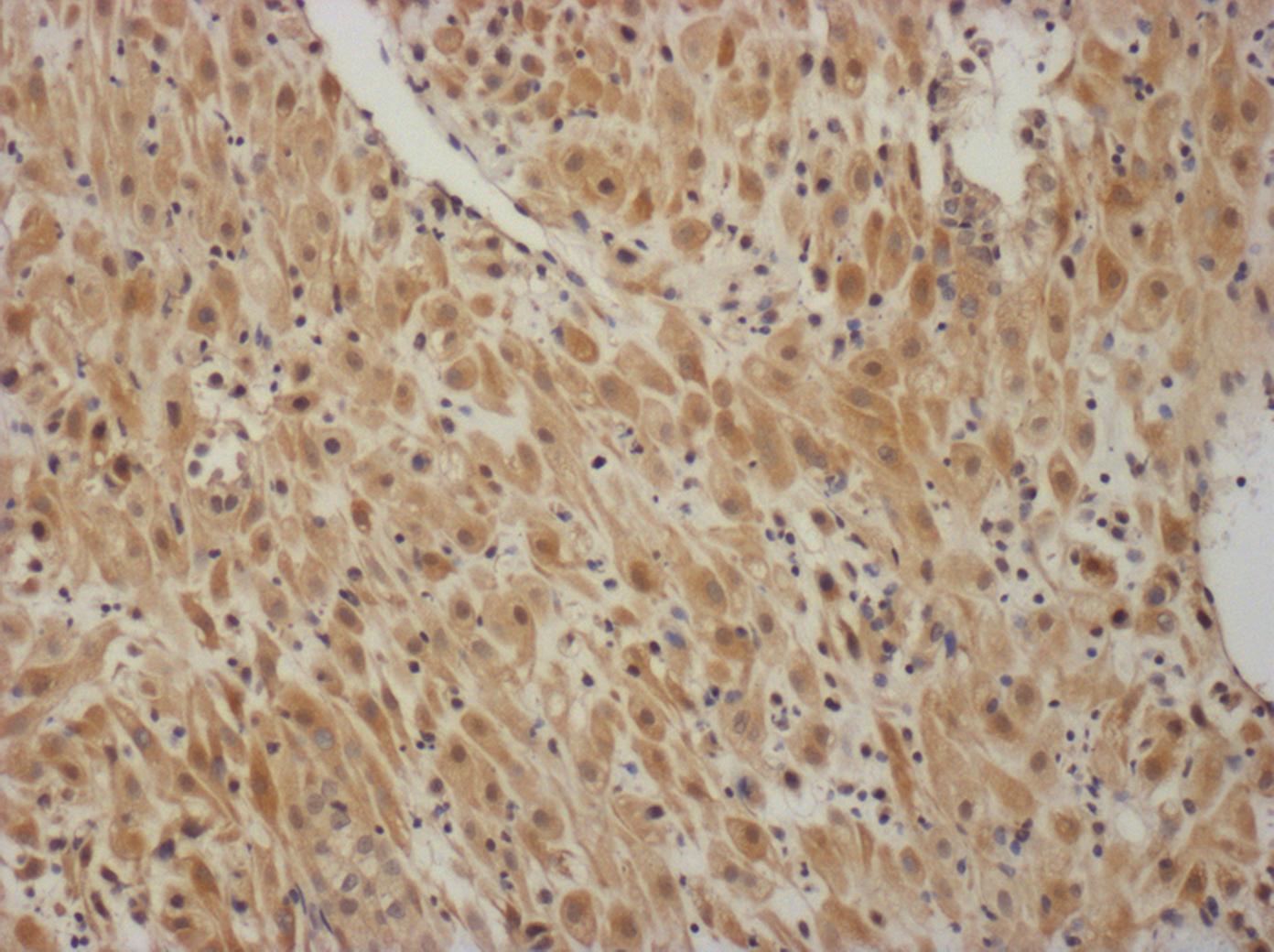
MMP-19 in Human Melanoma.
MMP-19 was detected in immersion fixed paraffin-embedded sections of human melanoma using Goat Anti-Human MMP-19 Antigen Affinity-purified Polyclonal Antibody (Catalog # AF6790) at 10 µg/mL overnight at 4 °C. Before incubation with the primary antibody, tissue was subjected to heat-induced epitope retrieval using Antigen Retrieval Reagent-Basic (Catalog # CTS013). Tissue was stained using the Anti-Goat HRP-DAB Cell & Tissue Staining Kit (brown; Catalog # CTS008) and counterstained with hematoxylin (blue). Specific staining was localized to cytoplasm and plasma membranes. View our protocol for Chromogenic IHC Staining of Paraffin-embedded Tissue Sections.Applications for Human MMP-19 Antibody
Application
Recommended Usage
Immunohistochemistry
5-15 µg/mL
Sample: Immersion fixed paraffin-embedded sections of human melanoma
Sample: Immersion fixed paraffin-embedded sections of human melanoma
Western Blot
1 µg/mL
Sample: HeLa human cervical epithelial carcinoma cell line and human small intestine tissue
Sample: HeLa human cervical epithelial carcinoma cell line and human small intestine tissue
Reviewed Applications
Read 2 reviews rated 4 using AF6790 in the following applications:
Formulation, Preparation, and Storage
Purification
Antigen Affinity-purified
Reconstitution
Sterile PBS to a final concentration of 0.2 mg/mL. For liquid material, refer to CoA for concentration.
Loading...
Formulation
Lyophilized from a 0.2 μm filtered solution in PBS with Trehalose. *Small pack size (SP) is supplied either lyophilized or as a 0.2 µm filtered solution in PBS.
Shipping
Lyophilized product is shipped at ambient temperature. Liquid small pack size (-SP) is shipped with polar packs. Upon receipt, store immediately at the temperature recommended below.
Stability & Storage
Use a manual defrost freezer and avoid repeated freeze-thaw cycles.
- 12 months from date of receipt, -20 to -70 °C as supplied.
- 1 month, 2 to 8 °C under sterile conditions after reconstitution.
- 6 months, -20 to -70 °C under sterile conditions after reconstitution.
Calculators
Background: MMP-19
Long Name
Matrix Metalloproteinase 19
Alternate Names
MMP-18, MMP19, RASI-1
Gene Symbol
MMP19
UniProt
Additional MMP-19 Products
Product Documents for Human MMP-19 Antibody
Certificate of Analysis
To download a Certificate of Analysis, please enter a lot or batch number in the search box below.
Note: Certificate of Analysis not available for kit components.
Product Specific Notices for Human MMP-19 Antibody
For research use only
Related Research Areas
Citations for Human MMP-19 Antibody
 Powered by Bioz
Customer Reviews for Human MMP-19 Antibody (2)
4 out of 5
2 Customer Ratings
Have you used Human MMP-19 Antibody?
Submit a review and receive an Amazon gift card!
$25/€18/£15/$25CAN/¥2500 Yen for a review with an image
$10/€7/£6/$10CAN/¥1110 Yen for a review without an image
Submit a review
Customer Images
Showing
1
-
2 of
2 reviews
Showing All
Filter By:
-
Application: Functional AssaySample Tested: Cell culture supernatant and PlasmaSpecies: HumanVerified Customer | Posted 04/21/2017in comparison: Animal models have become increasingly useful to examine the potential of MMP inhibitors to suppress aneurysm development and growth in vivo

-
Application: ImmunohistochemistrySample Tested: Human First trimester deciduaSpecies: GoatVerified Customer | Posted 01/23/2017sodium citrate antigen retrieval
There are no reviews that match your criteria.
Protocols
Find general support by application which include: protocols, troubleshooting, illustrated assays, videos and webinars.
- Antigen Retrieval Protocol (PIER)
- Antigen Retrieval for Frozen Sections Protocol
- Appropriate Fixation of IHC/ICC Samples
- Cellular Response to Hypoxia Protocols
- Chromogenic IHC Staining of Formalin-Fixed Paraffin-Embedded (FFPE) Tissue Protocol
- Chromogenic Immunohistochemistry Staining of Frozen Tissue
- ClariTSA™ Fluorophore Kits
- Detection & Visualization of Antibody Binding
- Fluorescent IHC Staining of Frozen Tissue Protocol
- Graphic Protocol for Heat-induced Epitope Retrieval
- Graphic Protocol for the Preparation and Fluorescent IHC Staining of Frozen Tissue Sections
- Graphic Protocol for the Preparation and Fluorescent IHC Staining of Paraffin-embedded Tissue Sections
- Graphic Protocol for the Preparation of Gelatin-coated Slides for Histological Tissue Sections
- IHC Sample Preparation (Frozen sections vs Paraffin)
- Immunofluorescent IHC Staining of Formalin-Fixed Paraffin-Embedded (FFPE) Tissue Protocol
- Immunohistochemistry (IHC) and Immunocytochemistry (ICC) Protocols
- Immunohistochemistry Frozen Troubleshooting
- Immunohistochemistry Paraffin Troubleshooting
- Preparing Samples for IHC/ICC Experiments
- Preventing Non-Specific Staining (Non-Specific Binding)
- Primary Antibody Selection & Optimization
- Protocol for Heat-Induced Epitope Retrieval (HIER)
- Protocol for Making a 4% Formaldehyde Solution in PBS
- Protocol for VisUCyte™ HRP Polymer Detection Reagent
- Protocol for the Preparation & Fixation of Cells on Coverslips
- Protocol for the Preparation and Chromogenic IHC Staining of Frozen Tissue Sections
- Protocol for the Preparation and Chromogenic IHC Staining of Frozen Tissue Sections - Graphic
- Protocol for the Preparation and Chromogenic IHC Staining of Paraffin-embedded Tissue Sections
- Protocol for the Preparation and Chromogenic IHC Staining of Paraffin-embedded Tissue Sections - Graphic
- Protocol for the Preparation and Fluorescent IHC Staining of Frozen Tissue Sections
- Protocol for the Preparation and Fluorescent IHC Staining of Paraffin-embedded Tissue Sections
- Protocol for the Preparation of Gelatin-coated Slides for Histological Tissue Sections
- R&D Systems Quality Control Western Blot Protocol
- TUNEL and Active Caspase-3 Detection by IHC/ICC Protocol
- The Importance of IHC/ICC Controls
- Troubleshooting Guide: Immunohistochemistry
- Troubleshooting Guide: Western Blot Figures
- Western Blot Conditions
- Western Blot Protocol
- Western Blot Protocol for Cell Lysates
- Western Blot Troubleshooting
- Western Blot Troubleshooting Guide
- View all Protocols, Troubleshooting, Illustrated assays and Webinars
Loading...