DCX is very conserved across species. Human and non-human primates DCX proteins are 98-99% identical at the aminoacid level, and 100% identical at the sequences used for immunogen of AF10025; human, mouse and rat DCX aminoacid sequences are 98% identical.
Human/Mouse Doublecortin/DCX Antibody
R&D Systems | Catalog # AF10025

Key Product Details
Species Reactivity
Validated:
Human, Mouse
Cited:
Mouse
Applications
Validated:
Immunohistochemistry, Western Blot
Cited:
Immunohistochemistry
Label
Unconjugated
Antibody Source
Polyclonal Sheep IgG
Loading...
Product Specifications
Immunogen
E.coli-derived recombinant human Doublecortin/DCX
Met116-Thr293
Accession # O43602
Met116-Thr293
Accession # O43602
Specificity
Detects human Doublecortin/DCX in direct ELISAs and Western blots. Detects mouse Doublecortin/DCX in immunohistochemistry.
Clonality
Polyclonal
Host
Sheep
Isotype
IgG
Scientific Data Images for Human/Mouse Doublecortin/DCX Antibody
Detection of Human Doublecortin/DCX by Western Blot.
Western blot shows lysates of human motor cortex tissue, human hypothalamus tissue, and human hippocampus tissue. PVDF membrane was probed with 1 µg/mL of Sheep Anti-Human/Mouse Doublecortin/DCX Antigen Affinity-purified Polyclonal Antibody (Catalog # AF10025) followed by HRP-conjugated Anti-Sheep IgG Secondary Antibody (Catalog # HAF016). A specific band was detected for Doublecortin/DCX at approximately 40 kDa (as indicated). This experiment was conducted under reducing conditions and using Immunoblot Buffer Group 1.
Doublecortin/DCX in Mouse Brain.
Doublecortin/DCX was detected in perfusion fixed frozen sections of mouse brain (hippocampus) using Sheep Anti-Human/Mouse Doublecortin/DCX Antigen Affinity-purified Polyclonal Antibody (Catalog # AF10025) at 8 µg/mL overnight at 4 °C. Tissue was stained using the NorthernLights™ 557-conjugated Anti-Sheep IgG Secondary Antibody (red; Catalog # NL010) and counterstained with DAPI (blue). Specific staining was localized to cytoplasm in neurons. View our protocol for Chromogenic IHC Staining of Frozen Tissue Sections.Applications for Human/Mouse Doublecortin/DCX Antibody
Application
Recommended Usage
Immunohistochemistry
8-25 µg/mL
Sample: Perfusion fixed frozen sections of mouse brain (hippocampus)
Sample: Perfusion fixed frozen sections of mouse brain (hippocampus)
Western Blot
1 µg/mL
Sample: Human motor cortex tissue, Human hypothalamus tissue, and Human hippocampus tissue
Sample: Human motor cortex tissue, Human hypothalamus tissue, and Human hippocampus tissue
Reviewed Applications
Read 3 reviews rated 4 using AF10025 in the following applications:
Formulation, Preparation, and Storage
Purification
Antigen Affinity-purified
Reconstitution
Reconstitute at 0.5 mg/mL in sterile PBS. For liquid material, refer to CoA for concentration.
Loading...
Formulation
Lyophilized from a 0.2 μm filtered solution in PBS with Trehalose. *Small pack size (SP) is supplied either lyophilized or as a 0.2 µm filtered solution in PBS.
Shipping
Lyophilized product is shipped at ambient temperature. Liquid small pack size (-SP) is shipped with polar packs. Upon receipt, store immediately at the temperature recommended below.
Stability & Storage
Use a manual defrost freezer and avoid repeated freeze-thaw cycles.
- 12 months from date of receipt, -20 to -70 °C as supplied.
- 1 month, 2 to 8 °C under sterile conditions after reconstitution.
- 6 months, -20 to -70 °C under sterile conditions after reconstitution.
Calculators
Background: Doublecortin/DCX
Long Name
Doublecortin
Alternate Names
DBCN, Doublecortex, Doublin, Lis-X, LISX
Gene Symbol
DCX
UniProt
Additional Doublecortin/DCX Products
Product Documents for Human/Mouse Doublecortin/DCX Antibody
Certificate of Analysis
To download a Certificate of Analysis, please enter a lot or batch number in the search box below.
Note: Certificate of Analysis not available for kit components.
Product Specific Notices for Human/Mouse Doublecortin/DCX Antibody
For research use only
Related Research Areas
Citations for Human/Mouse Doublecortin/DCX Antibody
 Powered by Bioz
Customer Reviews for Human/Mouse Doublecortin/DCX Antibody (3)
4 out of 5
3 Customer Ratings
Have you used Human/Mouse Doublecortin/DCX Antibody?
Submit a review and receive an Amazon gift card!
$25/€18/£15/$25CAN/¥2500 Yen for a review with an image
$10/€7/£6/$10CAN/¥1110 Yen for a review without an image
Submit a review
Customer Images
Showing
1
-
3 of
3 reviews
Showing All
Filter By:
-
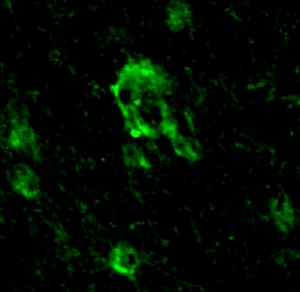
Application: Immunocytochemistry/ImmunofluorescenceSample Tested: Adult brainSpecies: PrimateVerified Customer | Posted 08/05/2025

-
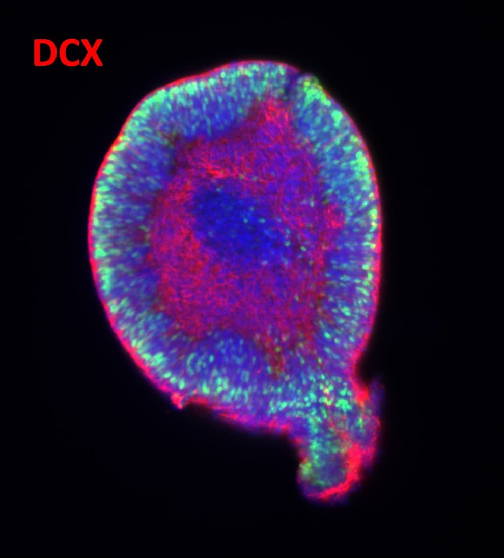
Application: Immunocytochemistry/ImmunofluorescenceSample Tested: Human iPSC-Derived Cerebral OrganoidsSpecies: HumanVerified Customer | Posted 09/21/2023
-
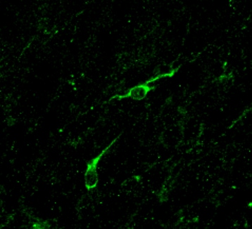
Application: Immunocytochemistry/ImmunofluorescenceSample Tested: Adult brainSpecies: PrimateVerified Customer | Posted 03/08/2022
There are no reviews that match your criteria.
Protocols
Find general support by application which include: protocols, troubleshooting, illustrated assays, videos and webinars.
- Antigen Retrieval Protocol (PIER)
- Antigen Retrieval for Frozen Sections Protocol
- Appropriate Fixation of IHC/ICC Samples
- Cellular Response to Hypoxia Protocols
- Chromogenic IHC Staining of Formalin-Fixed Paraffin-Embedded (FFPE) Tissue Protocol
- Chromogenic Immunohistochemistry Staining of Frozen Tissue
- ClariTSA™ Fluorophore Kits
- Detection & Visualization of Antibody Binding
- Fluorescent IHC Staining of Frozen Tissue Protocol
- Graphic Protocol for Heat-induced Epitope Retrieval
- Graphic Protocol for the Preparation and Fluorescent IHC Staining of Frozen Tissue Sections
- Graphic Protocol for the Preparation and Fluorescent IHC Staining of Paraffin-embedded Tissue Sections
- Graphic Protocol for the Preparation of Gelatin-coated Slides for Histological Tissue Sections
- IHC Sample Preparation (Frozen sections vs Paraffin)
- Immunofluorescent IHC Staining of Formalin-Fixed Paraffin-Embedded (FFPE) Tissue Protocol
- Immunohistochemistry (IHC) and Immunocytochemistry (ICC) Protocols
- Immunohistochemistry Frozen Troubleshooting
- Immunohistochemistry Paraffin Troubleshooting
- Preparing Samples for IHC/ICC Experiments
- Preventing Non-Specific Staining (Non-Specific Binding)
- Primary Antibody Selection & Optimization
- Protocol for Heat-Induced Epitope Retrieval (HIER)
- Protocol for Making a 4% Formaldehyde Solution in PBS
- Protocol for VisUCyte™ HRP Polymer Detection Reagent
- Protocol for the Preparation & Fixation of Cells on Coverslips
- Protocol for the Preparation and Chromogenic IHC Staining of Frozen Tissue Sections
- Protocol for the Preparation and Chromogenic IHC Staining of Frozen Tissue Sections - Graphic
- Protocol for the Preparation and Chromogenic IHC Staining of Paraffin-embedded Tissue Sections
- Protocol for the Preparation and Chromogenic IHC Staining of Paraffin-embedded Tissue Sections - Graphic
- Protocol for the Preparation and Fluorescent IHC Staining of Frozen Tissue Sections
- Protocol for the Preparation and Fluorescent IHC Staining of Paraffin-embedded Tissue Sections
- Protocol for the Preparation of Gelatin-coated Slides for Histological Tissue Sections
- R&D Systems Quality Control Western Blot Protocol
- TUNEL and Active Caspase-3 Detection by IHC/ICC Protocol
- The Importance of IHC/ICC Controls
- Troubleshooting Guide: Immunohistochemistry
- Troubleshooting Guide: Western Blot Figures
- Western Blot Conditions
- Western Blot Protocol
- Western Blot Protocol for Cell Lysates
- Western Blot Troubleshooting
- Western Blot Troubleshooting Guide
- View all Protocols, Troubleshooting, Illustrated assays and Webinars
Loading...