
Key Product Details
Species Reactivity
Validated:
Human, Mouse
Cited:
Human, Mouse, Rat, Chicken
Applications
Validated:
Immunohistochemistry, Western Blot, Neutralization
Cited:
Immunohistochemistry, Immunohistochemistry-Paraffin, Immunohistochemistry-Frozen, Western Blot, Neutralization, Simple Western, Bioassay, ELISA Capture, ELISA Development, Immuno-PCR
Label
Unconjugated
Antibody Source
Polyclonal Goat IgG
Loading...
Product Specifications
Immunogen
S. frugiperda insect ovarian cell line Sf 21-derived recombinant human (rh) Follistatin
Gly30-Asp329
Accession # P19883
Gly30-Asp329
Accession # P19883
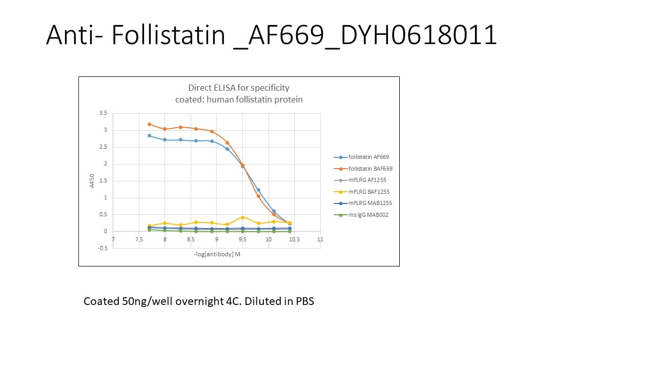
Specificity
Detects human and mouse Follistatin in direct ELISAs and Western blots.
Clonality
Polyclonal
Host
Goat
Isotype
IgG
Endotoxin Level
<0.10 EU per 1 μg of the antibody by the LAL method.
Scientific Data Images for Follistatin Antibody
Follistatin Inhibition of Activin A-induced Hemo-globin Expression and Neutralization by Human Follistatin Antibody.
Recombinant Human Follistatin 300 (Catalog # 669-FO) inhibits Recombinant Human/Mouse/Rat Activin A (Catalog # 338-AC) induced hemoglobin expression in the K562 human chronic myelogenous leukemia cell line in a dose-dependent manner (orange line), as measured by psuedoperoxidase activity. Inhibition of Recombinant Human/Mouse/Rat Activin A (7.5 ng/mL) activity elicited by Recombinant Human Follistatin 300 (0.4 µg/mL) is neutralized (green line) by increasing concentrations of Goat Anti-Human/Mouse Follistatin Antigen Affinity-purified Polyclonal Antibody (Catalog # AF669). At 6 µg/mL, this antibody will neutralize > 60% of rhFollistatin 300 bioactivity.
Follistatin in Human Breast.
Follistatin was detected in immersion fixed paraffin-embedded sections of human breast using Goat Anti-Human/Mouse Follistatin Antigen Affinity-purified Polyclonal Antibody (Catalog # AF669) at 10 µg/mL overnight at 4 °C. Tissue was stained using the Anti-Goat HRP-DAB Cell & Tissue Staining Kit (brown; Catalog # CTS008) and counterstained with hematoxylin (blue). Specific staining was localized to epithelial cells. View our protocol for Chromogenic IHC Staining of Paraffin-embedded Tissue Sections.
Detection of Mouse Follistatin by Immunohistochemistry
Follistatin (FST) expression is repressed in mouse mammary tumors and is restored using an Fst-expressing transgene. a Endogenous Fst expression in normal mouse mammary glands compared with human epidermal growth factor receptor 2, receptor tyrosine protein kinase erbB-2, proto-oncogene Neu (Her2/Neu)-induced mammary tumors (*p < 0.05). b Immunohistochemical analysis of endogenous FST expression in mouse mammary epithelia and tumors. cFst expression in Neu (single) and Neu/FST (bitransgenic) tumors. Fst messenger RNA expression was assessed by quantitative reverse transcription-polymerase chain reaction (**p < 0.01). d Hematoxylin and eosin (H&E)-stained mammary tumors from Neu (single) and Neu/FST (bitransgenic) mice are shown in the upper two panels. Immunohistochemistry for total (endogenous and transgenic) FST expression in lower two panels in single (left) and bitransgenic (right) tumors. Image collected and cropped by CiteAb from the following open publication (https://pubmed.ncbi.nlm.nih.gov/28583174), licensed under a CC-BY license. Not internally tested by R&D Systems.
Detection of Mouse Follistatin by Immunohistochemistry
Follistatin (FST) expression is repressed in mouse mammary tumors and is restored using an Fst-expressing transgene. a Endogenous Fst expression in normal mouse mammary glands compared with human epidermal growth factor receptor 2, receptor tyrosine protein kinase erbB-2, proto-oncogene Neu (Her2/Neu)-induced mammary tumors (*p < 0.05). b Immunohistochemical analysis of endogenous FST expression in mouse mammary epithelia and tumors. cFst expression in Neu (single) and Neu/FST (bitransgenic) tumors. Fst messenger RNA expression was assessed by quantitative reverse transcription-polymerase chain reaction (**p < 0.01). d Hematoxylin and eosin (H&E)-stained mammary tumors from Neu (single) and Neu/FST (bitransgenic) mice are shown in the upper two panels. Immunohistochemistry for total (endogenous and transgenic) FST expression in lower two panels in single (left) and bitransgenic (right) tumors Image collected and cropped by CiteAb from the following open publication (https://pubmed.ncbi.nlm.nih.gov/28583174), licensed under a CC-BY license. Not internally tested by R&D Systems.Applications for Follistatin Antibody
Application
Recommended Usage
Immunohistochemistry
5-15 µg/mL
Sample: Immersion fixed paraffin-embedded sections of human placenta and human breast
Sample: Immersion fixed paraffin-embedded sections of human placenta and human breast
Western Blot
0.1 µg/mL
Sample: Recombinant Human (rh) Follistatin 300 aa 30-329 (Catalog # 669-FO)
Sample: Recombinant Human (rh) Follistatin 300 aa 30-329 (Catalog # 669-FO)
Neutralization
Measured by its ability to neutralize Follistatin inhibition of Activin A-dependent hemoglobin expression in the K562 human chronic myelogenous leukemia cell line. At 6 μg/mL, this antibody will neutralize >60% of rhFollistatin 300 bioactivity on K562 cells.
Reviewed Applications
Read 1 review rated 5 using AF669 in the following applications:
Formulation, Preparation, and Storage
Purification
Antigen Affinity-purified
Reconstitution
Reconstitute at 0.2 mg/mL in sterile PBS. For liquid material, refer to CoA for concentration.
Loading...
Formulation
Lyophilized from a 0.2 μm filtered solution in PBS with Trehalose. *Small pack size (SP) is supplied either lyophilized or as a 0.2 µm filtered solution in PBS.
Shipping
Lyophilized product is shipped at ambient temperature. Liquid small pack size (-SP) is shipped with polar packs. Upon receipt, store immediately at the temperature recommended below.
Stability & Storage
Use a manual defrost freezer and avoid repeated freeze-thaw cycles.
- 12 months from date of receipt, -20 to -70 °C as supplied.
- 1 month, 2 to 8 °C under sterile conditions after reconstitution.
- 6 months, -20 to -70 °C under sterile conditions after reconstitution.
Calculators
Background: Follistatin
References
- Iemura, S. et al. (1998) Proc. Natl. Acad. Sci. USA 95:9337
- Guo, Q. (1998) Mol. Endocrinol. 12:96
- Hashimoto, O. et al. (1997) J. Biol. Chem. 272:13835
Alternate Names
FS, FST
Gene Symbol
FST
UniProt
Additional Follistatin Products
Product Documents for Follistatin Antibody
Certificate of Analysis
To download a Certificate of Analysis, please enter a lot or batch number in the search box below.
Note: Certificate of Analysis not available for kit components.
Product Specific Notices for Follistatin Antibody
For research use only
Related Research Areas
Citations for Follistatin Antibody
 Powered by Bioz
Customer Reviews for Follistatin Antibody (1)
5 out of 5
1 Customer Rating
Have you used Follistatin Antibody?
Submit a review and receive an Amazon gift card!
$25/€18/£15/$25CAN/¥2500 Yen for a review with an image
$10/€7/£6/$10CAN/¥1110 Yen for a review without an image
Submit a review
Customer Images
Showing
1
-
1 of
1 review
Showing All
Filter By:
-
Application: ELISASample Tested: Recombinant proteinSpecies: HumanVerified Customer | Posted 07/08/2020incubated 1hr at 37C. diluted in reagent diluent 2

There are no reviews that match your criteria.
Protocols
Find general support by application which include: protocols, troubleshooting, illustrated assays, videos and webinars.
- Antigen Retrieval Protocol (PIER)
- Antigen Retrieval for Frozen Sections Protocol
- Appropriate Fixation of IHC/ICC Samples
- Cellular Response to Hypoxia Protocols
- Chromogenic IHC Staining of Formalin-Fixed Paraffin-Embedded (FFPE) Tissue Protocol
- Chromogenic Immunohistochemistry Staining of Frozen Tissue
- ClariTSA™ Fluorophore Kits
- Detection & Visualization of Antibody Binding
- Fluorescent IHC Staining of Frozen Tissue Protocol
- Graphic Protocol for Heat-induced Epitope Retrieval
- Graphic Protocol for the Preparation and Fluorescent IHC Staining of Frozen Tissue Sections
- Graphic Protocol for the Preparation and Fluorescent IHC Staining of Paraffin-embedded Tissue Sections
- Graphic Protocol for the Preparation of Gelatin-coated Slides for Histological Tissue Sections
- IHC Sample Preparation (Frozen sections vs Paraffin)
- Immunofluorescent IHC Staining of Formalin-Fixed Paraffin-Embedded (FFPE) Tissue Protocol
- Immunohistochemistry (IHC) and Immunocytochemistry (ICC) Protocols
- Immunohistochemistry Frozen Troubleshooting
- Immunohistochemistry Paraffin Troubleshooting
- Preparing Samples for IHC/ICC Experiments
- Preventing Non-Specific Staining (Non-Specific Binding)
- Primary Antibody Selection & Optimization
- Protocol for Heat-Induced Epitope Retrieval (HIER)
- Protocol for Making a 4% Formaldehyde Solution in PBS
- Protocol for VisUCyte™ HRP Polymer Detection Reagent
- Protocol for the Preparation & Fixation of Cells on Coverslips
- Protocol for the Preparation and Chromogenic IHC Staining of Frozen Tissue Sections
- Protocol for the Preparation and Chromogenic IHC Staining of Frozen Tissue Sections - Graphic
- Protocol for the Preparation and Chromogenic IHC Staining of Paraffin-embedded Tissue Sections
- Protocol for the Preparation and Chromogenic IHC Staining of Paraffin-embedded Tissue Sections - Graphic
- Protocol for the Preparation and Fluorescent IHC Staining of Frozen Tissue Sections
- Protocol for the Preparation and Fluorescent IHC Staining of Paraffin-embedded Tissue Sections
- Protocol for the Preparation of Gelatin-coated Slides for Histological Tissue Sections
- R&D Systems Quality Control Western Blot Protocol
- TUNEL and Active Caspase-3 Detection by IHC/ICC Protocol
- The Importance of IHC/ICC Controls
- Troubleshooting Guide: Immunohistochemistry
- Troubleshooting Guide: Western Blot Figures
- Western Blot Conditions
- Western Blot Protocol
- Western Blot Protocol for Cell Lysates
- Western Blot Troubleshooting
- Western Blot Troubleshooting Guide
- View all Protocols, Troubleshooting, Illustrated assays and Webinars
Loading...