LRRN1 (Leucine-rich repeat neuronal protein 1; NLRR1) is a 95 kDa member of the neuronal LRR family of proteins. It is a cell-surface glycoprotein that is expressed on embryonic motor neurons and somatic myoblasts. Human LRRN1 is 716 amino acids (aa) in length. It is a type I transmembrane protein that contains a 606 aa extracellular region (aa 26-631) and a short 64 aa cytoplasmic tail. The extracellular region shows eleven LRRs (aa 94-384) that mediate protein-protein interactions, one C2-type Ig-like domain and one fibronectin type III domain. There is one potential alternate start site at Met286. Over aa 26-631, human LRRN1 shares 96% aa sequence identity with mouse LRRN1.

Key Product Details
Species Reactivity
Validated:
Human, Mouse
Cited:
Human, Mouse, Xenograft
Applications
Validated:
Immunohistochemistry, Western Blot
Cited:
Immunohistochemistry, Immunohistochemistry-Paraffin, Western Blot, Immunoprecipitation
Label
Unconjugated
Antibody Source
Polyclonal Sheep IgG
Loading...
Product Specifications
Immunogen
Mouse myeloma cell line NS0-derived recombinant human LRRN1
Ser26-Ala631
Accession # AAH34947
Ser26-Ala631
Accession # AAH34947
Specificity
Detects human LRRN1 in direct ELISAs and Western blots. In direct ELISAs and Western blots, approximately 5% cross-reactivity with recombinant human NLRR-3 is observed.
Clonality
Polyclonal
Host
Sheep
Isotype
IgG
Scientific Data Images for LRRN1/NLRR-1 Antibody
LRRN1/NLRR‑1 in Embryonic Mouse Somites.
LRRN1/NLRR-1 was detected in immersion fixed frozen sections of embryonic mouse somites (E9.5) using 10 µg/mL Sheep Anti-Human LRRN1/NLRR-1 Antigen Affinity-purified Polyclonal Antibody (Catalog # AF4990) overnight at 4 °C. Tissue was stained with the NorthernLights™ 557-conjugated Anti-Sheep IgG Secondary Antibody (red; Catalog # NL010) and counterstained with DAPI (blue). View our protocol for Fluorescent IHC Staining of Frozen Tissue Sections.
Detection of LRRN1/NLRR-1 by Western Blot
N1mAbs potentiates EGFR inhibitor-induced growth suppression. (A) NLRR1-stably expressing SH-SY5Y cells were treated with N1mAb 281 (25 μg/ml) and different concentrations of AG1478 (AG) for 72 h Data are represented as mean ± SD. Quantification of cell proliferation was performed by WST-8 assays. Data were normalized to the results for untreated cells and represented as percentage of control (mean ± SD). (B) AG1478-resistant A549 cells were treated with N1mAb 281 and different concentrations of AG1478 for 72 h (C) NLRR1-stably expressing SH-SY5Y cells were starved and treated with N1mAb 240 or 281 at 25 μg/ml for 3 h, followed by EGF treatment at the indicated concentration for 10 min. Cell lysates were subjected to western blot analyses. Arrowheads, glycosylated NLRR1. (D) MCF7 cells overexpressing NLRR1 were starved and treated with N1mAb 281 at 25 μg/ml for 3 h, followed by EGF treatment (1 ng/ml) for 10 min. The cell lysates were subjected to western blot analyses. (E) To check the effect of the long-term treatment of N1mAbs, SK-N-BE cells were incubated with N1mAbs (30 μg/ml) for 7 days with medium change every 2 days, and the cell lysates were subjected to western blot analyses. Image collected and cropped by CiteAb from the following open publication (https://pubmed.ncbi.nlm.nih.gov/34277416), licensed under a CC-BY license. Not internally tested by R&D Systems.Applications for LRRN1/NLRR-1 Antibody
Application
Recommended Usage
Immunohistochemistry
5-15 µg/mL
Sample: Immersion fixed frozen sections of embryonic mouse somites (E9.5)
Sample: Immersion fixed frozen sections of embryonic mouse somites (E9.5)
Western Blot
0.1 µg/mL
Sample: Recombinant Human LRRN1/NLRR‑1
Sample: Recombinant Human LRRN1/NLRR‑1
Reviewed Applications
Read 1 review rated 5 using AF4990 in the following applications:
Formulation, Preparation, and Storage
Purification
Antigen Affinity-purified
Reconstitution
Reconstitute at 0.2 mg/mL in sterile PBS. For liquid material, refer to CoA for concentration.
Loading...
Formulation
Lyophilized from a 0.2 μm filtered solution in PBS with Trehalose. *Small pack size (SP) is supplied either lyophilized or as a 0.2 µm filtered solution in PBS.
Shipping
Lyophilized product is shipped at ambient temperature. Liquid small pack size (-SP) is shipped with polar packs. Upon receipt, store immediately at the temperature recommended below.
Stability & Storage
Use a manual defrost freezer and avoid repeated freeze-thaw cycles.
- 12 months from date of receipt, -20 to -70 °C as supplied.
- 1 month, 2 to 8 °C under sterile conditions after reconstitution.
- 6 months, -20 to -70 °C under sterile conditions after reconstitution.
Calculators
Background: LRRN1/NLRR-1
Long Name
Leucine-rich Repeat Neuronal 1
Alternate Names
FIGLER3, LRN, NLRR-1, NLRR1
Gene Symbol
LRRN1
UniProt
Additional LRRN1/NLRR-1 Products
Product Documents for LRRN1/NLRR-1 Antibody
Certificate of Analysis
To download a Certificate of Analysis, please enter a lot or batch number in the search box below.
Note: Certificate of Analysis not available for kit components.
Product Specific Notices for LRRN1/NLRR-1 Antibody
For research use only
Related Research Areas
Citations for LRRN1/NLRR-1 Antibody
 Powered by Bioz
Customer Reviews for LRRN1/NLRR-1 Antibody (1)
5 out of 5
1 Customer Rating
Have you used LRRN1/NLRR-1 Antibody?
Submit a review and receive an Amazon gift card!
$25/€18/£15/$25CAN/¥2500 Yen for a review with an image
$10/€7/£6/$10CAN/¥1110 Yen for a review without an image
Submit a review
Customer Images
Showing
1
-
1 of
1 review
Showing All
Filter By:
-
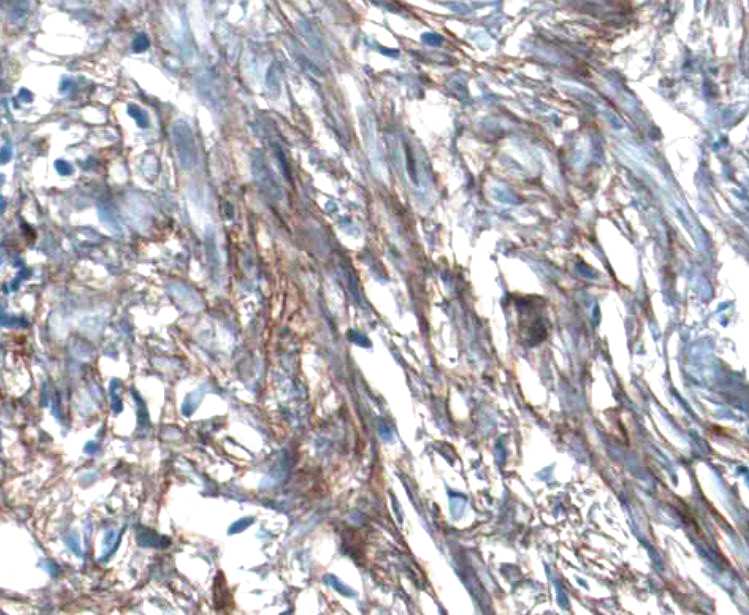
Application: ImmunohistochemistrySample Tested: Prostate cancer tissueSpecies: HumanVerified Customer | Posted 09/10/2021

There are no reviews that match your criteria.
Protocols
Find general support by application which include: protocols, troubleshooting, illustrated assays, videos and webinars.
- Antigen Retrieval Protocol (PIER)
- Antigen Retrieval for Frozen Sections Protocol
- Appropriate Fixation of IHC/ICC Samples
- Cellular Response to Hypoxia Protocols
- Chromogenic IHC Staining of Formalin-Fixed Paraffin-Embedded (FFPE) Tissue Protocol
- Chromogenic Immunohistochemistry Staining of Frozen Tissue
- ClariTSA™ Fluorophore Kits
- Detection & Visualization of Antibody Binding
- Fluorescent IHC Staining of Frozen Tissue Protocol
- Graphic Protocol for Heat-induced Epitope Retrieval
- Graphic Protocol for the Preparation and Fluorescent IHC Staining of Frozen Tissue Sections
- Graphic Protocol for the Preparation and Fluorescent IHC Staining of Paraffin-embedded Tissue Sections
- Graphic Protocol for the Preparation of Gelatin-coated Slides for Histological Tissue Sections
- IHC Sample Preparation (Frozen sections vs Paraffin)
- Immunofluorescent IHC Staining of Formalin-Fixed Paraffin-Embedded (FFPE) Tissue Protocol
- Immunohistochemistry (IHC) and Immunocytochemistry (ICC) Protocols
- Immunohistochemistry Frozen Troubleshooting
- Immunohistochemistry Paraffin Troubleshooting
- Preparing Samples for IHC/ICC Experiments
- Preventing Non-Specific Staining (Non-Specific Binding)
- Primary Antibody Selection & Optimization
- Protocol for Heat-Induced Epitope Retrieval (HIER)
- Protocol for Making a 4% Formaldehyde Solution in PBS
- Protocol for VisUCyte™ HRP Polymer Detection Reagent
- Protocol for the Preparation & Fixation of Cells on Coverslips
- Protocol for the Preparation and Chromogenic IHC Staining of Frozen Tissue Sections
- Protocol for the Preparation and Chromogenic IHC Staining of Frozen Tissue Sections - Graphic
- Protocol for the Preparation and Chromogenic IHC Staining of Paraffin-embedded Tissue Sections
- Protocol for the Preparation and Chromogenic IHC Staining of Paraffin-embedded Tissue Sections - Graphic
- Protocol for the Preparation and Fluorescent IHC Staining of Frozen Tissue Sections
- Protocol for the Preparation and Fluorescent IHC Staining of Paraffin-embedded Tissue Sections
- Protocol for the Preparation of Gelatin-coated Slides for Histological Tissue Sections
- R&D Systems Quality Control Western Blot Protocol
- TUNEL and Active Caspase-3 Detection by IHC/ICC Protocol
- The Importance of IHC/ICC Controls
- Troubleshooting Guide: Immunohistochemistry
- Troubleshooting Guide: Western Blot Figures
- Western Blot Conditions
- Western Blot Protocol
- Western Blot Protocol for Cell Lysates
- Western Blot Troubleshooting
- Western Blot Troubleshooting Guide
- View all Protocols, Troubleshooting, Illustrated assays and Webinars
Loading...