Parvalbumin (Parvalbumin alpha ) is a 12 kDa member of the parvalbumin family of Ca++-binding proteins. In human, it is expressed in intrafusal muscle fibers, plus GABAergic interneurons and cerebellar Purkinje and basket cells. It presumably acts as a Ca++ buffer that shortens the duration of fiber contraction. Human Parvalbumin is 110 amino acids (aa) in length. It contains two EF-hand domains (aa 39-74 and 78-110) that bind calcium. There are three potential isoform variants. One shows an alternate start site at Met33, a second shows a six aa substitution for the C-terminal nine amino acids and a third shows a deletion of Gly99-Val100. Human Parvalbumin alpha is 51% aa identical to human Parvalbumin beta and is 87% plus 92% aa identical to mouse and rat Parvalbumin, respectively.

Key Product Details
Species Reactivity
Validated:
Human, Mouse, Rat
Cited:
Human, Mouse, Rat, Primate - Macaca fascicularis (Crab-eating Monkey or Cynomolgus Macaque), Transgenic Mouse
Applications
Validated:
Immunohistochemistry, Western Blot, Simple Western
Cited:
Immunohistochemistry, Immunohistochemistry-Frozen, Immunocytochemistry
Label
Unconjugated
Antibody Source
Polyclonal Sheep IgG
Loading...
Product Specifications
Immunogen
E. coli-derived recombinant human Parvalbumin alpha
Ser2-Ser110
Accession # P20472
Ser2-Ser110
Accession # P20472
Specificity
Detects human, mouse, and rat Parvalbumin alpha in direct ELISAs and Western blots.
Clonality
Polyclonal
Host
Sheep
Isotype
IgG
Scientific Data Images for Parvalbumin alpha Antibody
Detection of Human/Mouse/Rat Parvalbumin alpha by Western Blot.
Western blot shows lysates of human, mouse, and rat brain tissue. PVDF membrane was probed with 1 µg/mL of Sheep Anti-Human/Mouse/Rat Parvalbumin a Antigen Affinity-purified Polyclonal Antibody (Catalog # AF5058) followed by HRP-conjugated Anti-Sheep IgG Secondary Antibody (Catalog # HAF016). A specific band was detected for Parvalbumin a at approximately 12 kDa (as indicated). This experiment was conducted under reducing conditions and using Immunoblot Buffer Group 8.
Parvalbumin alpha in Human Brain.
Parvalbumin a was detected in immersion fixed paraffin-embedded sections of human brain (cortex) using 5 µg/mL Sheep Anti-Human/Mouse/Rat Parvalbumin a Antigen Affinity-purified Polyclonal Antibody (Catalog # AF5058) overnight at 4 °C. Tissue was stained with the Anti-Sheep HRP-DAB Cell & Tissue Staining Kit (brown; Catalog # CTS019) and counterstained with hematoxylin (blue). View our protocol for Chromogenic IHC Staining of Paraffin-embedded Tissue Sections.
Detection of Rat Parvalbumin alpha by Simple WesternTM.
Simple Western lane view shows lysates of rat brain tissue, loaded at 0.2 mg/mL. A specific band was detected for Parvalbumin a at approximately 15 kDa (as indicated) using 50 µg/mL of Sheep Anti-Human/Mouse/Rat Parvalbumin a Antigen Affinity-purified Polyclonal Antibody (Catalog # AF5058) followed by 1:50 dilution of HRP-conjugated Anti-Sheep IgG Secondary Antibody (Catalog # HAF016). This experiment was conducted under reducing conditions and using the 12-230 kDa separation system.
Detection of Human and Mouse Parvalbumin alpha by Simple WesternTM.
Simple Western lane view shows lysates of mouse brain (cortex) and human brain (cerebellum), loaded at 0.2 mg/mL. A specific band was detected for Parvalbumin a at approximately 14 and 17 kDa (as indicated) using 50 µg/mL of Sheep Anti-Human/Mouse/Rat Parvalbumin a Antigen Affinity-purified Polyclonal Antibody (Catalog # AF5058) followed by 1:50 dilution of HRP-conjugated Anti-Sheep IgG Secondary Antibody (Catalog # HAF016). This experiment was conducted under reducing conditions and using the 12-230 kDa separation system.
Detection of Mouse Parvalbumin alpha by Simple Western
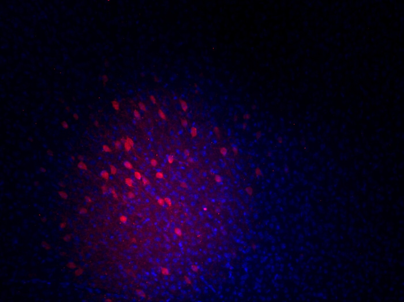
Altered NARP expression impacts the expression of parvalbumin in inhibitory neurons in socially isolated mice. (A) Schematic diagram of molecular & behavioral analyses. Molecular analysis was conducted at P35 & adulthood, & behavioral analysis was conducted in adulthood. (B) NARP mRNA levels in the medial PFC of jSI mice were lower than those in GH mice at P35, but not at adult [2-way ANOVA, age (P35 & adult) x housing (GH & jSI) interaction F(1. 24) = 6.677 p = 0.0163, age F(1. 24) = 3.505 p = 0.0734, housing F(1. 24) = 4.600 p = 0.0423; Tukey test p = 0.0135 (GH vs jSI at P35), n = 7 (GH, P35), n = 7 (jSI, P35), n = 7 (GH, adult), n = 7 (jSI, adult)]. (C) NARP protein levels in the medial PFC of jSI mice were lower than those in GH mice at P35 (two-tailed t-test, t12 = 2.348, p = 0.0369). (D) Representative images of NARP protein expression in the medial PFC of GH & jSI mice. (E) PV mRNA levels in the medial PFC of jSI mice were lower than those in GH mice at adult, but not at P35 [2-way ANOVA, age (P35 & adult) x housing (GH & jSI) interaction F(1. 24) = 0.4791, p = 0.4955; age F(1. 24) = 3.821, p = 0.0624; housing F(1. 24) = 11.71, p = 0.0022; Tukey test p = 0.0361 (GH vs jSI at adult), n = 7 (GH, P35), n = 7 (jSI, P35), n = 7 (GH, adult), n = 7 (jSI, adult)]. (F) PV protein levels in the medial PFC of jSI mice were lower than those in GH mice at adult (two-tailed t-test, t12 = 3.440, p = 0.0049). (G) Representative images of PV protein expression in the medial PFC of GH & jSI mice. NARP, Neuronal Activity-Regulated Pentraxin; PV, Parvalbumin; PFC, Prefrontal Cortex; GH, Group-Housed; jSI, Juvenile Social Isolation; P35, Postnatal Day 35; Adult/Adulthood, Postnatal Day 63–70; ANOVA, Analysis of Variance. * 0.01 ≤ p < 0.05; ** 0.001 ≤ p < 0.01. Image collected & cropped by CiteAb from the following open publication (https://www.frontiersin.org/articles/10.3389/fpsyt.2024.1403476/full), licensed under a CC-BY license. Not internally tested by R&D Systems.Applications for Parvalbumin alpha Antibody
Application
Recommended Usage
Immunohistochemistry
5-15 µg/mL
Sample: Immersion fixed paraffin-embedded sections of human brain (cortex)
Sample: Immersion fixed paraffin-embedded sections of human brain (cortex)
Simple Western
50 µg/mL
Sample: Rat brain tissue, mouse brain (cortex), and human brain (cerebellum)
Sample: Rat brain tissue, mouse brain (cortex), and human brain (cerebellum)
Western Blot
1 µg/mL
Sample: Human, mouse and rat brain tissue
Sample: Human, mouse and rat brain tissue
Reviewed Applications
Read 1 review rated 5 using AF5058 in the following applications:
Formulation, Preparation, and Storage
Purification
Antigen Affinity-purified
Reconstitution
Reconstitute at 0.2 mg/mL in sterile PBS. For liquid material, refer to CoA for concentration.
Loading...
Formulation
Lyophilized from a 0.2 μm filtered solution in PBS with Trehalose. *Small pack size (SP) is supplied either lyophilized or as a 0.2 µm filtered solution in PBS.
Shipping
Lyophilized product is shipped at ambient temperature. Liquid small pack size (-SP) is shipped with polar packs. Upon receipt, store immediately at the temperature recommended below.
Stability & Storage
Use a manual defrost freezer and avoid repeated freeze-thaw cycles.
- 12 months from date of receipt, -20 to -70 °C as supplied.
- 1 month, 2 to 8 °C under sterile conditions after reconstitution.
- 6 months, -20 to -70 °C under sterile conditions after reconstitution.
Calculators
Background: Parvalbumin alpha
Alternate Names
PVALB
Gene Symbol
PVALB
UniProt
Additional Parvalbumin alpha Products
Product Documents for Parvalbumin alpha Antibody
Certificate of Analysis
To download a Certificate of Analysis, please enter a lot or batch number in the search box below.
Note: Certificate of Analysis not available for kit components.
Product Specific Notices for Parvalbumin alpha Antibody
For research use only
Related Research Areas
Citations for Parvalbumin alpha Antibody
 Powered by Bioz
Customer Reviews for Parvalbumin alpha Antibody (1)
5 out of 5
1 Customer Rating
Have you used Parvalbumin alpha Antibody?
Submit a review and receive an Amazon gift card!
$25/€18/£15/$25CAN/¥2500 Yen for a review with an image
$10/€7/£6/$10CAN/¥1110 Yen for a review without an image
Submit a review
Customer Images
Showing
1
-
1 of
1 review
Showing All
Filter By:
-
Application: Immunocytochemistry/ImmunofluorescenceSample Tested: Adult brain and Adult brain (hippocampus)Species: MouseVerified Customer | Posted 07/25/2019

There are no reviews that match your criteria.
Protocols
Find general support by application which include: protocols, troubleshooting, illustrated assays, videos and webinars.
- Antigen Retrieval Protocol (PIER)
- Antigen Retrieval for Frozen Sections Protocol
- Appropriate Fixation of IHC/ICC Samples
- Cellular Response to Hypoxia Protocols
- Chromogenic IHC Staining of Formalin-Fixed Paraffin-Embedded (FFPE) Tissue Protocol
- Chromogenic Immunohistochemistry Staining of Frozen Tissue
- ClariTSA™ Fluorophore Kits
- Detection & Visualization of Antibody Binding
- Fluorescent IHC Staining of Frozen Tissue Protocol
- Graphic Protocol for Heat-induced Epitope Retrieval
- Graphic Protocol for the Preparation and Fluorescent IHC Staining of Frozen Tissue Sections
- Graphic Protocol for the Preparation and Fluorescent IHC Staining of Paraffin-embedded Tissue Sections
- Graphic Protocol for the Preparation of Gelatin-coated Slides for Histological Tissue Sections
- IHC Sample Preparation (Frozen sections vs Paraffin)
- Immunofluorescent IHC Staining of Formalin-Fixed Paraffin-Embedded (FFPE) Tissue Protocol
- Immunohistochemistry (IHC) and Immunocytochemistry (ICC) Protocols
- Immunohistochemistry Frozen Troubleshooting
- Immunohistochemistry Paraffin Troubleshooting
- Preparing Samples for IHC/ICC Experiments
- Preventing Non-Specific Staining (Non-Specific Binding)
- Primary Antibody Selection & Optimization
- Protocol for Heat-Induced Epitope Retrieval (HIER)
- Protocol for Making a 4% Formaldehyde Solution in PBS
- Protocol for VisUCyte™ HRP Polymer Detection Reagent
- Protocol for the Preparation & Fixation of Cells on Coverslips
- Protocol for the Preparation and Chromogenic IHC Staining of Frozen Tissue Sections
- Protocol for the Preparation and Chromogenic IHC Staining of Frozen Tissue Sections - Graphic
- Protocol for the Preparation and Chromogenic IHC Staining of Paraffin-embedded Tissue Sections
- Protocol for the Preparation and Chromogenic IHC Staining of Paraffin-embedded Tissue Sections - Graphic
- Protocol for the Preparation and Fluorescent IHC Staining of Frozen Tissue Sections
- Protocol for the Preparation and Fluorescent IHC Staining of Paraffin-embedded Tissue Sections
- Protocol for the Preparation of Gelatin-coated Slides for Histological Tissue Sections
- R&D Systems Quality Control Western Blot Protocol
- TUNEL and Active Caspase-3 Detection by IHC/ICC Protocol
- The Importance of IHC/ICC Controls
- Troubleshooting Guide: Immunohistochemistry
- Troubleshooting Guide: Western Blot Figures
- Western Blot Conditions
- Western Blot Protocol
- Western Blot Protocol for Cell Lysates
- Western Blot Troubleshooting
- Western Blot Troubleshooting Guide
- View all Protocols, Troubleshooting, Illustrated assays and Webinars
Loading...