Human/Mouse/Rat PEDFR/PNPLA2 Antibody
R&D Systems | Catalog # AF5365

Key Product Details
Species Reactivity
Validated:
Human, Mouse, Rat
Cited:
Human, Mouse, Rat
Applications
Validated:
Immunohistochemistry, Western Blot, Simple Western
Cited:
Immunohistochemistry, Western Blot, Immunocytochemistry
Label
Unconjugated
Antibody Source
Polyclonal Sheep IgG
Loading...
Product Specifications
Immunogen
E. coli-derived recombinant mouse PEDF R/PNPLA2
Val162-Gly253
Accession # Q8BJ56
Val162-Gly253
Accession # Q8BJ56
Specificity
Detects human, mouse, and rat PEDF R/PNPLA2 in direct ELISAs and Western blots.
Clonality
Polyclonal
Host
Sheep
Isotype
IgG
Scientific Data Images for Human/Mouse/Rat PEDFR/PNPLA2 Antibody
Detection of Human and Mouse PEDF R/PNPLA2 by Western Blot.
Western blot shows lysates of human adipose tissue and mouse adipose tissue. PVDF membrane was probed with 1 µg/mL of Sheep Anti-Human/Mouse/Rat PEDF R/PNPLA2 Antigen Affinity-purified Polyclonal Antibody (Catalog # AF5365) followed by HRP-conjugated Anti-Sheep IgG Secondary Antibody (Catalog # HAF016). A specific band was detected for PEDF R/PNPLA2 at approximately 55 kDa (as indicated). This experiment was conducted under reducing conditions and using Immunoblot Buffer Group 8.
PEDF R/PNPLA2 in Mouse Kidney.
PEDF R/PNPLA2 was detected in perfusion fixed frozen sections of mouse kidney using 15 µg/mL Sheep Anti-Human/Mouse/Rat PEDF R/PNPLA2 Antigen Affinity-purified Polyclonal Antibody (Catalog # AF5365) overnight at 4 °C. Tissue was stained with the Anti-Sheep HRP-DAB Cell & Tissue Staining Kit (brown; Catalog # CTS019) and counterstained with hematoxylin (blue). Specific labeling was localized to the plasma membrane of epithelial cells in tubules. View our protocol for Chromogenic IHC Staining of Frozen Tissue Sections.
Detection of Mouse PEDF R/PNPLA2 by Simple WesternTM.
Simple Western lane view shows lysates of mouse adipose tissue, loaded at 0.2 mg/mL. A specific band was detected for PEDF R/PNPLA2 at approximately 59 kDa (as indicated) using 10 µg/mL of Sheep Anti-Human/Mouse/Rat PEDF R/PNPLA2 Antigen Affinity-purified Polyclonal Antibody (Catalog # AF5365) followed by 1:50 dilution of HRP-conjugated Anti-Sheep IgG Secondary Antibody (Catalog # HAF016). This experiment was conducted under reducing conditions and using the 12-230 kDa separation system.
Detection of Human PEDFR/PNPLA2/ATGL by Western Blot
Loss-of-function analyses of PEDF in Bkid cells. (A) PEDF micro RNA (miR) expressing vector was transfected into Bkid cells. Immunoblot by anti-PEDF antibody showed the reduction of PEDF proteins by miRs. (B) The renal and the hepatic lesions of the results of the intracardiac injections of Bkid cells or Bkid+PEDFmiR cells. The adrenal metastasis was observed. Also, by Bkid cell injections, the renal metastasis was observed frequently (left, 12-positive/12), while no renal nodule was observed in Bkid+PEDFmiR injected mouse (right, 17-negative/18). The ruler is 1mm square. (C) The hepatic metastases of both cells were not inhibited with or without PEDF. (D) The section Hematoxylin/Eosin (H&E) staining of the similar samples to (B). The adrenal hypertrophy was observed both in Bkid- and Bkid+PEDFmiR-injected mouse organs. The renal metastatic lesion (met) was seen as the lighter color. The scale is 2 mm. (E) The hepatic metastasis. Both tumors had the cavity. (F) PEDF-immunohistochemistry (IHC) of the section (D). PEDF staining was observed inside of the adrenal gland cystic vesicle of Bkid tumors. The overall staining level of PEDF in Bkid tumor was higher than Bkid+PEDFmiR cells. The scale is 1 mm. (G) IHC for PEDF on paraffin sections of collagen gel-embedded 143B and 143B+PEDF cells. (H-L) The renal section IHC by various antibodies. (H) Anti-SPARC (osteonectin)-IHC indicates that the lesions are derived from the injected osteosarcoma. In the section of kidney from Bkid-injected mouse, there is SPARC-positive tumor both in kidney and adrenal gland sides. On the right, the kidney from Bkid+PEDFmiR-injected mouse had SPARC-positive cells only in adrenal gland. The scale indicates 100 µm. (I–L) The scale indicates 20 µm. (I) The higher magnified picture of the tissue surrounded glomerulus. There is SPARC-positive cells were seen around the glomerulus of Bkid-injected mouse (arrow), while no signal was seen in Bkid+PEDFmiR-injected mouse, (J) PEDF-IHC. PEDF was accumu
Detection of PEDFR/PNPLA2 by Western Blot
Effect of the 29-mer on the expression of MMP-9 in EDE. The schedule of induction of EDE is described in Fig. 2. (A) Representative immunofluorescence images showing MMP-9 in corneal and conjunctival epithelia after 14 days of EDE induction (n = 6 per group). (B) qPCR evaluates the levels of MMP-9 in the ocular surface epithelia (n = 6 per group). (C and D) Representative Western blots and densitometric analyses of MMP-9 and PEDF-R of ocular surface epithelia are shown from three independent experiments. Image collected and cropped by CiteAb from the following open publication (https://pubmed.ncbi.nlm.nih.gov/36201200), licensed under a CC-BY license. Not internally tested by R&D Systems.
Detection of PEDFR/PNPLA2 by Western Blot
Effect of the 29-mer on the expression of MMP-9 in EDE. The schedule of induction of EDE is described in Fig. 2. (A) Representative immunofluorescence images showing MMP-9 in corneal and conjunctival epithelia after 14 days of EDE induction (n = 6 per group). (B) qPCR evaluates the levels of MMP-9 in the ocular surface epithelia (n = 6 per group). (C and D) Representative Western blots and densitometric analyses of MMP-9 and PEDF-R of ocular surface epithelia are shown from three independent experiments. Image collected and cropped by CiteAb from the following open publication (https://pubmed.ncbi.nlm.nih.gov/36201200), licensed under a CC-BY license. Not internally tested by R&D Systems.Applications for Human/Mouse/Rat PEDFR/PNPLA2 Antibody
Application
Recommended Usage
Immunohistochemistry
5-15 µg/mL
Sample: Perfusion fixed frozen sections of mouse kidney
Sample: Perfusion fixed frozen sections of mouse kidney
Simple Western
10 µg/mL
Sample: Mouse adipose tissue
Sample: Mouse adipose tissue
Western Blot
1 µg/mL
Sample: Human adipose tissue and mouse adipose tissue
Sample: Human adipose tissue and mouse adipose tissue
Reviewed Applications
Read 1 review rated 3 using AF5365 in the following applications:
Formulation, Preparation, and Storage
Purification
Antigen Affinity-purified
Reconstitution
Reconstitute at 0.2 mg/mL in sterile PBS. For liquid material, refer to CoA for concentration.
Loading...
Formulation
Lyophilized from a 0.2 μm filtered solution in PBS with Trehalose. *Small pack size (SP) is supplied either lyophilized or as a 0.2 µm filtered solution in PBS.
Shipping
Lyophilized product is shipped at ambient temperature. Liquid small pack size (-SP) is shipped with polar packs. Upon receipt, store immediately at the temperature recommended below.
Stability & Storage
Use a manual defrost freezer and avoid repeated freeze-thaw cycles.
- 12 months from date of receipt, -20 to -70 °C as supplied.
- 1 month, 2 to 8 °C under sterile conditions after reconstitution.
- 6 months, -20 to -70 °C under sterile conditions after reconstitution.
Calculators
Background: PEDFR/PNPLA2
Long Name
Pigment Epithelium-derived Factor Receptor/Patatin-like Phospholipase Domain-containing Protein 2
Alternate Names
ATGL, Desnutrin, IPLA2-zeta, PEDF R, PNPLA2, TTS2
Gene Symbol
PNPLA2
UniProt
Additional PEDFR/PNPLA2 Products
Product Documents for Human/Mouse/Rat PEDFR/PNPLA2 Antibody
Certificate of Analysis
To download a Certificate of Analysis, please enter a lot or batch number in the search box below.
Note: Certificate of Analysis not available for kit components.
Product Specific Notices for Human/Mouse/Rat PEDFR/PNPLA2 Antibody
For research use only
Related Research Areas
Citations for Human/Mouse/Rat PEDFR/PNPLA2 Antibody
 Powered by Bioz
Customer Reviews for Human/Mouse/Rat PEDFR/PNPLA2 Antibody (1)
3 out of 5
1 Customer Rating
Have you used Human/Mouse/Rat PEDFR/PNPLA2 Antibody?
Submit a review and receive an Amazon gift card!
$25/€18/£15/$25CAN/¥2500 Yen for a review with an image
$10/€7/£6/$10CAN/¥1110 Yen for a review without an image
Submit a review
Customer Images
Showing
1
-
1 of
1 review
Showing All
Filter By:
-
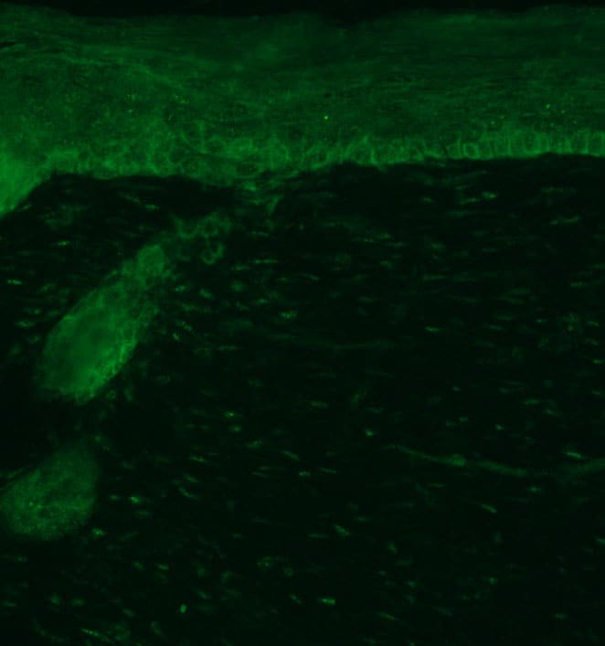
Application: Immunocytochemistry/ImmunofluorescenceSample Tested: Skin tissueSpecies: MouseVerified Customer | Posted 01/17/2018

There are no reviews that match your criteria.
Protocols
Find general support by application which include: protocols, troubleshooting, illustrated assays, videos and webinars.
- Antigen Retrieval Protocol (PIER)
- Antigen Retrieval for Frozen Sections Protocol
- Appropriate Fixation of IHC/ICC Samples
- Cellular Response to Hypoxia Protocols
- Chromogenic IHC Staining of Formalin-Fixed Paraffin-Embedded (FFPE) Tissue Protocol
- Chromogenic Immunohistochemistry Staining of Frozen Tissue
- ClariTSA™ Fluorophore Kits
- Detection & Visualization of Antibody Binding
- Fluorescent IHC Staining of Frozen Tissue Protocol
- Graphic Protocol for Heat-induced Epitope Retrieval
- Graphic Protocol for the Preparation and Fluorescent IHC Staining of Frozen Tissue Sections
- Graphic Protocol for the Preparation and Fluorescent IHC Staining of Paraffin-embedded Tissue Sections
- Graphic Protocol for the Preparation of Gelatin-coated Slides for Histological Tissue Sections
- IHC Sample Preparation (Frozen sections vs Paraffin)
- Immunofluorescent IHC Staining of Formalin-Fixed Paraffin-Embedded (FFPE) Tissue Protocol
- Immunohistochemistry (IHC) and Immunocytochemistry (ICC) Protocols
- Immunohistochemistry Frozen Troubleshooting
- Immunohistochemistry Paraffin Troubleshooting
- Preparing Samples for IHC/ICC Experiments
- Preventing Non-Specific Staining (Non-Specific Binding)
- Primary Antibody Selection & Optimization
- Protocol for Heat-Induced Epitope Retrieval (HIER)
- Protocol for Making a 4% Formaldehyde Solution in PBS
- Protocol for VisUCyte™ HRP Polymer Detection Reagent
- Protocol for the Preparation & Fixation of Cells on Coverslips
- Protocol for the Preparation and Chromogenic IHC Staining of Frozen Tissue Sections
- Protocol for the Preparation and Chromogenic IHC Staining of Frozen Tissue Sections - Graphic
- Protocol for the Preparation and Chromogenic IHC Staining of Paraffin-embedded Tissue Sections
- Protocol for the Preparation and Chromogenic IHC Staining of Paraffin-embedded Tissue Sections - Graphic
- Protocol for the Preparation and Fluorescent IHC Staining of Frozen Tissue Sections
- Protocol for the Preparation and Fluorescent IHC Staining of Paraffin-embedded Tissue Sections
- Protocol for the Preparation of Gelatin-coated Slides for Histological Tissue Sections
- R&D Systems Quality Control Western Blot Protocol
- TUNEL and Active Caspase-3 Detection by IHC/ICC Protocol
- The Importance of IHC/ICC Controls
- Troubleshooting Guide: Immunohistochemistry
- Troubleshooting Guide: Western Blot Figures
- Western Blot Conditions
- Western Blot Protocol
- Western Blot Protocol for Cell Lysates
- Western Blot Troubleshooting
- Western Blot Troubleshooting Guide
- View all Protocols, Troubleshooting, Illustrated assays and Webinars
Loading...