Human Nestin Antibody (196908)
R&D Systems | Catalog # MAB1259

Key Product Details
Validated by
Biological Validation
Species Reactivity
Validated:
Human
Cited:
Human, Mouse, Rat, Drosophila, Xenograft
Applications
Validated:
Intracellular Staining by Flow Cytometry, Immunocytochemistry, CyTOF-reported
Cited:
Immunohistochemistry, Immunohistochemistry-Paraffin, Immunohistochemistry-Frozen, Western Blot, Flow Cytometry, Immunofluorescence, Immunocytochemistry, Bioassay, Cell Culture
Label
Unconjugated
Antibody Source
Monoclonal Mouse IgG1 Clone # 196908
Loading...
Product Specifications
Immunogen
NS0 mouse myeloma cell line transfected with human Nestin
Specificity
Detects human Nestin.
Clonality
Monoclonal
Host
Mouse
Isotype
IgG1
Scientific Data Images for Human Nestin Antibody (196908)
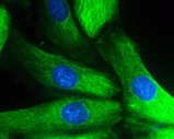
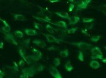
Nestin in Human Neural Progenitor Cells.
Nestin was detected in immersion fixed human fetal neural progenitor cells using 10 µg/mL Human Nestin Monoclonal Antibody (Catalog # MAB1259) for 3 hours at room temperature. Cells were stained (green) and counterstained with DAPI (blue). View our protocol for Fluorescent ICC Staining of Cells on Coverslips.
Detection of Nestin in A172 cells by Flow Cytometry.
A172 cells were stained with Mouse Anti-Human Nestin Monoclonal Antibody (Catalog # MAB1259, filled histogram) or isotype control antibody (Catalog # MAB002, open histogram), followed by Phycoerythrin-conjugated Anti-Mouse IgG Secondary Antibody (Catalog # F0102B). To facilitate intracellular staining, cells were fixed and permeabilized with FlowX FoxP3 Fixation & Permeabilization Buffer Kit (Catalog # FC012). View our protocol for Staining Intracellular Molecules.
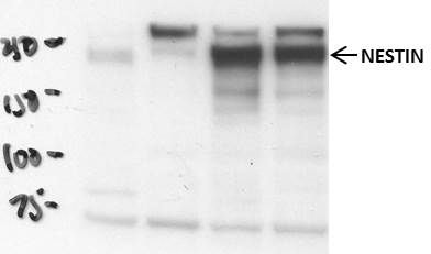
Detection of Mouse Nestin by Western Blot
Cardiolipin is externalized to the mitochondrial surface in alpha -syn mutant hNs and transgenic mice in response to alpha -syn accumulation. a Western analysis of lysates from corrected and A53T cells at DIV 14 and DIV 60 labeled for total alpha -syn, PS129, TH (DA neuronal marker), nestin (NPC marker) or GAPDH show that levels of PS129-modified alpha -syn are elevated in A53T hNs at DIV 14 and remain elevated at DIV 60 relative to corrected cells. b, c Mitochondria were purified from A53T cells and genetically corrected cells at both DIV 14 and DIV 60. Lysates were then separated based on TX-100 solubility (soluble) or urea solubility (insoluble). Western analysis of A53T cells shows elevated soluble alpha -syn at the mitochondria relative to corrected cells at DIV 14 and DIV 60 (denoted by ** and * respectively). Quantification of soluble and insoluble alpha -syn levels in mitochondrial fractions normalized to Coomassie (c). Data represent mean ± s.e.m. *P < 0.05, **P < 0.01 by ANOVA followed by Tukey’s post hoc test, n = 4. d, e Micrographs of corrected and A53T-mutant NPCs expressing the negative charge probe RPRE-RFP and CL-GFP show RPRE-RFP translocation from a primarily plasma membrane localization in isogenic-corrected hNs to a punctate intracellular localization in A53T hNs in tight cellular localization with CL-GFP (d). Quantification of percent total cell number with colocalized RPRE-RFP and CL-GFP (e). Data represent mean ± s.e.m. **P < 0.0001 by t-test, n = 3 coverslips over 2 independent differentiations, DIV: 14; scale bar: 10 μm. f, g Fluorescence resonance energy transfer (FRET) from CL-GFP to RPRE-RFP in hiPSC-derived A53T and corrected hNs or hESC-derived WT, A53T and E46K hNs was assessed (f) and mean FRET intensity was quantified (g). Data represent mean ± s.e.m. **P < 0.01 by ANOVA followed by Tukey’s post hoc test, for hiPSCs, n = 6, for hESCs, n = 8 coverslips over two independent differentiations, DIV: 14. Scale bar: 10 µm. h Mitochondria were isolated from the SNpc of
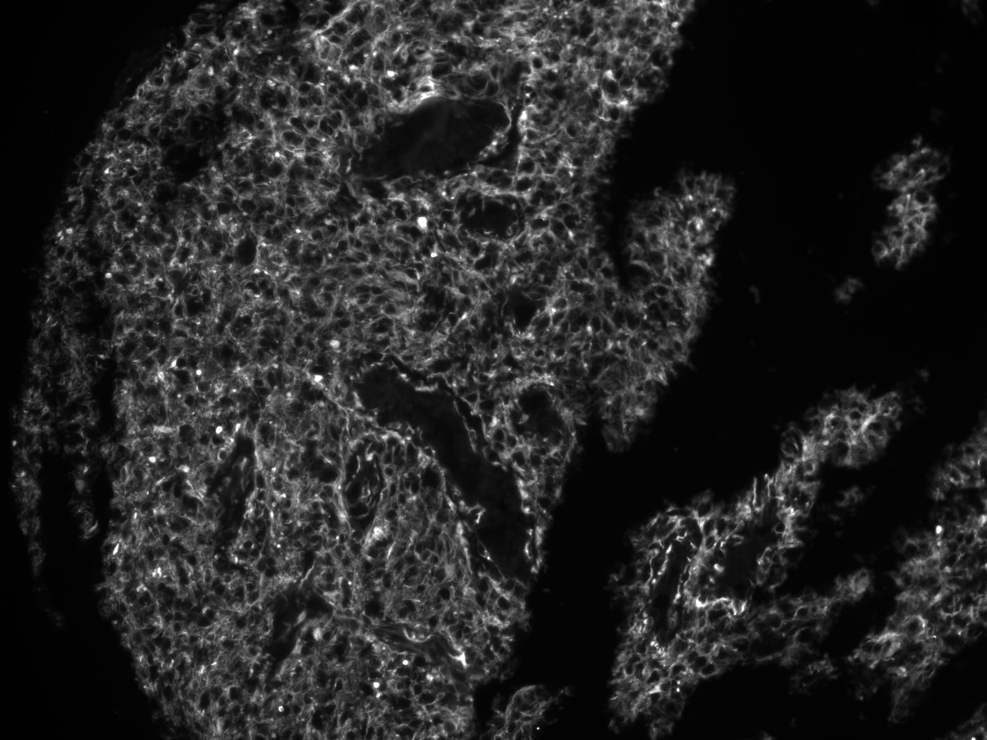
Detection of Human Nestin by Immunohistochemistry
MET and TMZ effects in in vivo glioma models(A) Variation of U251 tumor volume measured at contrast-enhanced T1 MRI after treatment expressed as percentage of volume change. Error bars indicate SEM. (B) U251 derived orthotopic brain tumors from vehicle (CTRL), TMZ or TMZ/metformin (Combo) treated mice were analyzed for Ki67, Nestin and CD133 markers by immunohistochemistry. No difference between the tumor edge and bulk areas in IHC staining intensities or percentages of positive cells was observed for any of the investigated markers. H&E was performed for morphological evaluation of the tumors. Representative images are shown with original magnification x200. (C) Variation of T98G tumor volume measured using caliper after treatment expressed as percentage of volume variation. In T98G xenograft model already at 7 days, the combined treatment significantly slowed down tumor growth compared to control (p value = 0.005), moreover a transient reduction was observed with MET (p value = 0.03). After 3 weeks, only Combo-treated mice displayed tumor smaller than vehicle (p value = 0.04). Error bars indicate SEM. (D) T98G tumors from vehicle (CTRL), TMZ, MET or TMZ/metformin (Combo) treated mice, were analyzed for Ki67, Nestin and CD133 markers expression by immunohistochemistry. No difference between the tumor edge and bulk areas in IHC staining intensities or percentages of positive cells was observed for any of the investigated markers. Hematoxylin and eosin staining (H&E) was performed for morphological evaluation of the tumors. Representative images are shown with Original magnification x200. Image collected and cropped by CiteAb from the following publication (https://www.oncotarget.com/lookup/doi/10.18632/oncotarget.23028), licensed under a CC-BY license. Not internally tested by R&D Systems.
Detection of Rat Nestin by Immunocytochemistry/Immunofluorescence
Neural stem cells could proliferate and differentiate into neurons and astrocytes.(A) Representative photomicrograph of neurospheres in culture. (B) Immunocytochemical staining of purified NSCs with Nestin. (C) Immunocytochemical staining of purified protoplasmic astrocytes with GFAP. (D) Immunocytochemical staining of purified neurons with beta -tubulin-III. (E) Nucleus staining of differentiated cells from NSCs with DAPI. Image collected and cropped by CiteAb from the following publication (https://pubmed.ncbi.nlm.nih.gov/26431046), licensed under a CC-BY license. Not internally tested by R&D Systems.
Detection of Rat Nestin by Western Blot
miR–381 promoted neural stem cells proliferation.(A) The expression of miR–381 was measured by qRT-PCR. (B) CCK–8 was performed to detect the neural stem cells proliferation. (C) The mRNA expression of nestin was detected by qRT-PCR. (D) The protein expression of nestin was measured by Western blot. **p<0.01 and ***p<0.001. Image collected and cropped by CiteAb from the following publication (https://pubmed.ncbi.nlm.nih.gov/26431046), licensed under a CC-BY license. Not internally tested by R&D Systems.
Detection of Mouse Nestin by Immunohistochemistry
Dissimilar tumorigenesis in the in vivo animal model.(a) Kaplan-Meier survival plots; 3 mice were used for cells of each clone. (b) Representative brain tumors of NOD-SCID mice harboring xenografts of the clones #2 (upper left), #3 (lower left), #4 (upper right) and #5 (lower right); H.E. staining, arrows: tumor, scale bar = 5 mm. (c) A representative xenograft of the clone #4; nestin-positive cells infiltrated the contralateral hemisphere (arrows). Scale bars = 5 mm (left), 1 mm (middle), 50 μm (right). Image collected and cropped by CiteAb from the following publication (https://pubmed.ncbi.nlm.nih.gov/25623281), licensed under a CC-BY license. Not internally tested by R&D Systems.
Detection of Mouse Human Nestin Antibody by Western Blot
Cardiolipin is externalized to the mitochondrial surface in alpha -syn mutant hNs and transgenic mice in response to alpha -syn accumulation. a Western analysis of lysates from corrected and A53T cells at DIV 14 and DIV 60 labeled for total alpha -syn, PS129, TH (DA neuronal marker), nestin (NPC marker) or GAPDH show that levels of PS129-modified alpha -syn are elevated in A53T hNs at DIV 14 and remain elevated at DIV 60 relative to corrected cells. Image collected and cropped by CiteAb from the following publication (https://pubmed.ncbi.nlm.nih.gov/29483518), licensed under a CC-BY license. Not internally tested by R&D Systems.
Detection of Human Nestin by Western Blot
Validation of the phosphorylation status regarding the representative phosphoproteins by western blot analyses.Glioblastoma initiating cells were treated with 20 ng/ml EGF for 15 min and subjected to immunoblotting with the corresponding antibodies. Image collected and cropped by CiteAb from the following open publication (https://pubmed.ncbi.nlm.nih.gov/22912867), licensed under a CC-BY license. Not internally tested by R&D Systems.
Detection of Nestin by Western Blot
VAMP5 KO doesn’t impair the stemness of ESCs.a–d Representative western blot images of VAMP5 KO potentially impair the related markers expression in hESCs (a) and also after differentiated into mesoderm (MES) (b), endoderm (END), (c) and ectoderm (ECT) (d). Data are represented as mean ± SEM, n = 3 biological replicates in each group. Student’s two tailed t test was used. No adjustments for multiple comparisons were made, as only two groups were compared. *p < 0.05, **p < 0.01, ***p < 0.001, ****p < 0.0001. e PCA of the transcriptomic comparison of VAMP5 WT and KO in hESCs and followed differentiated three germ layers, n = 3 in each cell type. f Representative heatmap of hESCs and three germ layers related markers comparison of VAMP5 WT and KO in hESCs and followed differentiated three germ layers. g Immunofluorescence staining for lineage differentiation from VAMP5 WT and KO hESCs to mesoderm derived smooth muscles cells (markers of alpha -SMA and Transgelin), endoderm derived alveolar type II cells (markers of EpCAM and SP-B), and ectoderm derived neuron cells ( beta 3 tubulin and MAP2). The results are representative of three independent experiments. Scale bars, 50 μm. KO, knock-out; WT, wild-type. Image collected and cropped by CiteAb from the following open publication (https://pubmed.ncbi.nlm.nih.gov/40624080), licensed under a CC-BY license. Not internally tested by R&D Systems.Applications for Human Nestin Antibody (196908)
Application
Recommended Usage
CyTOF-reported
This clone has been commercially reported for use in CyTOF®. Ready to be labeled using established conjugation methods. No BSA or other carrier proteins that could interfere with conjugation.
Immunocytochemistry
8-25 µg/mL
Sample: Immersion fixed human neural progenitor cells
Sample: Immersion fixed human neural progenitor cells
Intracellular Staining by Flow Cytometry
0.25 µg/106 cells
Sample: A172 human glioblastoma cells fixed and permeabilized with FlowX FoxP3 Fixation & Permeabilization Buffer Kit (Catalog # FC012).
Sample: A172 human glioblastoma cells fixed and permeabilized with FlowX FoxP3 Fixation & Permeabilization Buffer Kit (Catalog # FC012).
Reviewed Applications
Read 7 reviews rated 4.3 using MAB1259 in the following applications:
Flow Cytometry Panel Builder
Bio-Techne Knows Flow Cytometry
Save time and reduce costly mistakes by quickly finding compatible reagents using the Panel Builder Tool.
Advanced Features
- Spectra Viewer - Custom analysis of spectra from multiple fluorochromes
- Spillover Popups - Visualize the spectra of individual fluorochromes
- Antigen Density Selector - Match fluorochrome brightness with antigen density

Formulation, Preparation, and Storage
Purification
Protein A or G purified from hybridoma culture supernatant
Reconstitution
Reconstitute at 0.5 mg/mL in sterile PBS. For liquid material, refer to CoA for concentration.
Loading...
Formulation
Lyophilized from a 0.2 μm filtered solution in PBS with Trehalose. *Small pack size (SP) is supplied either lyophilized or as a 0.2 µm filtered solution in PBS.
Shipping
Lyophilized product is shipped at ambient temperature. Liquid small pack size (-SP) is shipped with polar packs. Upon receipt, store immediately at the temperature recommended below.
Stability & Storage
Use a manual defrost freezer and avoid repeated freeze-thaw cycles.
- 12 months from date of receipt, -20 to -70 °C as supplied.
- 1 month, 2 to 8 °C under sterile conditions after reconstitution.
- 6 months, -20 to -70 °C under sterile conditions after reconstitution.
Calculators
Background: Nestin
References
- Hockfield, S. and R.D. McKay (1985) J. Neurosci. 5:3310.
- Lendahl, U. et al. (1990) Cell 60:585.
- Frederiksen, K. and R.D. McKay (1988) J. Neurosci. 8:1144.
- Tohyama, T. et. al. (1992) Lab. Invest. 66:303.
- Uchida, N. et al. (2000) Proc. Natl. Acad.Sci. USA 97:14720.
- Frederiksen, K. et al. (1988) Neuron 1:439.
- Cattaneo, E. et al. (1990) Nature 347:762.
- Reynolds, B.A. and S. Weiss (1992) Science 255:1707.
- Rietze, R.L. et al. (2001) Nature 412:736.
- Carpenter, M.K. et al. (2001) Exp. Neurol 172:383.
- Zulewski, H. et al. (2001) Diabetes 50:521.
- Lumelsky, N. et al. (2001) Science 292:1389.
- Lechner, A. et al. (2002) Biochem.Biophys. Res. Commun. 293:670.
- Shih, C.C. et al. (2001) Blood 98:2412.
Alternate Names
NES
Gene Symbol
NES
Additional Nestin Products
Product Documents for Human Nestin Antibody (196908)
Certificate of Analysis
To download a Certificate of Analysis, please enter a lot or batch number in the search box below.
Note: Certificate of Analysis not available for kit components.
Product Specific Notices for Human Nestin Antibody (196908)
For research use only
Citations for Human Nestin Antibody (196908)
 Powered by Bioz
Customer Reviews for Human Nestin Antibody (196908) (7)
4.3 out of 5
7 Customer Ratings
Have you used Human Nestin Antibody (196908)?
Submit a review and receive an Amazon gift card!
$25/€18/£15/$25CAN/¥2500 Yen for a review with an image
$10/€7/£6/$10CAN/¥1110 Yen for a review without an image
Submit a review
Customer Images
Showing
1
-
5 of
7 reviews
Showing All
Filter By:
-
Application: Immunocytochemistry/ImmunofluorescenceSample Tested: Neuroblastoma cellsSpecies: HumanVerified Customer | Posted 06/17/2022

-
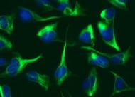
Application: ImmunocytochemistrySample Tested: Neural stem cellsSpecies: HumanVerified Customer | Posted 02/10/2022Human neural stem cells
-
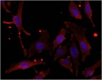
Application: Immunofluorescence - paraffinSample Tested: human glioblastomaSpecies: HumanVerified Customer | Posted 03/09/2019Glioblastoma multiforme stained with anti-nestin antibody 1:100pH9 heat induced antigen retrieval
-
Application: Immunocytochemistry/ImmunofluorescenceSample Tested: HeLa cellsSpecies: HumanVerified Customer | Posted 10/26/2015Specificity: Reasonably specific<br />Sensitivity: Reasonably sensitive<br />Buffer: 1% BSA + 0.1% Triton X-100 in PBS<br />Dilution: 1/100
-
Application: Immunocytochemistry/ImmunofluorescenceSample Tested: Human cell lineSpecies: HumanVerified Customer | Posted 10/26/2015Specificity: Specific<br />Sensitivity: Reasonably sensitive<br />Buffer: 1% BSA + 0.3% Triton X-100 in PBS<br />Dilution: 1/100
-
Application: Western BlotSample Tested: Cell LysatesSpecies: HumanVerified Customer | Posted 10/26/2015Specificity: Reasonably specific<br />Sensitivity: Reasonably sensitive<br />Buffer: 5% non-fat milk in TBST<br />Dilution: 1/1000
-
Application: ImmunofluorescenceSample Tested: See PMID 24213561Species: HumanVerified Customer | Posted 02/04/2015
There are no reviews that match your criteria.
Protocols
Find general support by application which include: protocols, troubleshooting, illustrated assays, videos and webinars.
- 7-Amino Actinomycin D (7-AAD) Cell Viability Flow Cytometry Protocol
- Appropriate Fixation of IHC/ICC Samples
- Cellular Response to Hypoxia Protocols
- ClariTSA™ Fluorophore Kits
- Detection & Visualization of Antibody Binding
- Extracellular Membrane Flow Cytometry Protocol
- Flow Cytometry Protocol for Cell Surface Markers
- Flow Cytometry Protocol for Staining Membrane Associated Proteins
- Flow Cytometry Staining Protocols
- Flow Cytometry Troubleshooting Guide
- ICC Cell Smear Protocol for Suspension Cells
- ICC Immunocytochemistry Protocol Videos
- ICC for Adherent Cells
- Immunocytochemistry (ICC) Protocol
- Immunocytochemistry Troubleshooting
- Immunofluorescence of Organoids Embedded in Cultrex Basement Membrane Extract
- Immunohistochemistry (IHC) and Immunocytochemistry (ICC) Protocols
- Intracellular Flow Cytometry Protocol Using Alcohol (Methanol)
- Intracellular Flow Cytometry Protocol Using Detergents
- Intracellular Nuclear Staining Flow Cytometry Protocol Using Detergents
- Intracellular Staining Flow Cytometry Protocol Using Alcohol Permeabilization
- Intracellular Staining Flow Cytometry Protocol Using Detergents to Permeabilize Cells
- Preparing Samples for IHC/ICC Experiments
- Preventing Non-Specific Staining (Non-Specific Binding)
- Primary Antibody Selection & Optimization
- Propidium Iodide Cell Viability Flow Cytometry Protocol
- Protocol for Liperfluo
- Protocol for VisUCyte™ HRP Polymer Detection Reagent
- Protocol for the Characterization of Human Th22 Cells
- Protocol for the Characterization of Human Th9 Cells
- Protocol for the Fluorescent ICC Staining of Cell Smears - Graphic
- Protocol for the Fluorescent ICC Staining of Cultured Cells on Coverslips - Graphic
- Protocol for the Preparation and Fluorescent ICC Staining of Cells on Coverslips
- Protocol for the Preparation and Fluorescent ICC Staining of Non-adherent Cells
- Protocol for the Preparation and Fluorescent ICC Staining of Stem Cells on Coverslips
- Protocol for the Preparation of a Cell Smear for Non-adherent Cell ICC - Graphic
- Protocol: Annexin V and PI Staining by Flow Cytometry
- Protocol: Annexin V and PI Staining for Apoptosis by Flow Cytometry
- TUNEL and Active Caspase-3 Detection by IHC/ICC Protocol
- The Importance of IHC/ICC Controls
- Troubleshooting Guide: Fluorokine Flow Cytometry Kits
- View all Protocols, Troubleshooting, Illustrated assays and Webinars

