
Key Product Details
Species Reactivity
Human
Applications
Immunohistochemistry, Western Blot
Label
Unconjugated
Antibody Source
Polyclonal Sheep IgG
Loading...
Product Specifications
Immunogen
Chinese hamster ovary cell line CHO-derived recombinant human Neuroglycan C/CSPG5
Val31-Gln420
Accession # AAQ04776
Val31-Gln420
Accession # AAQ04776
Specificity
Detects human Neuroglycan C/CSPG5 in direct ELISAs and Western blots. In direct ELISAs, approximately 50% cross‑reactivity with recombinant mouse Neuroglycan C is observed.
Clonality
Polyclonal
Host
Sheep
Isotype
IgG
Scientific Data Images for Human Neuroglycan C/CSPG5 Antibody
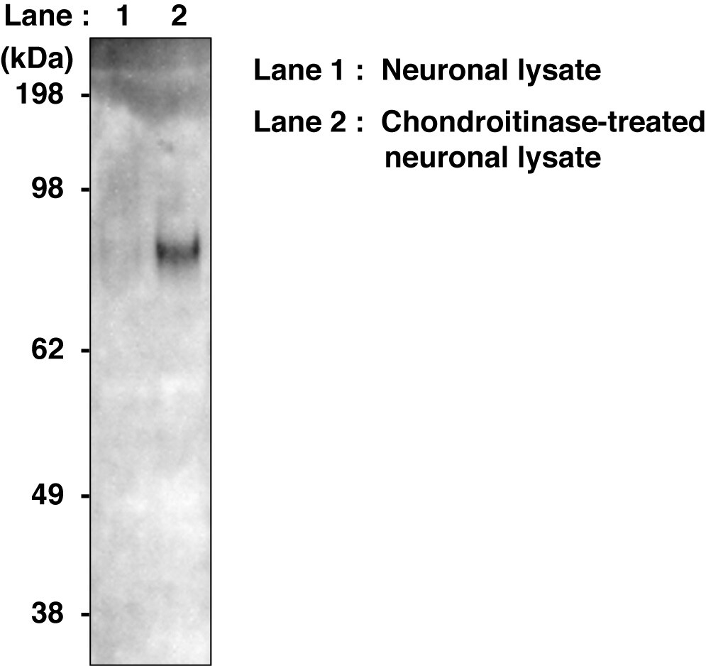
Detection of Human Neuroglycan C/CSPG5 by Western Blot.
Western blot shows lysates of SH-SY5Y human neuroblastoma cell line. PVDF membrane was probed with 1 µg/mL of Sheep Anti-Human Neuroglycan C/CSPG5 Antigen Affinity-purified Polyclonal Antibody (Catalog # AF5685) followed by HRP-conjugated Anti-Sheep IgG Secondary Antibody (Catalog # HAF016). Specific bands were detected for Neuroglycan C/CSPG5 at approximately 120 and 150 kDa (as indicated). This experiment was conducted under reducing conditions and using Immunoblot Buffer Group 8.Applications for Human Neuroglycan C/CSPG5 Antibody
Application
Recommended Usage
Immunohistochemistry
5-15 µg/mL
Sample: Perfusion fixed frozen sections of mouse brain (cerebellum)
Sample: Perfusion fixed frozen sections of mouse brain (cerebellum)
Western Blot
1 µg/mL
Sample: SH‑SY5Y human neuroblastoma cell line
Sample: SH‑SY5Y human neuroblastoma cell line
Reviewed Applications
Read 1 review rated 5 using AF5685 in the following applications:
Formulation, Preparation, and Storage
Purification
Antigen Affinity-purified
Reconstitution
Reconstitute at 0.2 mg/mL in sterile PBS. For liquid material, refer to CoA for concentration.
Loading...
Formulation
Lyophilized from a 0.2 μm filtered solution in PBS with Trehalose. *Small pack size (SP) is supplied either lyophilized or as a 0.2 µm filtered solution in PBS.
Shipping
Lyophilized product is shipped at ambient temperature. Liquid small pack size (-SP) is shipped with polar packs. Upon receipt, store immediately at the temperature recommended below.
Stability & Storage
Use a manual defrost freezer and avoid repeated freeze-thaw cycles.
- 12 months from date of receipt, -20 to -70 °C as supplied.
- 1 month, 2 to 8 °C under sterile conditions after reconstitution.
- 6 months, -20 to -70 °C under sterile conditions after reconstitution.
Calculators
Background: Neuroglycan C/CSPG5
References
- Kinugasa, Y. et al. (2004) Biochem. Biophys. Res. Commun 321:1045.
- Yasuda, Y. et al. (1998) Neurosci. Res. 32:313.
- Ichihara-Tanaka, K. et al. (2006) J. Biol. Chem. 281:30857.
- Aono, S. et al. (2004) J. Biol. Chem. 279:46536.
- Shuo, T. et al. (2007) J. Neurochem. 102:1561.
- Aono, S. et al. (2006) J. Neurosci. Res.83:110.
- Nakanishi, K. et al. (2006) J. Biol. Chem. 281:24970.
Alternate Names
CALEB, CSPG5, NGC
Gene Symbol
CSPG5
UniProt
Additional Neuroglycan C/CSPG5 Products
Product Documents for Human Neuroglycan C/CSPG5 Antibody
Certificate of Analysis
To download a Certificate of Analysis, please enter a lot or batch number in the search box below.
Note: Certificate of Analysis not available for kit components.
Product Specific Notices for Human Neuroglycan C/CSPG5 Antibody
For research use only
Related Research Areas
Customer Reviews for Human Neuroglycan C/CSPG5 Antibody (1)
5 out of 5
1 Customer Rating
Have you used Human Neuroglycan C/CSPG5 Antibody?
Submit a review and receive an Amazon gift card!
$25/€18/£15/$25CAN/¥2500 Yen for a review with an image
$10/€7/£6/$10CAN/¥1110 Yen for a review without an image
Submit a review
Customer Images
Showing
1
-
1 of
1 review
Showing All
Filter By:
-
Application: Western BlotSample Tested: Cultured iPS cells derived neuron and iPS cells-derived neuronSpecies: HumanVerified Customer | Posted 06/23/2020

There are no reviews that match your criteria.
Protocols
Find general support by application which include: protocols, troubleshooting, illustrated assays, videos and webinars.
- Antigen Retrieval Protocol (PIER)
- Antigen Retrieval for Frozen Sections Protocol
- Appropriate Fixation of IHC/ICC Samples
- Cellular Response to Hypoxia Protocols
- Chromogenic IHC Staining of Formalin-Fixed Paraffin-Embedded (FFPE) Tissue Protocol
- Chromogenic Immunohistochemistry Staining of Frozen Tissue
- ClariTSA™ Fluorophore Kits
- Detection & Visualization of Antibody Binding
- Fluorescent IHC Staining of Frozen Tissue Protocol
- Graphic Protocol for Heat-induced Epitope Retrieval
- Graphic Protocol for the Preparation and Fluorescent IHC Staining of Frozen Tissue Sections
- Graphic Protocol for the Preparation and Fluorescent IHC Staining of Paraffin-embedded Tissue Sections
- Graphic Protocol for the Preparation of Gelatin-coated Slides for Histological Tissue Sections
- IHC Sample Preparation (Frozen sections vs Paraffin)
- Immunofluorescent IHC Staining of Formalin-Fixed Paraffin-Embedded (FFPE) Tissue Protocol
- Immunohistochemistry (IHC) and Immunocytochemistry (ICC) Protocols
- Immunohistochemistry Frozen Troubleshooting
- Immunohistochemistry Paraffin Troubleshooting
- Preparing Samples for IHC/ICC Experiments
- Preventing Non-Specific Staining (Non-Specific Binding)
- Primary Antibody Selection & Optimization
- Protocol for Heat-Induced Epitope Retrieval (HIER)
- Protocol for Making a 4% Formaldehyde Solution in PBS
- Protocol for VisUCyte™ HRP Polymer Detection Reagent
- Protocol for the Preparation & Fixation of Cells on Coverslips
- Protocol for the Preparation and Chromogenic IHC Staining of Frozen Tissue Sections
- Protocol for the Preparation and Chromogenic IHC Staining of Frozen Tissue Sections - Graphic
- Protocol for the Preparation and Chromogenic IHC Staining of Paraffin-embedded Tissue Sections
- Protocol for the Preparation and Chromogenic IHC Staining of Paraffin-embedded Tissue Sections - Graphic
- Protocol for the Preparation and Fluorescent IHC Staining of Frozen Tissue Sections
- Protocol for the Preparation and Fluorescent IHC Staining of Paraffin-embedded Tissue Sections
- Protocol for the Preparation of Gelatin-coated Slides for Histological Tissue Sections
- R&D Systems Quality Control Western Blot Protocol
- TUNEL and Active Caspase-3 Detection by IHC/ICC Protocol
- The Importance of IHC/ICC Controls
- Troubleshooting Guide: Immunohistochemistry
- Troubleshooting Guide: Western Blot Figures
- Western Blot Conditions
- Western Blot Protocol
- Western Blot Protocol for Cell Lysates
- Western Blot Troubleshooting
- Western Blot Troubleshooting Guide
- View all Protocols, Troubleshooting, Illustrated assays and Webinars
Loading...