Axl (Ufo, Ark), Dtk (Sky, Tyro3, Rse, Brt), and Mer (human and mouse homologues of chicken c-Eyk) constitute a subfamily of the receptor tyrosine kinases (1, 2). The extracellular domains of these proteins contain two Ig-like motifs and two fibronectin type III motifs. This characteristic topology is also found in neural cell adhesion molecules and in receptor tyrosine phosphatases. The human Axl cDNA encodes an 887 amino acid (aa) precursor that includes an 18 aa signal sequence, a 426 aa extracellular domain, a 21 aa transmembrane segment, and a 422 aa cytoplasmic domain. The extracellular domains of human and mouse Axl share 81% aa sequence identity. A short alternately spliced form of human Axl is distinguished by a 9 aa deletion in the extracellular juxtamembrane region. These receptors bind the vitamin K‑dependent protein growth arrest specific gene 6 (Gas6) which is structurally related to the anticoagulation factor protein S. Binding of Gas6 induces receptor autophosphorylation and downstream signaling pathways that can lead to cell proliferation, migration, or the prevention of apoptosis (3). This family of tyrosine kinase receptors is involved in hematopoiesis, embryonic development, tumorigenesis, and regulation of testicular functions.
Human Phospho-Axl (Y779) Antibody
R&D Systems | Catalog # AF2228
 in Human Stomach Cancer Tissue.")
Key Product Details
Species Reactivity
Validated:
Human
Cited:
Human, Mouse, Rat, Xenograft
Applications
Validated:
Immunohistochemistry, Immunocytochemistry
Cited:
Immunohistochemistry, Immunohistochemistry-Paraffin, Immunohistochemistry-Frozen, Western Blot, Flow Cytometry, Immunoprecipitation
Label
Unconjugated
Antibody Source
Polyclonal Rabbit IgG
Loading...
Product Specifications
Immunogen
Phosphopeptide containing human Axl Y779 site
Specificity
Detects human and mouse Axl when phosphorylated at Y779.
Clonality
Polyclonal
Host
Rabbit
Isotype
IgG
Scientific Data Images for Human Phospho-Axl (Y779) Antibody
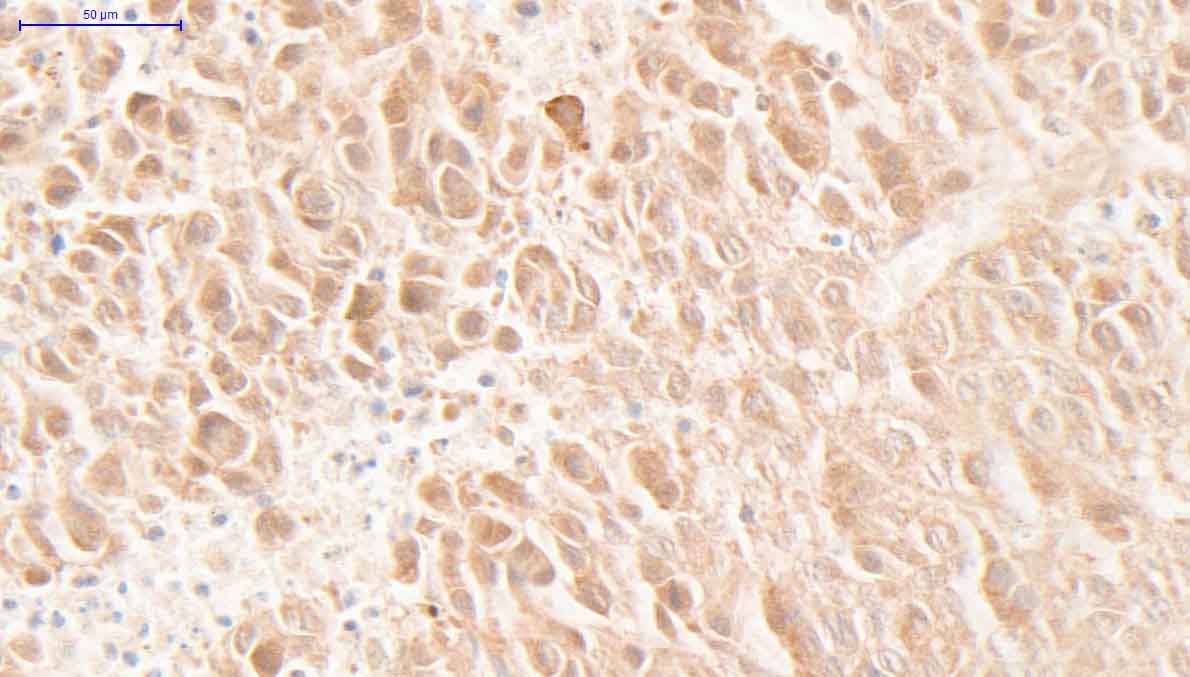
Phospho-Axl (Y779) in Human Stomach Cancer Tissue.
Axl phosphorylated at site Y779 was detected in immersion fixed paraffin-embedded sections of human stomach cancer tissue using Human Phospho-Axl (Y779) Antigen Affinity-purified Polyclonal Antibody (Catalog # AF2228) at 15 µg/mL overnight at 4 °C. Tissue was stained using the Anti-Rabbit HRP-DAB Cell & Tissue Staining Kit (brown; CTS005) and counterstained with hematoxylin (blue). Specific labeling was localized to the cytoplasm of epithelial cells. View our protocol for Chromogenic IHC Staining of Paraffin-embedded Tissue Sections. in A172 Human Cell Line.")
Phospho-Axl (Y779) in A172 Human Cell Line.
Axl phosphorylated at Y779 (panels B, D) and total Axl (panels A, C) were assessed in immersion fixed A172 human glioblastoma cells incubated with (panels C, D) or without (panels A, B) pervanadate. Phospho-Axl was detected using Rabbit Anti-Human Phospho-Axl (Y779) Antigen Affinity-purified Polyclonal Antibody (Catalog # AF2228) at 10 µg/mL for 3 hours at room temperature. Cells were stained using the NorthernLights™ 557-conjugated Anti-Rabbit IgG Secondary Antibody (red, panels B, D); NL004) and counterstained using DAPI (blue). Total Axl was detected using Goat Anti-Human Axl Antigen Affinity-purified Polyclonal Antibody (AF154). Cells were stained using the NorthernLights™ 493-conjugated Anti-Goat IgG Secondary Antibody (green, panels A, C); NL003). Specific staining was localized to cytoplasm. View our protocol for Fluorescent ICC Staining of Cells on Coverslips.
Detection of Rat Axl by Immunohistochemistry
GAS6 and p-AXL are expressed in the regenerative cavernous nerve of rats subjected to BCNI(A) Representative ICP tracing responses of each group (n=5 per group) to the stimulation of the cavernous nerve at 14 days and 28 days. The green bar represents an electrical stimulus duration of 60 seconds. (B) Immunohistochemistry images demonstrated that GAS6 and p-AXL expression levels increased in the cavernous nerve after BCNI. (C) The quantitative analysis results showed a significantly higher expression of GAS6 and p-AXL in the cavernous nerve of animals subjected to injury after 28 days. Image collected and cropped by CiteAb from the following publication (https://www.oncotarget.com/lookup/doi/10.18632/oncotarget.23978), licensed under a CC-BY license. Not internally tested by R&D Systems. Antibody by Western Blot")
Detection of Rat Human Phospho-Axl (Y779) Antibody by Western Blot
GAS6 triggered Schwann cell proliferation primarily through CIP2A and DAPK(A) RSC96 cells were transfected with either control or DAPK siRNA for 48 h and then exposed to GAS6 (100 ng/ml) for 30 min. Immunoblotting evaluations of pAxl, Axl, CIP2A, DAPK, pERK1/2, ERK1/2, pAKT, AKT, Myc and Survivin. (B) RSC96 cells were transfected with either control or CIP2A siRNA for 48 h and then exposed to GAS6 (100 ng/ml) for 30 min. Immunoblotting evaluations of pAxl, Axl, DAPK, pDAPK, pERK1/2, ERK1/2, pAKT, AKT, Myc and Survivin. (C) DAPK or CIP2A was knocked down for 48 h then RSC96 cells were incubated with GAS6 (100 ng/ml) at the indicated hours. Cell viability was analysed via the WST-1 assay. Data are the mean ± SD, and n = 3 for each time point. * p < 0.05, ** p < 0.01 vs. scramble. Image collected and cropped by CiteAb from the following publication (https://pubmed.ncbi.nlm.nih.gov/29464081), licensed under a CC-BY license. Not internally tested by R&D Systems.
Detection of Human Axl by Immunohistochemistry
DCC-2036 inhibits CSCs in vivo according to the limited dilution assay by pretreating 4T1 cells with DCC-2036 or DMSO for 48 h.A In vivo tumorigenicity assay with limited dilution using DMSO or DCC-2036 treated 4T1 cells respectively: 200,000 (n = 7 and 8), 20,000 (n = 9 and 8), or 2000 (n = 10 and 10) cells per injection site. The frequency of BCSCs was calculated by ELDA. B Weight variation of mice following transplant of 4T1 cells into BALB/C mice after 7 days. C Images of tumor formation in BALB/C mice. D, E Transplanted tumors were harvested, and the tumor size and weight were measured at the end of the experiment. The statistical significance was determined by Student’s t-test. F Tumor-free survival curve of BALB/C mice is shown. G Hematoxylin and eosin (H&E) staining indicates the histology of tumor tissues. Scale bars, 20 μm. H Immunohistochemical analysis using p-AXL, AXL, and KLF5 antibodies in xenograft tissues from BALB/C mice. Scale bars, 20 μm. I Immunoblot of transplanted tumors from BALB/C mice posterior to the experiments. C means Control, T means DCC-2036. We chose the tumor tissues randomly for H&E staining, immunohistochemical analysis, and immunoblotting. Image collected and cropped by CiteAb from the following open publication (https://pubmed.ncbi.nlm.nih.gov/36042208), licensed under a CC-BY license. Not internally tested by R&D Systems.
Detection of Human Axl by Immunohistochemistry
The expression of p-AXL&AXL positively correlates with the expression of KLF5 in human TNBC specimens. Moreover, DCC-2036 increases the sensitivity of TNBC chemotherapy by decreasing BCSCs.A Representative immunohistochemical staining images of p-AXL, AXL,&KLF5 protein in TNBC specimens, in which the expression of p-AXL, AXL,&KLF5 proteins was indicated by mild positive (+), moderate positive (++),&strong positive (+++), respectively. Left: Scale bars, 200 μm (magnification 40×); Right: Scale bars, 50 μm (magnification 100×). Image collected & cropped by CiteAb from the following open publication (https://pubmed.ncbi.nlm.nih.gov/36042208), licensed under a CC-BY license. Not internally tested by R&D Systems.
Detection of Human Axl by Western Blot
TPC‐EV‐induced EPC vasculogenic properties are regulated by Gas6 in vitro. (f) The phosphorylated&total forms of Axl, Akt&Erk1/2 in EPCs after the indicated treatments determined. Representative blots&quantification of the ratio of p‐Axl/Axl (n = 3). TPC‐EV‐(siNC), EVs derived from TPCs transfected with NC siRNA. TPC‐EV‐(siGas6), EVs derived from TPCs transfected with Gas6 siRNA. Image collected & cropped by CiteAb from the following open publication (https://pubmed.ncbi.nlm.nih.gov/34035882), licensed under a CC-BY license. Not internally tested by R&D Systems.
Detection of Human Axl by Western Blot
Migration of H1299 NSCLC cells enhanced by ligand-dependent Axl activation. (A) Western blotting to assess Gas6 expression in H1299 cells. Expression of Gas6 in LCAFhTERT cells was used as a positive control. (B) Phosphorylation of Axl was analyzed by Western blotting of whole cell lysates using different antibodies. GAPDH was used as an internal control. H1299 cells were stimulated for 15 min with 400 nM recombinant human Gas6 (rGas6). H1299 cells were treated with or without TP-0903 (0.2 µmol/L) for 24 h. (C) Migration of H1299 cells was analyzed using rGas6 (400 nM) added to the lower chamber. H1299 cells were treated with or without TP-0903 (0.2 µmol/L) for 24 h. (D) Western blotting of conditioned medium from LCAFhTERT transfected with siGas6 or siScr (control) to assess whether they contains Gas6 secreted by CAFs. (E) Phosphorylation of Axl in H1299 cells analyzed by Western blotting after stimulation with conditioned medium from siRNA-transfected LCAFhTERT. H1299 cells were treated with or without TP-0903 (0.2 µmol/L) for 24 h. The medium (DMEM containing 10% FBS) was used as control. (F) Migration of H1299 cells analyzed using conditioned medium of siRNA-transfected LCAFhTERT. H1299 cells were treated with or without TP-0903 (0.2 µmol/L) for 24 h. The medium (DMEM containing 10% FBS) was used as control. The relative number of migrated H1299 cells is indicated on the y-axis. Data show the mean ± SEM (n = 3); **p < 0.01. Image collected and cropped by CiteAb from the following open publication (https://pubmed.ncbi.nlm.nih.gov/28878389), licensed under a CC-BY license. Not internally tested by R&D Systems.
Detection of Human Axl by Western Blot
DHA treatment inhibits Axl expression and proteins involved in the Axl signaling pathway, reduces cell migration and invasion and induces apoptosis in mCRPCa cell lines.a qRT-PCR for Axl in PCa cells treated with 5 μM of DHA for 24 h. Values were normalized to Gapdh levels and to DMSO control. The experiment was performed in triplicate. Data are representative of 3 independent experiments; *p < 0.05 two-tailed Student’s t-test. b Immunoblot analysis of protein extracts obtained from DU145 and PC-3 treated with 5 μM DHA for 24 h using anti-phospho-Axl, anti-phospho-Akt and anti-phospho-Stat3 and anti-Axl, anti-Akt, anti-Stat3 and anti- GAPDH. c Migration and d invasion analysis of mCRPC cell lines 24 h post treatment with 5 μM of DHA. Cells were fixed and stained, and 3–5 random microscopic fields were counted. Data are representative of three independent experiments and all values shown are mean ± SD from a representative experiment; **p < 0.01, two-tailed Student’s t-test. e Apoptosis assay for mCRPCa cell lines treated with 5 μM of DHA for 8 h. DMSO 0.05% was used as a control. Data are representative of three independent experiments and all values shown are mean ± SD from a representative experiment; *p < 0.05, two-tailed Student’s t-test Image collected and cropped by CiteAb from the following open publication (https://pubmed.ncbi.nlm.nih.gov/30783079), licensed under a CC-BY license. Not internally tested by R&D Systems. by Western Blot")
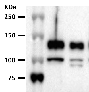
Detection of Phospho-Axl (Y779) by Western Blot
The effects of ProS1, Gas6 and chimeras on TAM receptor and coupled downstream signalling molecule activation in SCC-25 cells. (a) Schematic representation of recombinant TAM ligand constructs used in this study. These included human ProS1, Gas6, and three ProS1/Gas6 chimeras. All of the chimeras contained the Gla domain and EGF-like domains of ProS1. Light grey colour denotes regions corresponding to ProS1 amino acid sequence, whereas dark grey denotes regions corresponding to Gas6 amino acid sequence. (b) Western blot showing phosphorylated Tyro3 (pTyro3) and Erk (pErk) levels after stimulation with recombinant Gas6, ProS1 and three chimeras (7.5 nM) for 9 min. (c) Western blot showing phosphorylated Axl (pAxl) and Akt (pAkt) levels under the same experimental conditions as in (b). Each representative blot image is followed by accompanying graphs of densitometric quantification of bands (n = 3 separate experiments). Data as mean ± SEM expression for each phosphoprotein was normalized against the total protein/loading control (tTyro3, tERK, GAPDH, actin). ANOVA with Tukey's multiple comparison post-hoc analysis; ***p < 0.001, **p < 0.01, *p < 0.05 versus control (untreated). While the sample loading order is different in the pAxl blot, the quantification bar charts are presented in the same order for consistency. Image collected and cropped by CiteAb from the following open publication (https://pubmed.ncbi.nlm.nih.gov/35518197), licensed under a CC-BY license. Not internally tested by R&D Systems.Applications for Human Phospho-Axl (Y779) Antibody
Application
Recommended Usage
Immunocytochemistry
5-15 µg/mL
Sample: Immersion fixed A172 human glioblastoma cell line with and without pervanadate co-staining with total Axl (Catalog # AF154)
Sample: Immersion fixed A172 human glioblastoma cell line with and without pervanadate co-staining with total Axl (Catalog # AF154)
Immunohistochemistry
5-15 µg/mL
Sample: Immersion fixed paraffin-embedded sections of human stomach
Sample: Immersion fixed paraffin-embedded sections of human stomach
Reviewed Applications
Read 6 reviews rated 4.2 using AF2228 in the following applications:
Formulation, Preparation, and Storage
Purification
Antigen and protein A Affinity-purified
Reconstitution
Reconstitute at 0.2 mg/mL in sterile PBS. For liquid material, refer to CoA for concentration.
Loading...
Formulation
Lyophilized from a 0.2 μm filtered solution in PBS with Trehalose. *Small pack size (SP) is supplied either lyophilized or as a 0.2 µm filtered solution in PBS.
Shipping
Lyophilized product is shipped at ambient temperature. Liquid small pack size (-SP) is shipped with polar packs. Upon receipt, store immediately at the temperature recommended below.
Stability & Storage
Use a manual defrost freezer and avoid repeated freeze-thaw cycles.
- 12 months from date of receipt, -20 to -70 °C as supplied.
- 1 month, 2 to 8 °C under sterile conditions after reconstitution.
- 6 months, -20 to -70 °C under sterile conditions after reconstitution.
Calculators
Background: Axl
References
- Yanagita, M. (2004) Curr. Opin. Nephrol. Hypertens. 13:465.
- Nagata, K. et al. (1996) J. Biol. Chem. 22:30022.
- Holland, S. et al. (2005) Canc. Res. 65:9294.
Long Name
Axl Receptor Tyrosine Kinase
Alternate Names
AI323647, ARK, JTK11, Tyro7, UFO
Gene Symbol
AXL
Additional Axl Products
Product Documents for Human Phospho-Axl (Y779) Antibody
Certificate of Analysis
To download a Certificate of Analysis, please enter a lot or batch number in the search box below.
Note: Certificate of Analysis not available for kit components.
Product Specific Notices for Human Phospho-Axl (Y779) Antibody
For research use only
Related Research Areas
Citations for Human Phospho-Axl (Y779) Antibody
 Powered by Bioz
Customer Reviews for Human Phospho-Axl (Y779) Antibody (6)
4.2 out of 5
6 Customer Ratings
Have you used Human Phospho-Axl (Y779) Antibody?
Submit a review and receive an Amazon gift card!
$25/€18/£15/$25CAN/¥2500 Yen for a review with an image
$10/€7/£6/$10CAN/¥1110 Yen for a review without an image
Submit a review
Customer Images
Showing
1
-
5 of
6 reviews
Showing All
Filter By:
-
Application: Western BlotSample Tested: DU145 human prostate carcinoma cell lineSpecies: HumanVerified Customer | Posted 11/14/2020

-
Application: ImmunohistochemistrySample Tested: B16F0 melanoma tumourSpecies: MouseVerified Customer | Posted 01/03/2019
-
Application: Western BlotSample Tested: Brain tissueSpecies: MouseVerified Customer | Posted 06/23/2017
-
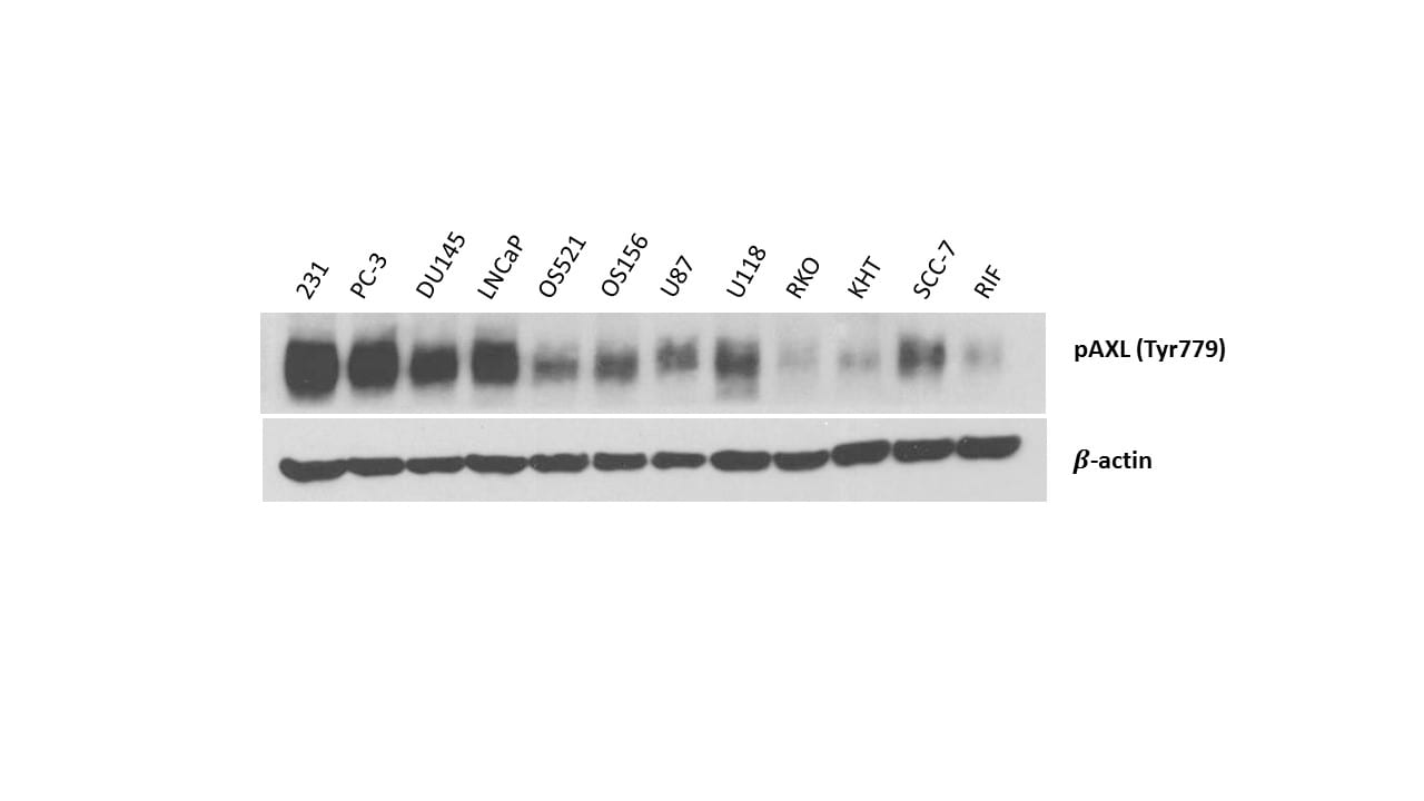
Application: Western BlotSample Tested: MDA-MB-231 human breast cancer cell line, PC-3 human prostate cancer cell line, DU145 human prostate carcinoma cell line, LNCaP human prostate cancer cell line, S521, S156, U-87 MG human glioblastoma/astrocytoma cell line, U-118-MG human glioblastoma/astrocytoma cell line, RKO, KHT and SCC-7Species: Mouse and HumanVerified Customer | Posted 10/06/2016
-
Application: Western BlotSample Tested: MCF 10A human breast epithelial cell line, mcf10aDCIS.com and MDA-MB-231 human breast cancer cell lineSpecies: HumanVerified Customer | Posted 04/25/2016
-
Application: Immunohistochemistry-ParaffinSample Tested: See Pmid 24687913Species: HumanVerified Customer | Posted 01/06/2015
There are no reviews that match your criteria.
Protocols
Find general support by application which include: protocols, troubleshooting, illustrated assays, videos and webinars.
- Antigen Retrieval Protocol (PIER)
- Antigen Retrieval for Frozen Sections Protocol
- Appropriate Fixation of IHC/ICC Samples
- Cellular Response to Hypoxia Protocols
- Chromogenic IHC Staining of Formalin-Fixed Paraffin-Embedded (FFPE) Tissue Protocol
- Chromogenic Immunohistochemistry Staining of Frozen Tissue
- ClariTSA™ Fluorophore Kits
- Detection & Visualization of Antibody Binding
- Fluorescent IHC Staining of Frozen Tissue Protocol
- Graphic Protocol for Heat-induced Epitope Retrieval
- Graphic Protocol for the Preparation and Fluorescent IHC Staining of Frozen Tissue Sections
- Graphic Protocol for the Preparation and Fluorescent IHC Staining of Paraffin-embedded Tissue Sections
- Graphic Protocol for the Preparation of Gelatin-coated Slides for Histological Tissue Sections
- ICC Cell Smear Protocol for Suspension Cells
- ICC Immunocytochemistry Protocol Videos
- ICC for Adherent Cells
- IHC Sample Preparation (Frozen sections vs Paraffin)
- Immunocytochemistry (ICC) Protocol
- Immunocytochemistry Troubleshooting
- Immunofluorescence of Organoids Embedded in Cultrex Basement Membrane Extract
- Immunofluorescent IHC Staining of Formalin-Fixed Paraffin-Embedded (FFPE) Tissue Protocol
- Immunohistochemistry (IHC) and Immunocytochemistry (ICC) Protocols
- Immunohistochemistry Frozen Troubleshooting
- Immunohistochemistry Paraffin Troubleshooting
- Preparing Samples for IHC/ICC Experiments
- Preventing Non-Specific Staining (Non-Specific Binding)
- Primary Antibody Selection & Optimization
- Protocol for Heat-Induced Epitope Retrieval (HIER)
- Protocol for Making a 4% Formaldehyde Solution in PBS
- Protocol for VisUCyte™ HRP Polymer Detection Reagent
- Protocol for the Fluorescent ICC Staining of Cell Smears - Graphic
- Protocol for the Fluorescent ICC Staining of Cultured Cells on Coverslips - Graphic
- Protocol for the Preparation & Fixation of Cells on Coverslips
- Protocol for the Preparation and Chromogenic IHC Staining of Frozen Tissue Sections
- Protocol for the Preparation and Chromogenic IHC Staining of Frozen Tissue Sections - Graphic
- Protocol for the Preparation and Chromogenic IHC Staining of Paraffin-embedded Tissue Sections
- Protocol for the Preparation and Chromogenic IHC Staining of Paraffin-embedded Tissue Sections - Graphic
- Protocol for the Preparation and Fluorescent ICC Staining of Cells on Coverslips
- Protocol for the Preparation and Fluorescent ICC Staining of Non-adherent Cells
- Protocol for the Preparation and Fluorescent ICC Staining of Stem Cells on Coverslips
- Protocol for the Preparation and Fluorescent IHC Staining of Frozen Tissue Sections
- Protocol for the Preparation and Fluorescent IHC Staining of Paraffin-embedded Tissue Sections
- Protocol for the Preparation of Gelatin-coated Slides for Histological Tissue Sections
- Protocol for the Preparation of a Cell Smear for Non-adherent Cell ICC - Graphic
- TUNEL and Active Caspase-3 Detection by IHC/ICC Protocol
- The Importance of IHC/ICC Controls
- Troubleshooting Guide: Immunohistochemistry
- View all Protocols, Troubleshooting, Illustrated assays and Webinars
Loading...