alpha ‑Fetoprotein is a major plasma protein in the fetus. Its concentration is normally low in the adult except when produced by certain tumors.

Key Product Details
Validated by
Knockout/Knockdown
Species Reactivity
Validated:
Human, Rat
Cited:
Human
Applications
Validated:
Knockout Validated, Immunohistochemistry, Western Blot, Immunocytochemistry, Simple Western
Cited:
Western Blot, Immunocytochemistry, ELISA Capture
Label
Unconjugated
Antibody Source
Polyclonal Chicken IgY
Loading...
Product Specifications
Immunogen
Human umbilical cord serum-derived alpha -Fetoprotein
Specificity
Detects human alpha -Fetoprotein in direct ELISAs and Western blots.
Clonality
Polyclonal
Host
Chicken
Isotype
IgY
Scientific Data Images for alpha-Fetoprotein/AFP Antibody
Detection of Human and Rat alpha ‑Fetoprotein/AFP by Western Blot.
Western blot shows lysates of human liver tissue and H4‑II‑E‑C3 rat hepatoma cell line. PVDF membrane was probed with 0.2 µg/mL of Chicken Anti-Human/Rat alpha ‑Fetoprotein/ AFP Antigen Affinity-purified Polyclonal Antibody (Catalog # AF1369) followed by HRP-conjugated Anti-Chicken IgY Secondary Antibody. A specific band was detected for alpha ‑Fetoprotein/AFP at approximately 70 kDa (as indicated). This experiment was conducted under reducing conditions and using Immunoblot Buffer Group 1.
alpha ‑Fetoprotein/AFP in BG01V Human Embryonic Stem Cells.
a-Fetoprotein/AFP was detected in immersion fixed BG01V human embryonic stem cells using Chicken Anti-Human/Rat a-Fetoprotein/AFP Antigen Affinity-purified Polyclonal Antibody (Catalog # AF1369) at 10 µg/mL for 3 hours at room temperature. Cells were stained using the NorthernLights™ 557-conjugated Anti-Chicken IgY Secondary Antibody (red; Catalog # NL016) and counterstained with DAPI(blue). Specific staining was localized to cytoplasm. View our protocol for Fluorescent ICC Staining of Cells on Coverslips.
alpha ‑Fetoprotein/AFP in Human Liver cancer
alpha ‑Fetoprotein/AFP was detected in immersion fixed paraffin-embedded sections of human liver cancer using 15 µg/mL Chicken anti-Human/ Rat alpha ‑Fetoprotein/AFP Antigen Affinity-purified Polyclonal Antibody (Catalog # AF1369) overnight at 4 °C. Tissue was stained (brown) and counterstained with hematoxylin (blue). View our protocol for Chromogenic IHC Staining of Paraffin-embedded Tissue Sections.
Detection of Human alpha ‑Fetoprotein/AFP by Simple WesternTM.
Simple Western lane view shows lysates of HepG2 human hepatocellular carcinoma cell line, loaded at 0.2 mg/mL. A specific band was detected for alpha ‑Fetoprotein/AFP at approximately 70 kDa (as indicated) using 2 µg/mL of Chicken Anti-Human/Rat alpha ‑Fetoprotein/AFP Antigen Affinity-purified Polyclonal Antibody (Catalog # AF1369) followed by HRP-conjugated Anti-Chicken IgY Secondary Antibody. This experiment was conducted under reducing conditions and using the 12-230 kDa separation system.
alpha ‑Fetoprotein/AFP Specificity is Shown by Immunocytochemistry in Knockout Cell Line.
a-Fetoprotein/AFP was detected in immersion fixed HepG2 human hepatocellular carcinoma cell line but is not detected in a-Fetoprotein/AFP knockout (KO) HepG2 Human Cell Line cell line using Chicken Anti-Human/Rat a-Fetoprotein/AFP Antigen Affinity-purified Polyclonal Antibody (Catalog # AF1369) at 10 µg/mL for 3 hours at room temperature. Cells were stained using the NorthernLights™ 493-conjugated Anti-Chicken IgY Secondary Antibody (green; Catalog # NL018) and counterstained with DAPI (blue). Specific staining was localized to cytoplasm. View our protocol for Fluorescent ICC Staining of Cells on Coverslips.Applications for alpha-Fetoprotein/AFP Antibody
Application
Recommended Usage
Immunocytochemistry
8-25 µg/mL
Sample: Immersion fixed BG01V human embryonic stem cells
Sample: Immersion fixed BG01V human embryonic stem cells
Immunohistochemistry
5-15 µg/mL
Sample: Immersion fixed paraffin-embedded sections of human liver cancer
Sample: Immersion fixed paraffin-embedded sections of human liver cancer
Knockout Validated
alpha ‑Fetoprotein is specifically detected in HepG2 human hepatocellular carcinoma cell line but is not detectable in alpha ‑Fetoprotein knockout HepG2 cell line.
Simple Western
2 µg/mL
Sample: HepG2 human hepatocellular carcinoma cell line
Sample: HepG2 human hepatocellular carcinoma cell line
Western Blot
0.2 µg/mL
Sample: Human liver tissue and H4‑II‑E‑C3 rat hepatoma cell line
Sample: Human liver tissue and H4‑II‑E‑C3 rat hepatoma cell line
Reviewed Applications
Read 2 reviews rated 5 using AF1369 in the following applications:
Formulation, Preparation, and Storage
Purification
Antigen Affinity-purified from egg yolks
Reconstitution
Reconstitute at 0.2 mg/mL in sterile PBS. For liquid material, refer to CoA for concentration.
Loading...
Formulation
Lyophilized from a 0.2 μm filtered solution in PBS with Trehalose. *Small pack size (SP) is supplied either lyophilized or as a 0.2 µm filtered solution in PBS.
Shipping
Lyophilized product is shipped at ambient temperature. Liquid small pack size (-SP) is shipped with polar packs. Upon receipt, store immediately at the temperature recommended below.
Stability & Storage
Use a manual defrost freezer and avoid repeated freeze-thaw cycles.
- 12 months from date of receipt, -20 to -70 °C as supplied.
- 1 month, 2 to 8 °C under sterile conditions after reconstitution.
- 6 months, -20 to -70 °C under sterile conditions after reconstitution.
Calculators
Background: alpha-Fetoprotein/AFP
Additional alpha-Fetoprotein/AFP Products
Product Documents for alpha-Fetoprotein/AFP Antibody
Certificate of Analysis
To download a Certificate of Analysis, please enter a lot or batch number in the search box below.
Note: Certificate of Analysis not available for kit components.
Product Specific Notices for alpha-Fetoprotein/AFP Antibody
For research use only
Related Research Areas
Citations for alpha-Fetoprotein/AFP Antibody
 Powered by Bioz
Customer Reviews for alpha-Fetoprotein/AFP Antibody (2)
5 out of 5
2 Customer Ratings
Have you used alpha-Fetoprotein/AFP Antibody?
Submit a review and receive an Amazon gift card!
$25/€18/£15/$25CAN/¥2500 Yen for a review with an image
$10/€7/£6/$10CAN/¥1110 Yen for a review without an image
Submit a review
Customer Images
Showing
1
-
2 of
2 reviews
Showing All
Filter By:
-
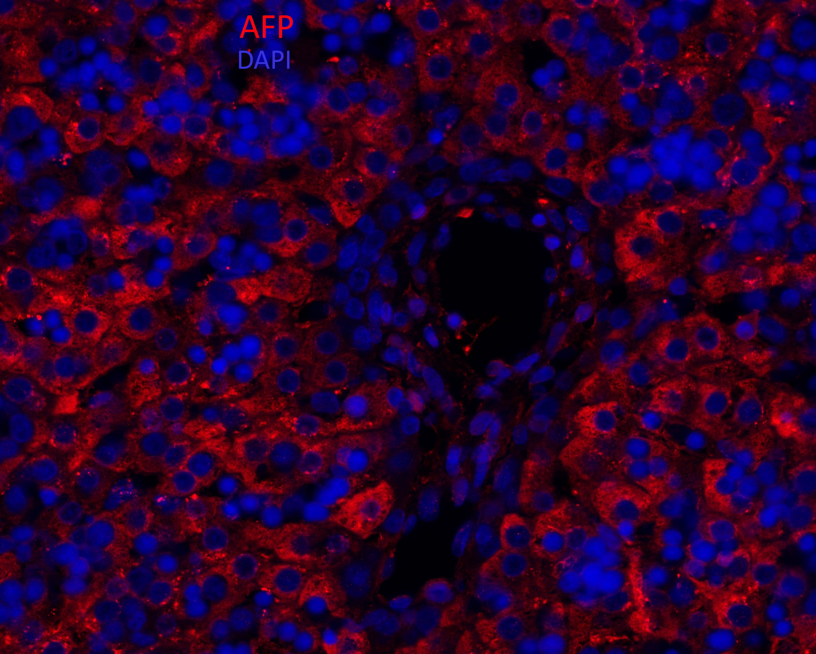
Application: Immunohistochemistry-ParaffinSample Tested: human fetal liverSpecies: HumanVerified Customer | Posted 03/26/2018Normal Human Fetal Liver-Paraffin SectionFormalin/PFA-fixed paraffin-embedded sections Heat mediated - Buffer/Enzyme Used: Tris/EDTA buffer pH 6.0 Sea Block as blocking agent for 30 minutes Incubation is overnight at 4degC AFP antibody 1/100 in Sea Block Goat Anti-Chicken IgY H&L (Alexa Fluor® 555) preadsorbed secondary DAPI counterstain

-
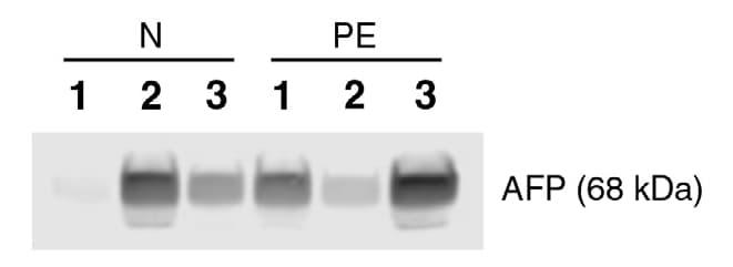
Application: Western BlotSample Tested: PlasmaSpecies: HumanVerified Customer | Posted 06/13/2017Plasma sample 10 µg / well Antigen: alpha -Fetoprotein (AFP) Primary antibody: Anti-Human AFP antibody (Polyclonal Chicken IgY) from R&D systems; Cat No. AF1369 Secondary antibody_1: Goat anti-chicken IgY-unconjugated (Dilution: 1:5000) from R&D systems; Cat No. AF010 Secondary antibody_2: Donkey anti-goat IgG-HRP from Santa Cruz; Cat no. sc-2020) Molecular weight of the antigen: 68 kDa Percentage of SDS-PAGE gel: 12.5 % Run time: 3.5 hours Wet transfer @ 100 V for 1 hr
There are no reviews that match your criteria.
Protocols
Find general support by application which include: protocols, troubleshooting, illustrated assays, videos and webinars.
- Antigen Retrieval Protocol (PIER)
- Antigen Retrieval for Frozen Sections Protocol
- Appropriate Fixation of IHC/ICC Samples
- Cellular Response to Hypoxia Protocols
- Chromogenic IHC Staining of Formalin-Fixed Paraffin-Embedded (FFPE) Tissue Protocol
- Chromogenic Immunohistochemistry Staining of Frozen Tissue
- ClariTSA™ Fluorophore Kits
- Detection & Visualization of Antibody Binding
- Fluorescent IHC Staining of Frozen Tissue Protocol
- Graphic Protocol for Heat-induced Epitope Retrieval
- Graphic Protocol for the Preparation and Fluorescent IHC Staining of Frozen Tissue Sections
- Graphic Protocol for the Preparation and Fluorescent IHC Staining of Paraffin-embedded Tissue Sections
- Graphic Protocol for the Preparation of Gelatin-coated Slides for Histological Tissue Sections
- ICC Cell Smear Protocol for Suspension Cells
- ICC Immunocytochemistry Protocol Videos
- ICC for Adherent Cells
- IHC Sample Preparation (Frozen sections vs Paraffin)
- Immunocytochemistry (ICC) Protocol
- Immunocytochemistry Troubleshooting
- Immunofluorescence of Organoids Embedded in Cultrex Basement Membrane Extract
- Immunofluorescent IHC Staining of Formalin-Fixed Paraffin-Embedded (FFPE) Tissue Protocol
- Immunohistochemistry (IHC) and Immunocytochemistry (ICC) Protocols
- Immunohistochemistry Frozen Troubleshooting
- Immunohistochemistry Paraffin Troubleshooting
- Preparing Samples for IHC/ICC Experiments
- Preventing Non-Specific Staining (Non-Specific Binding)
- Primary Antibody Selection & Optimization
- Protocol for Heat-Induced Epitope Retrieval (HIER)
- Protocol for Making a 4% Formaldehyde Solution in PBS
- Protocol for VisUCyte™ HRP Polymer Detection Reagent
- Protocol for the Fluorescent ICC Staining of Cell Smears - Graphic
- Protocol for the Fluorescent ICC Staining of Cultured Cells on Coverslips - Graphic
- Protocol for the Preparation & Fixation of Cells on Coverslips
- Protocol for the Preparation and Chromogenic IHC Staining of Frozen Tissue Sections
- Protocol for the Preparation and Chromogenic IHC Staining of Frozen Tissue Sections - Graphic
- Protocol for the Preparation and Chromogenic IHC Staining of Paraffin-embedded Tissue Sections
- Protocol for the Preparation and Chromogenic IHC Staining of Paraffin-embedded Tissue Sections - Graphic
- Protocol for the Preparation and Fluorescent ICC Staining of Cells on Coverslips
- Protocol for the Preparation and Fluorescent ICC Staining of Non-adherent Cells
- Protocol for the Preparation and Fluorescent ICC Staining of Stem Cells on Coverslips
- Protocol for the Preparation and Fluorescent IHC Staining of Frozen Tissue Sections
- Protocol for the Preparation and Fluorescent IHC Staining of Paraffin-embedded Tissue Sections
- Protocol for the Preparation of Gelatin-coated Slides for Histological Tissue Sections
- Protocol for the Preparation of a Cell Smear for Non-adherent Cell ICC - Graphic
- R&D Systems Quality Control Western Blot Protocol
- TUNEL and Active Caspase-3 Detection by IHC/ICC Protocol
- The Importance of IHC/ICC Controls
- Troubleshooting Guide: Immunohistochemistry
- Troubleshooting Guide: Western Blot Figures
- Western Blot Conditions
- Western Blot Protocol
- Western Blot Protocol for Cell Lysates
- Western Blot Troubleshooting
- Western Blot Troubleshooting Guide
- View all Protocols, Troubleshooting, Illustrated assays and Webinars
Loading...
Associated Pathways
