CBFA1, also called runt-related transcription factor 2 (RUNX2), is an essential transcription factor for the regulation of osteoblast differentiation (1). The CBFA1 gene potentially encodes several proteins that differ in their N-terminal sequences and transactivation capacities (2).
Human RUNX2/CBFA1 Antibody (232902)
R&D Systems | Catalog # MAB2006

Key Product Details
Species Reactivity
Validated:
Human
Cited:
Human, Mouse, Transgenic Mouse
Applications
Validated:
Immunocytochemistry
Cited:
Immunohistochemistry, Immunohistochemistry-Paraffin, Immunohistochemistry-Frozen, Western Blot, Flow Cytometry, Immunocytochemistry
Label
Unconjugated
Antibody Source
Monoclonal Rat IgG2B Clone # 232902
Loading...
Product Specifications
Immunogen
E. coli-derived recombinant human RUNX2/CBFA1 isoform 2
Lys233-Tyr418
Accession # Q13950
Lys233-Tyr418
Accession # Q13950
Specificity
Detects human RUNX2/CBFA1.
Clonality
Monoclonal
Host
Rat
Isotype
IgG2B
Scientific Data Images for Human RUNX2/CBFA1 Antibody (232902)
RUNX2/CBFA1 in U2OS Human Cell Line.
RUNX2/CBFA1 was detected in immersion fixed U2OS human osteosarcoma cell line using Rat Anti-Human RUNX2/CBFA1 Monoclonal Antibody (Catalog # MAB2006) at 10 µg/mL for 3 hours at room temperature. Cells were stained using the NorthernLights™ 557-conjugated Anti-Rat IgG Secondary Antibody (red, upper panel; Catalog # NL013) and counterstained with DAPI (blue, lower panel). Specific staining was localized to nuclei. View our protocol for Fluorescent ICC Staining of Cells on Coverslips.
Detection of Human RUNX2/CBFA1 by Immunohistochemistry
Proteomic differences of common SCCs&rare SCCs.j Immunohistochemistry staining for RUNX2, FOXO1,&PLIN1 expression in rare SCCs (one case of thyroid SCC&one case of pancreatic SCC) was concordant with the mass spectrometry findings. Scale bar, 100 μm. Image collected & cropped by CiteAb from the following open publication (https://pubmed.ncbi.nlm.nih.gov/35851595), licensed under a CC-BY license. Not internally tested by R&D Systems.
Detection of RUNX2/CBFA1 by Western Blot
The activated Wnt signalling pathway affects chondrogenic differentiation of Cesca KEGG analysis of bulk RNA-seq at different induction points. b The transcription factor SOX9, condylar chondrocyte anabolism markers COL2A1 and COL1A1, and the catabolism marker MMP13 were obviously upregulated 3 days after the removal of CHIR99021. c An obvious downregulation of beta -catenin was detected by western blotting 7 days after the removal of CHIR99021. d The chondrogenic transcription factors SOX9 and RUNX2 were decreased, as well as COL2A1 and OPN, while MMP13 was enhanced in the presence of CHIR99021 in N2B27. e A scheme for the Wnt signalling pathway involved in chondrogenic differentiation of ECCs. Image collected and cropped by CiteAb from the following open publication (https://pubmed.ncbi.nlm.nih.gov/36477591), licensed under a CC-BY license. Not internally tested by R&D Systems.
Detection of RUNX2/CBFA1 by Western Blot
CSSEDF-expanded cells stably self-renewed and shared markers of CPCs/C.S.S. A single CSSEDF-expanded cell (passage 5) colony on a Matrigel-coated surface. Scale bars, 50 µm. b–d Immunocytochemistry showing that CSSEDF-expanded cells expressed genes identified as neural ectodermal markers, including SIX1, NESTIN and ETS1. Scale bars, 50 µm. e–i Immunocytochemistry showing that CSSEDF-expanded cells (passage 5) expressed genes identified as CPC/CSC markers, including SOX9, RUNX2, SOX5, TWIST1, and CD29. Scale bars, 50 µm. j–l Immunocytochemistry showing that CSSEDF-expanded cells (passage 5) express genes recently identified as TMJ condylar cartilage markers, including FOXC1, FOXC2 and MSX1. Scale bars, 50 µm. m Flow cytometry analysis showing that CSSEDF-expanded cells stably express cell proliferation and CPC/CSC markers after long-term in vitro expansion. n Gene expression of chondrocytic lineage markers by CSSEDF-expanded cells at different passages and patient TMJ condylar cartilage were analysed by PCR. CPCs/CSCs: cartilaginous progenitor cells/cartilaginous stem cells. Image collected and cropped by CiteAb from the following open publication (https://pubmed.ncbi.nlm.nih.gov/36477591), licensed under a CC-BY license. Not internally tested by R&D Systems.
Detection of RUNX2/CBFA1 by Western Blot
The activated Wnt signalling pathway affects chondrogenic differentiation of Cesca KEGG analysis of bulk RNA-seq at different induction points. b The transcription factor SOX9, condylar chondrocyte anabolism markers COL2A1 and COL1A1, and the catabolism marker MMP13 were obviously upregulated 3 days after the removal of CHIR99021. c An obvious downregulation of beta -catenin was detected by western blotting 7 days after the removal of CHIR99021. d The chondrogenic transcription factors SOX9 and RUNX2 were decreased, as well as COL2A1 and OPN, while MMP13 was enhanced in the presence of CHIR99021 in N2B27. e A scheme for the Wnt signalling pathway involved in chondrogenic differentiation of ECCs. Image collected and cropped by CiteAb from the following open publication (https://pubmed.ncbi.nlm.nih.gov/36477591), licensed under a CC-BY license. Not internally tested by R&D Systems.Applications for Human RUNX2/CBFA1 Antibody (232902)
Application
Recommended Usage
Immunocytochemistry
8-25 µg/mL
Sample: Immersion fixed U2OS human osteosarcoma cell line
Sample: Immersion fixed U2OS human osteosarcoma cell line
Reviewed Applications
Read 3 reviews rated 4 using MAB2006 in the following applications:
Formulation, Preparation, and Storage
Purification
Protein A or G purified from hybridoma culture supernatant
Reconstitution
Reconstitute at 0.5 mg/mL in sterile PBS. For liquid material, refer to CoA for concentration.
Loading...
Formulation
Lyophilized from a 0.2 μm filtered solution in PBS with Trehalose. *Small pack size (SP) is supplied either lyophilized or as a 0.2 µm filtered solution in PBS.
Shipping
Lyophilized product is shipped at ambient temperature. Liquid small pack size (-SP) is shipped with polar packs. Upon receipt, store immediately at the temperature recommended below.
Stability & Storage
Use a manual defrost freezer and avoid repeated freeze-thaw cycles.
- 12 months from date of receipt, -20 to -70 °C as supplied.
- 1 month, 2 to 8 °C under sterile conditions after reconstitution.
- 6 months, -20 to -70 °C under sterile conditions after reconstitution.
Calculators
Background: RUNX2/CBFA1
References
- Ducy, P. et al. (1997) Cell 89:747.
- Xiao, Z.S. et al. (1998) Gene 214:187.
- Sato, M. et al. (1998) Oncogene 17:1517.
Long Name
Runt-related Transcription Factor 2
Alternate Names
CBFA1
Entrez Gene IDs
860 (Human)
Gene Symbol
RUNX2
UniProt
Additional RUNX2/CBFA1 Products
Product Documents for Human RUNX2/CBFA1 Antibody (232902)
Certificate of Analysis
To download a Certificate of Analysis, please enter a lot or batch number in the search box below.
Note: Certificate of Analysis not available for kit components.
Product Specific Notices for Human RUNX2/CBFA1 Antibody (232902)
For research use only
Citations for Human RUNX2/CBFA1 Antibody (232902)
 Powered by Bioz
Customer Reviews for Human RUNX2/CBFA1 Antibody (232902) (3)
4 out of 5
3 Customer Ratings
Have you used Human RUNX2/CBFA1 Antibody (232902)?
Submit a review and receive an Amazon gift card!
$25/€18/£15/$25CAN/¥2500 Yen for a review with an image
$10/€7/£6/$10CAN/¥1110 Yen for a review without an image
Submit a review
Customer Images
Showing
1
-
3 of
3 reviews
Showing All
Filter By:
-
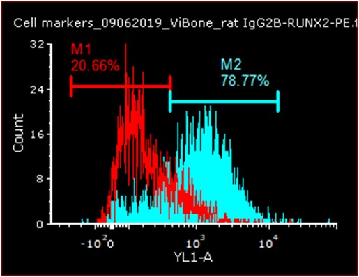
Application: Flow CytometrySample Tested: Bone ExtractsSpecies: HumanVerified Customer | Posted 02/13/2020

-
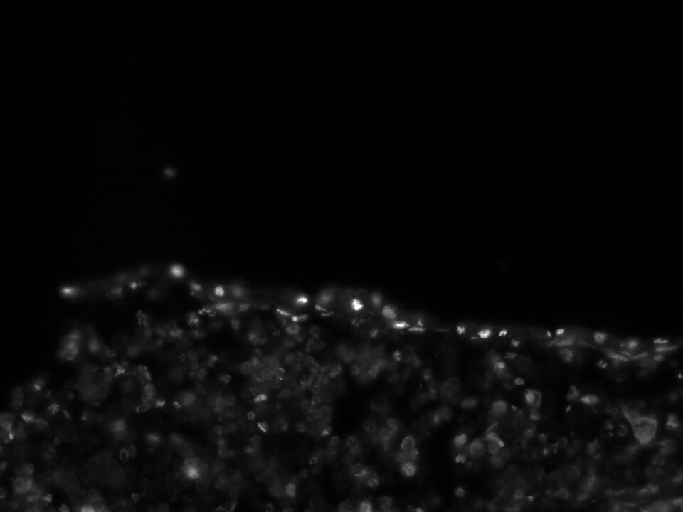
Application: Immunocytochemistry/ImmunofluorescenceSample Tested: bone marrowSpecies: MouseVerified Customer | Posted 08/31/2018
-
Application: Flow CytometrySample Tested: See PMID 22034088Species: HumanVerified Customer | Posted 02/16/2015
There are no reviews that match your criteria.
Protocols
Find general support by application which include: protocols, troubleshooting, illustrated assays, videos and webinars.
- Appropriate Fixation of IHC/ICC Samples
- Cellular Response to Hypoxia Protocols
- ClariTSA™ Fluorophore Kits
- Detection & Visualization of Antibody Binding
- ICC Cell Smear Protocol for Suspension Cells
- ICC Immunocytochemistry Protocol Videos
- ICC for Adherent Cells
- Immunocytochemistry (ICC) Protocol
- Immunocytochemistry Troubleshooting
- Immunofluorescence of Organoids Embedded in Cultrex Basement Membrane Extract
- Immunohistochemistry (IHC) and Immunocytochemistry (ICC) Protocols
- Preparing Samples for IHC/ICC Experiments
- Preventing Non-Specific Staining (Non-Specific Binding)
- Primary Antibody Selection & Optimization
- Protocol for VisUCyte™ HRP Polymer Detection Reagent
- Protocol for the Fluorescent ICC Staining of Cell Smears - Graphic
- Protocol for the Fluorescent ICC Staining of Cultured Cells on Coverslips - Graphic
- Protocol for the Preparation and Fluorescent ICC Staining of Cells on Coverslips
- Protocol for the Preparation and Fluorescent ICC Staining of Non-adherent Cells
- Protocol for the Preparation and Fluorescent ICC Staining of Stem Cells on Coverslips
- Protocol for the Preparation of a Cell Smear for Non-adherent Cell ICC - Graphic
- TUNEL and Active Caspase-3 Detection by IHC/ICC Protocol
- The Importance of IHC/ICC Controls
- View all Protocols, Troubleshooting, Illustrated assays and Webinars


