T-box expressed in T cells (T-bet), also known as T-box transcription factor TBX21, is a 62 kDa member of the T-box family of transcription factors and the Tbr1 subfamily. Human T-bet is 535 amino acids in length and contains a T-box DNA binding domain (aa 136-327). Human T-bet shares 88% aa sequence identity with mouse T-bet. T-bet is a nuclear protein highly apparent in Th1-cells. Northern blot analysis revealed that it is also expressed in lung, thymus and spleen. Functionally, T-bet controls the expression of the Th1 cytokine, IFN gamma, and initiates Th1 lineage development from naïve Th precursor cells by both activating Th1 genetic programs and by repressing the opposing Th2 programs.
Human T-bet/TBX21 Antibody (525803)
R&D Systems | Catalog # MAB5385

Discontinued Product
MAB5385 has been discontinued.
View all T-bet/TBX21 products.
Key Product Details
Species Reactivity
Validated:
Human
Cited:
Human
Applications
Validated:
Western Blot, Intracellular Staining by Flow Cytometry, Immunocytochemistry
Cited:
Immunohistochemistry, Immunohistochemistry-Paraffin, Flow Cytometry
Label
Unconjugated
Antibody Source
Monoclonal Mouse IgG1 Clone # 525803
Loading...
Product Specifications
Immunogen
E. coli-derived recombinant human T-bet
Glu326-Asn535
Accession # Q9UL17
Glu326-Asn535
Accession # Q9UL17
Specificity
Detects human T-bet.
Clonality
Monoclonal
Host
Mouse
Isotype
IgG1
Scientific Data Images for Human T-bet/TBX21 Antibody (525803)
Detection of T‑bet/TBX21 in Jurkat Human Cell Line by Flow Cytometry.
Jurkat human acute T cell leukemia cell line was stained with Human T‑bet/TBX21 Monoclonal Antibody (Catalog # MAB5385, filled histogram) or isotype control antibody (Catalog # MAB002, open histogram), followed by Phycoerythrin-conjugated Anti-Mouse IgG F(ab')2Secondary Antibody (Catalog # F0102B). To facilitate intracellular staining, cells were fixed with paraformaldehyde and permeabilized with ice-cold methanol.
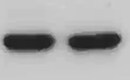
Detection of Human T-bet/TBX21 by Western Blot.
Western blot shows lysates of Raji human Burkitt's lymphoma cell line and Daudi human Burkitt's lymphoma cell line. PVDF membrane was probed with 2 µg/mL of Human T-bet/TBX21 Monoclonal Antibody (Catalog # MAB5385) followed by HRP-conjugated Anti-Mouse IgG Secondary Antibody (Catalog # HAF007). A specific band was detected for T-bet/TBX21 at approximately 55 kDa (as indicated). This experiment was conducted under reducing conditions and using Immunoblot Buffer Group 2.
Detection of T‑bet/TBX21 in CD45RO+/CD8+ PBMC lymphocytes by Flow Cytometry
CD45RO+/CD8+ PBMC lymphocytes were stained with either (A) Mouse Anti-Human T‑bet/TBX21 Monoclonal Antibody (Catalog # MAB5385) or (B) isotype control antibody (Catalog # MAB002) followed by Fluorescein-conjugated Anti-Mouse IgG Secondary Antibody (Catalog # F0103B). Cells were then stained with Mouse Anti-Human CD8 alpha APC‑conjugated Monoclonal Antibody (Catalog # FAB1509A) and Mouse Anti-Human CD45RO PE‑conjugated Monoclonal Antibody (Catalog # FAB10642P). To facilitate intracellular staining, cells were fixed and permeabilized with FlowX FoxP3 Fixation & Permeabilization Buffer Kit (Catalog # FC012). View our protocol for Staining Intracellular Molecules.Applications for Human T-bet/TBX21 Antibody (525803)
Application
Recommended Usage
Immunocytochemistry
8-25 µg/mL
Sample: Immersion fixed Jurkat human acute T cell leukemia cell line
Sample: Immersion fixed Jurkat human acute T cell leukemia cell line
Intracellular Staining by Flow Cytometry
2.5 µg/106 cells
Sample: Jurkat human acute T cell leukemia cell line fixed with paraformaldehyde and permeabilized with ice-cold methanol
Sample: Jurkat human acute T cell leukemia cell line fixed with paraformaldehyde and permeabilized with ice-cold methanol
Western Blot
2 µg/mL
Sample: Raji human Burkitt's lymphoma cell line and Daudi human Burkitt's lymphoma cell line
Sample: Raji human Burkitt's lymphoma cell line and Daudi human Burkitt's lymphoma cell line
Reviewed Applications
Read 1 review rated 5 using MAB5385 in the following applications:
Flow Cytometry Panel Builder
Bio-Techne Knows Flow Cytometry
Save time and reduce costly mistakes by quickly finding compatible reagents using the Panel Builder Tool.
Advanced Features
- Spectra Viewer - Custom analysis of spectra from multiple fluorochromes
- Spillover Popups - Visualize the spectra of individual fluorochromes
- Antigen Density Selector - Match fluorochrome brightness with antigen density

Formulation, Preparation, and Storage
Purification
Protein A or G purified from hybridoma culture supernatant
Reconstitution
Reconstitute at 0.5 mg/mL in sterile PBS. For liquid material, refer to CoA for concentration.
Formulation
Lyophilized from a 0.2 μm filtered solution in PBS with Trehalose. *Small pack size (SP) is supplied either lyophilized or as a 0.2 µm filtered solution in PBS.
Shipping
Lyophilized product is shipped at ambient temperature. Liquid small pack size (-SP) is shipped with polar packs. Upon receipt, store immediately at the temperature recommended below.
Stability & Storage
Use a manual defrost freezer and avoid repeated freeze-thaw cycles.
- 12 months from date of receipt, -20 to -70 °C as supplied.
- 1 month, 2 to 8 °C under sterile conditions after reconstitution.
- 6 months, -20 to -70 °C under sterile conditions after reconstitution.
Calculators
Background: T-bet/TBX21
Long Name
T-box transcription factor TBX21
Alternate Names
T-PET, Tbet, TBLYM, TBX21
Gene Symbol
TBX21
UniProt
Additional T-bet/TBX21 Products
Product Documents for Human T-bet/TBX21 Antibody (525803)
Certificate of Analysis
To download a Certificate of Analysis, please enter a lot or batch number in the search box below.
Note: Certificate of Analysis not available for kit components.
Product Specific Notices for Human T-bet/TBX21 Antibody (525803)
For research use only
Citations for Human T-bet/TBX21 Antibody (525803)
 Powered by Bioz
Customer Reviews for Human T-bet/TBX21 Antibody (525803) (1)
5 out of 5
1 Customer Rating
Have you used Human T-bet/TBX21 Antibody (525803)?
Submit a review and receive an Amazon gift card!
$25/€18/£15/$25CAN/¥2500 Yen for a review with an image
$10/€7/£6/$10CAN/¥1110 Yen for a review without an image
Submit a review
Customer Images
Showing
1
-
1 of
1 review
Showing All
Filter By:
-
Application: Western BlotSample Tested: Daudi human Burkitt's lymphoma cell lineSpecies: HumanVerified Customer | Posted 06/11/2022

There are no reviews that match your criteria.
Protocols
Find general support by application which include: protocols, troubleshooting, illustrated assays, videos and webinars.
- 7-Amino Actinomycin D (7-AAD) Cell Viability Flow Cytometry Protocol
- Appropriate Fixation of IHC/ICC Samples
- Cellular Response to Hypoxia Protocols
- ClariTSA™ Fluorophore Kits
- Detection & Visualization of Antibody Binding
- Extracellular Membrane Flow Cytometry Protocol
- Flow Cytometry Protocol for Cell Surface Markers
- Flow Cytometry Protocol for Staining Membrane Associated Proteins
- Flow Cytometry Staining Protocols
- Flow Cytometry Troubleshooting Guide
- ICC Cell Smear Protocol for Suspension Cells
- ICC Immunocytochemistry Protocol Videos
- ICC for Adherent Cells
- Immunocytochemistry (ICC) Protocol
- Immunocytochemistry Troubleshooting
- Immunofluorescence of Organoids Embedded in Cultrex Basement Membrane Extract
- Immunohistochemistry (IHC) and Immunocytochemistry (ICC) Protocols
- Intracellular Flow Cytometry Protocol Using Alcohol (Methanol)
- Intracellular Flow Cytometry Protocol Using Detergents
- Intracellular Nuclear Staining Flow Cytometry Protocol Using Detergents
- Intracellular Staining Flow Cytometry Protocol Using Alcohol Permeabilization
- Intracellular Staining Flow Cytometry Protocol Using Detergents to Permeabilize Cells
- Preparing Samples for IHC/ICC Experiments
- Preventing Non-Specific Staining (Non-Specific Binding)
- Primary Antibody Selection & Optimization
- Propidium Iodide Cell Viability Flow Cytometry Protocol
- Protocol for Liperfluo
- Protocol for VisUCyte™ HRP Polymer Detection Reagent
- Protocol for the Characterization of Human Th22 Cells
- Protocol for the Characterization of Human Th9 Cells
- Protocol for the Fluorescent ICC Staining of Cell Smears - Graphic
- Protocol for the Fluorescent ICC Staining of Cultured Cells on Coverslips - Graphic
- Protocol for the Preparation and Fluorescent ICC Staining of Cells on Coverslips
- Protocol for the Preparation and Fluorescent ICC Staining of Non-adherent Cells
- Protocol for the Preparation and Fluorescent ICC Staining of Stem Cells on Coverslips
- Protocol for the Preparation of a Cell Smear for Non-adherent Cell ICC - Graphic
- Protocol: Annexin V and PI Staining by Flow Cytometry
- Protocol: Annexin V and PI Staining for Apoptosis by Flow Cytometry
- R&D Systems Quality Control Western Blot Protocol
- TUNEL and Active Caspase-3 Detection by IHC/ICC Protocol
- The Importance of IHC/ICC Controls
- Troubleshooting Guide: Fluorokine Flow Cytometry Kits
- Troubleshooting Guide: Western Blot Figures
- Western Blot Conditions
- Western Blot Protocol
- Western Blot Protocol for Cell Lysates
- Western Blot Troubleshooting
- Western Blot Troubleshooting Guide
- View all Protocols, Troubleshooting, Illustrated assays and Webinars
Loading...
Associated Pathways

