Human Trypsin 1, encoded by the PRSS1 gene, is also known as Cationic Trypsinogen (1). Constituting approximately two-thirds of the total trypsin content in normal pancreatic juice, it is the most abundant trypsin isoform produced by the pancreas. It contains a signal peptide (residues 1‑15), a pro region (residues 16‑23), and a mature chain (residues 24‑247). Trypsin 1 is synthesized in the pancreas and secreted into the duodenum lumen, where it is activated by enterokinase. Its major physiologic function is to digest food and to activate other pro-enzymes (2). Mutations in the PRSS1 gene can cause hereditary pancreatitis (3).

Key Product Details
Species Reactivity
Validated:
Human
Cited:
Human, Mouse
Applications
Validated:
Immunohistochemistry, Western Blot, Immunoprecipitation
Cited:
Immunohistochemistry, Immunohistochemistry-Frozen, Western Blot
Label
Unconjugated
Antibody Source
Polyclonal Sheep IgG
Loading...
Product Specifications
Immunogen
Mouse myeloma cell line NS0-derived recombinant human Trypsin 1/PRSS1
Ala16-Ser247
Accession # P07477
Ala16-Ser247
Accession # P07477
Specificity
Detects human Trypsin 1/PRSS1 in direct ELISAs and Western blots. In direct ELISAs, approximately 50% cross-reactivity with recombinant human (rh) Trypsin 2 and rhTrypsin 3 is observed.
Clonality
Polyclonal
Host
Sheep
Isotype
IgG
Scientific Data Images for Human Trypsin 1/PRSS1 Antibody
Trypsin 1/PRSS1 in Human Pancreas.
Trypsin 1/PRSS1 was detected in immersion fixed paraffin-embedded sections of human pancreas using Mouse Anti-Human Trypsin 1/PRSS1 Monoclonal Antibody (Catalog # MAB3848) at 15 µg/mL overnight at 4 °C. Tissue was stained using the Anti-Sheep HRP-DAB Cell & Tissue Staining Kit (brown; Catalog # CTS019) and counterstained with hematoxylin (blue). Specific staining was localized to exocrine cells. View our protocol for Chromogenic IHC Staining of Paraffin-embedded Tissue Sections.Applications for Human Trypsin 1/PRSS1 Antibody
Application
Recommended Usage
Immunohistochemistry
5-15 µg/mL
Sample: Immersion fixed paraffin-embedded sections of human pancreas
Sample: Immersion fixed paraffin-embedded sections of human pancreas
Immunoprecipitation
25 µg/mL
Sample: Conditioned cell culture medium spiked with Recombinant Human Trypsin 1/PRSS1 (Catalog # 3848-SE), see our available Western blot detection antibodies
Sample: Conditioned cell culture medium spiked with Recombinant Human Trypsin 1/PRSS1 (Catalog # 3848-SE), see our available Western blot detection antibodies
Western Blot
0.1 µg/mL
Sample: Recombinant Human Trypsin 1/PRSS1 (Catalog # 3848-SE)
Sample: Recombinant Human Trypsin 1/PRSS1 (Catalog # 3848-SE)
Reviewed Applications
Read 2 reviews rated 5 using AF3848 in the following applications:
Formulation, Preparation, and Storage
Purification
Antigen Affinity-purified
Reconstitution
Reconstitute at 0.2 mg/mL in sterile PBS. For liquid material, refer to CoA for concentration.
Loading...
Formulation
Lyophilized from a 0.2 μm filtered solution in PBS with Trehalose. *Small pack size (SP) is supplied either lyophilized or as a 0.2 µm filtered solution in PBS.
Shipping
Lyophilized product is shipped at ambient temperature. Liquid small pack size (-SP) is shipped with polar packs. Upon receipt, store immediately at the temperature recommended below.
Stability & Storage
Use a manual defrost freezer and avoid repeated freeze-thaw cycles.
- 12 months from date of receipt, -20 to -70 °C as supplied.
- 1 month, 2 to 8 °C under sterile conditions after reconstitution.
- 6 months, -20 to -70 °C under sterile conditions after reconstitution.
Calculators
Background: Trypsin 1/PRSS1
References
- Emi, M. et al. (1986) Gene 41:305.
- Halfon, S. et al. (2004) in Handbook of Proteolytic Enzymes (ed. Barrett, et al.) p. 1483, Academic Press, San Diego.
- Teich, N. et al. (2006) Hum. Mutat. 27:721.
Alternate Names
PRSS1, PTRYI, TRY4, Trygn16, Trypsinogen 1
Gene Symbol
PRSS1
UniProt
Additional Trypsin 1/PRSS1 Products
Product Documents for Human Trypsin 1/PRSS1 Antibody
Certificate of Analysis
To download a Certificate of Analysis, please enter a lot or batch number in the search box below.
Note: Certificate of Analysis not available for kit components.
Product Specific Notices for Human Trypsin 1/PRSS1 Antibody
For research use only
Related Research Areas
Citations for Human Trypsin 1/PRSS1 Antibody
 Powered by Bioz
Customer Reviews for Human Trypsin 1/PRSS1 Antibody (2)
5 out of 5
2 Customer Ratings
Have you used Human Trypsin 1/PRSS1 Antibody?
Submit a review and receive an Amazon gift card!
$25/€18/£15/$25CAN/¥2500 Yen for a review with an image
$10/€7/£6/$10CAN/¥1110 Yen for a review without an image
Submit a review
Customer Images
Showing
1
-
2 of
2 reviews
Showing All
Filter By:
-
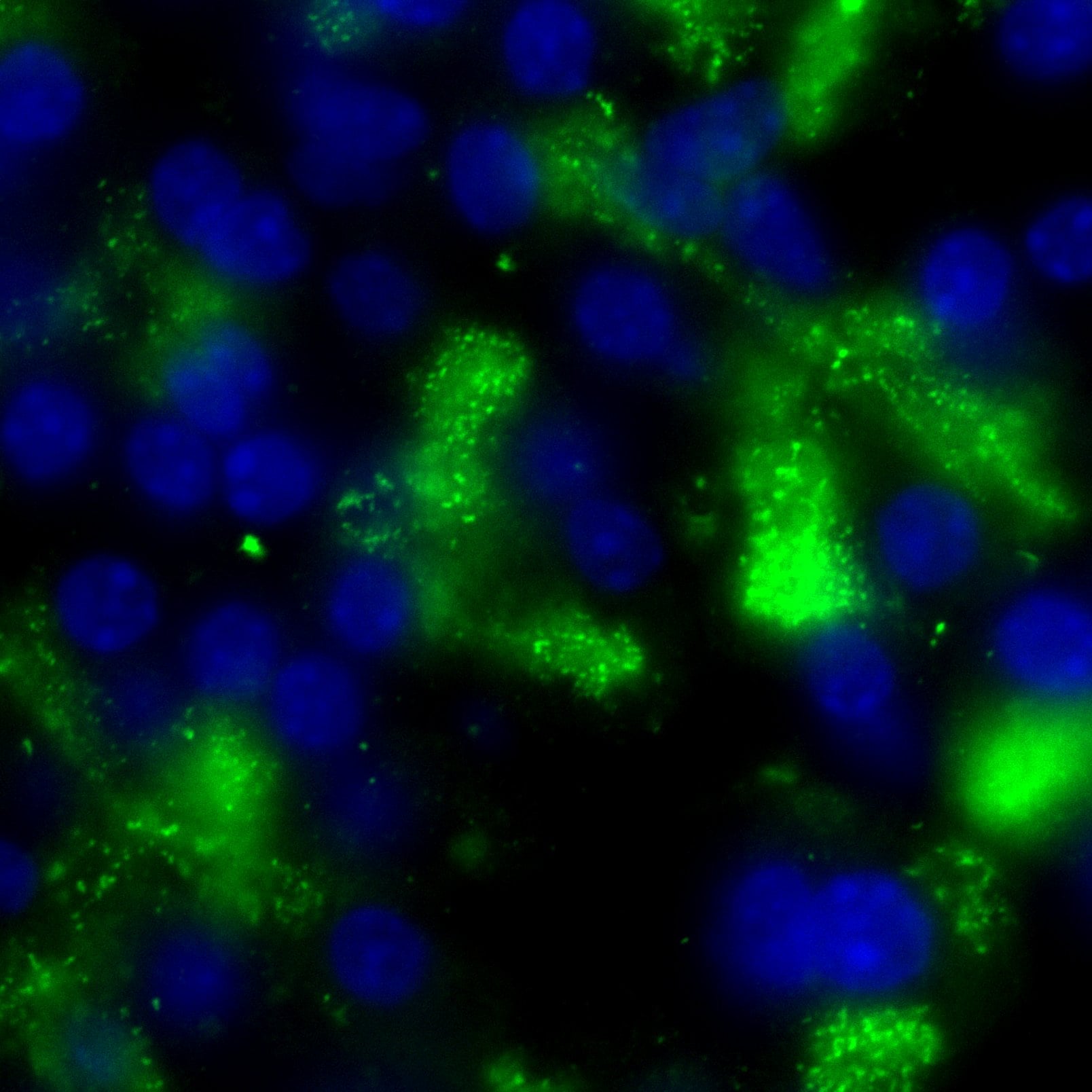
Application: Immunohistochemistry-FrozenSample Tested: Mouse pancreas in OCTSpecies: MouseVerified Customer | Posted 12/15/2017mouse pancreas cryosections labeled using antibody diluted 1/100. DAPI to label nuclei. imaged 60x epi-fluorescence.

-
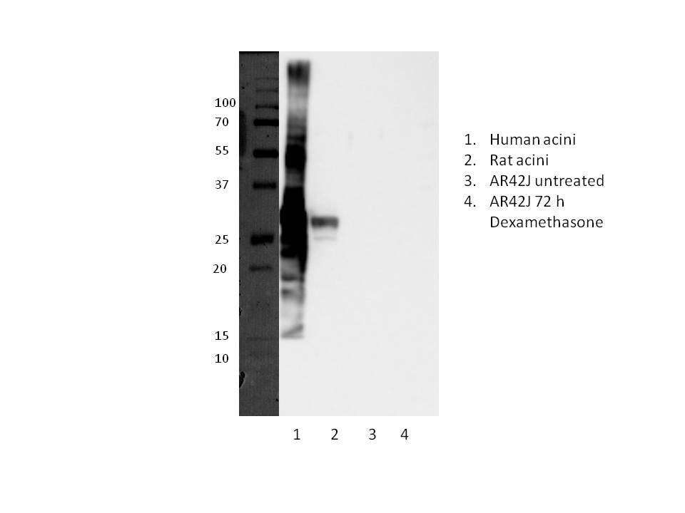
Application: Western BlotSample Tested: huamn acini, rat acini and AR42J rat acinar-like cellSpecies: Human and RatVerified Customer | Posted 05/19/2017Overexposed to determine rat reactivity.whole cell lysates 50 ug. 12% gel. 1/1000 dilution of primary antibody
There are no reviews that match your criteria.
Protocols
Find general support by application which include: protocols, troubleshooting, illustrated assays, videos and webinars.
- Antigen Retrieval Protocol (PIER)
- Antigen Retrieval for Frozen Sections Protocol
- Appropriate Fixation of IHC/ICC Samples
- Cellular Response to Hypoxia Protocols
- Chromogenic IHC Staining of Formalin-Fixed Paraffin-Embedded (FFPE) Tissue Protocol
- Chromogenic Immunohistochemistry Staining of Frozen Tissue
- ClariTSA™ Fluorophore Kits
- Detection & Visualization of Antibody Binding
- Fluorescent IHC Staining of Frozen Tissue Protocol
- Graphic Protocol for Heat-induced Epitope Retrieval
- Graphic Protocol for the Preparation and Fluorescent IHC Staining of Frozen Tissue Sections
- Graphic Protocol for the Preparation and Fluorescent IHC Staining of Paraffin-embedded Tissue Sections
- Graphic Protocol for the Preparation of Gelatin-coated Slides for Histological Tissue Sections
- IHC Sample Preparation (Frozen sections vs Paraffin)
- Immunofluorescent IHC Staining of Formalin-Fixed Paraffin-Embedded (FFPE) Tissue Protocol
- Immunohistochemistry (IHC) and Immunocytochemistry (ICC) Protocols
- Immunohistochemistry Frozen Troubleshooting
- Immunohistochemistry Paraffin Troubleshooting
- Immunoprecipitation Protocol
- Preparing Samples for IHC/ICC Experiments
- Preventing Non-Specific Staining (Non-Specific Binding)
- Primary Antibody Selection & Optimization
- Protocol for Heat-Induced Epitope Retrieval (HIER)
- Protocol for Making a 4% Formaldehyde Solution in PBS
- Protocol for VisUCyte™ HRP Polymer Detection Reagent
- Protocol for the Preparation & Fixation of Cells on Coverslips
- Protocol for the Preparation and Chromogenic IHC Staining of Frozen Tissue Sections
- Protocol for the Preparation and Chromogenic IHC Staining of Frozen Tissue Sections - Graphic
- Protocol for the Preparation and Chromogenic IHC Staining of Paraffin-embedded Tissue Sections
- Protocol for the Preparation and Chromogenic IHC Staining of Paraffin-embedded Tissue Sections - Graphic
- Protocol for the Preparation and Fluorescent IHC Staining of Frozen Tissue Sections
- Protocol for the Preparation and Fluorescent IHC Staining of Paraffin-embedded Tissue Sections
- Protocol for the Preparation of Gelatin-coated Slides for Histological Tissue Sections
- R&D Systems Quality Control Western Blot Protocol
- TUNEL and Active Caspase-3 Detection by IHC/ICC Protocol
- The Importance of IHC/ICC Controls
- Troubleshooting Guide: Immunohistochemistry
- Troubleshooting Guide: Western Blot Figures
- Western Blot Conditions
- Western Blot Protocol
- Western Blot Protocol for Cell Lysates
- Western Blot Troubleshooting
- Western Blot Troubleshooting Guide
- View all Protocols, Troubleshooting, Illustrated assays and Webinars
Loading...