Uromodulin (also Tamm-Horsfall glycoprotein or THP) is an 85‑95 kDa urinary glycoprotein. It is secreted by renal tubule epithelium, acts as a binding protein for IL‑1, TNF-alpha and C1q, activates resting monocytes and promotes neutrophil phagocytosis. Uromodulin forms high molecular weight oligomers that line the kidnay tubules. Human Uromodulin is GPI‑linked. Its proprecursor is 616 amino acids (aa) in length. It contains three EGF-like domains (aa 28‑149), a ZP domain that mediates oligomerization (aa 334‑589) and a cleavable C‑terminal propeptide (aa 615‑640). There are multiple splice variants. One shows a deletion of aa 67‑199, a second shows a nine aa substitution for aa 609‑640, a third shows a Pro substitution for aa 205‑234 and a fourth shows a 66 aa substitution for aa 613‑640. Over aa 25‑614, human Uromodulin is 78% aa identical to mouse Uromodulin.

Key Product Details
Species Reactivity
Validated:
Human
Cited:
Human
Applications
Validated:
Immunohistochemistry, Western Blot, Simple Western
Cited:
Immunohistochemistry, IF/IHC
Label
Unconjugated
Antibody Source
Polyclonal Sheep IgG
Loading...
Product Specifications
Immunogen
Mouse myeloma cell line NS0-derived recombinant human Uromodulin isoform 1
Asp25-Ser614
Accession # P07911
Asp25-Ser614
Accession # P07911
Specificity
Detects human Uromodulin in direct ELISAs and Western blots. In direct ELISAs and Western blots, approximately 25% cross-reactivity with recombinant mouse Uromodulin is observed.
Clonality
Polyclonal
Host
Sheep
Isotype
IgG
Scientific Data Images for Human Uromodulin Antibody
Detection of Human Uromodulin by Western Blot.
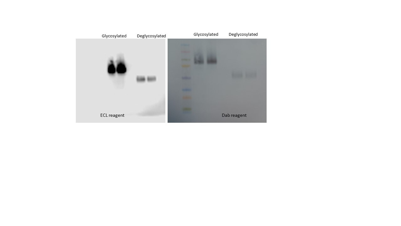
Western blot shows lysates of human kidney tissue. PVDF membrane was probed with 0.5 µg/mL of Sheep Anti-Human Uromodulin Antigen Affinity-purified Polyclonal Antibody (Catalog # AF5144) followed by HRP-conjugated Anti-Sheep IgG Secondary Antibody (Catalog # HAF016). A specific band was detected for Uromodulin at approximately 80-90 kDa (as indicated). This experiment was conducted under reducing conditions and using Immunoblot Buffer Group 1.
Detection of Human Uromodulin by Simple WesternTM.
Simple Western shows lysates of Exosome Standards (Human Urine) (NBP2-49840) and human kidney tissue, loaded at 0.5 mg/ml. A specific band was detected for Uromodulin at approximately 149 kDa (as indicated) using 50 µg/mL of Sheep Anti-Human Uromodulin Antigen Affinity-purified Polyclonal Antibody (Catalog # AF5144). This experiment was conducted under reducing conditions and using the 12-230kDa separation system.
Uromodulin in Human Kidney.
Uromodulin was detected in immersion fixed paraffin-embedded sections of human kidney using 1.7 µg/mL Sheep Anti-Human Uromodulin Antigen Affinity-purified Polyclonal Antibody (Catalog # AF5144) overnight at 4 °C. Tissue was stained with the Anti-Sheep HRP-DAB Cell & Tissue Staining Kit (brown; Catalog # CTS019) and counterstained with hematoxylin (blue). Specific labeling was localized to epithelial cells in tubules. View our protocol for Chromogenic IHC Staining of Paraffin-embedded Tissue Sections.Applications for Human Uromodulin Antibody
Application
Recommended Usage
Immunohistochemistry
2-15 µg/mL
Sample: Immersion fixed paraffin-embedded sections of human kidney
Sample: Immersion fixed paraffin-embedded sections of human kidney
Simple Western
50 µg/mL
Sample: Exosome Standards (Human Urine) (Catalog # NBP2-49840) and human kidney tissue
Sample: Exosome Standards (Human Urine) (Catalog # NBP2-49840) and human kidney tissue
Western Blot
0.5 µg/mL
Sample: Human kidney tissue
Sample: Human kidney tissue
Reviewed Applications
Read 1 review rated 5 using AF5144 in the following applications:
Formulation, Preparation, and Storage
Purification
Antigen Affinity-purified
Reconstitution
Reconstitute at 0.2 mg/mL in sterile PBS. For liquid material, refer to CoA for concentration.
Loading...
Formulation
Lyophilized from a 0.2 μm filtered solution in PBS with Trehalose. See Certificate of Analysis for details.
*Small pack size (-SP) is supplied either lyophilized or as a 0.2 µm filtered solution in PBS.
*Small pack size (-SP) is supplied either lyophilized or as a 0.2 µm filtered solution in PBS.
Shipping
Lyophilized product is shipped at ambient temperature. Liquid small pack size (-SP) is shipped with polar packs. Upon receipt, store immediately at the temperature recommended below.
Stability & Storage
Use a manual defrost freezer and avoid repeated freeze-thaw cycles.
- 12 months from date of receipt, -20 to -70 °C as supplied.
- 1 month, 2 to 8 °C under sterile conditions after reconstitution.
- 6 months, -20 to -70 °C under sterile conditions after reconstitution.
Calculators
Background: Uromodulin
Alternate Names
ADMCKD2, FJHN, HNFJ, MCKD2, THGP, THP, UMOD, Uromucoid
Gene Symbol
UMOD
UniProt
Additional Uromodulin Products
Product Documents for Human Uromodulin Antibody
Certificate of Analysis
To download a Certificate of Analysis, please enter a lot or batch number in the search box below.
Note: Certificate of Analysis not available for kit components.
Product Specific Notices for Human Uromodulin Antibody
For research use only
Citations for Human Uromodulin Antibody
 Powered by Bioz
Customer Reviews for Human Uromodulin Antibody (1)
5 out of 5
1 Customer Rating
Have you used Human Uromodulin Antibody?
Submit a review and receive an Amazon gift card!
$25/€18/£15/$25CAN/¥2500 Yen for a review with an image
$10/€7/£6/$10CAN/¥1110 Yen for a review without an image
Submit a review
Customer Images
Showing
1
-
1 of
1 review
Showing All
Filter By:
-
Application: Western BlotSample Tested: UrineSpecies: RatVerified Customer | Posted 07/06/2018

There are no reviews that match your criteria.
Protocols
Find general support by application which include: protocols, troubleshooting, illustrated assays, videos and webinars.
- Antigen Retrieval Protocol (PIER)
- Antigen Retrieval for Frozen Sections Protocol
- Appropriate Fixation of IHC/ICC Samples
- Cellular Response to Hypoxia Protocols
- Chromogenic IHC Staining of Formalin-Fixed Paraffin-Embedded (FFPE) Tissue Protocol
- Chromogenic Immunohistochemistry Staining of Frozen Tissue
- ClariTSA™ Fluorophore Kits
- Detection & Visualization of Antibody Binding
- Fluorescent IHC Staining of Frozen Tissue Protocol
- Graphic Protocol for Heat-induced Epitope Retrieval
- Graphic Protocol for the Preparation and Fluorescent IHC Staining of Frozen Tissue Sections
- Graphic Protocol for the Preparation and Fluorescent IHC Staining of Paraffin-embedded Tissue Sections
- Graphic Protocol for the Preparation of Gelatin-coated Slides for Histological Tissue Sections
- IHC Sample Preparation (Frozen sections vs Paraffin)
- Immunofluorescent IHC Staining of Formalin-Fixed Paraffin-Embedded (FFPE) Tissue Protocol
- Immunohistochemistry (IHC) and Immunocytochemistry (ICC) Protocols
- Immunohistochemistry Frozen Troubleshooting
- Immunohistochemistry Paraffin Troubleshooting
- Preparing Samples for IHC/ICC Experiments
- Preventing Non-Specific Staining (Non-Specific Binding)
- Primary Antibody Selection & Optimization
- Protocol for Heat-Induced Epitope Retrieval (HIER)
- Protocol for Making a 4% Formaldehyde Solution in PBS
- Protocol for VisUCyte™ HRP Polymer Detection Reagent
- Protocol for the Preparation & Fixation of Cells on Coverslips
- Protocol for the Preparation and Chromogenic IHC Staining of Frozen Tissue Sections
- Protocol for the Preparation and Chromogenic IHC Staining of Frozen Tissue Sections - Graphic
- Protocol for the Preparation and Chromogenic IHC Staining of Paraffin-embedded Tissue Sections
- Protocol for the Preparation and Chromogenic IHC Staining of Paraffin-embedded Tissue Sections - Graphic
- Protocol for the Preparation and Fluorescent IHC Staining of Frozen Tissue Sections
- Protocol for the Preparation and Fluorescent IHC Staining of Paraffin-embedded Tissue Sections
- Protocol for the Preparation of Gelatin-coated Slides for Histological Tissue Sections
- R&D Systems Quality Control Western Blot Protocol
- TUNEL and Active Caspase-3 Detection by IHC/ICC Protocol
- The Importance of IHC/ICC Controls
- Troubleshooting Guide: Immunohistochemistry
- Troubleshooting Guide: Western Blot Figures
- Western Blot Conditions
- Western Blot Protocol
- Western Blot Protocol for Cell Lysates
- Western Blot Troubleshooting
- Western Blot Troubleshooting Guide
- View all Protocols, Troubleshooting, Illustrated assays and Webinars
Loading...