Mouse IgG1 Kappa Isotype Control (P3.6.2.8.1)
Novus Biologicals | Catalog # NBP1-43319
![Flow Cytometry: Mouse IgG1 Kappa Isotype Control (P3.6.2.8.1) [NBP1-43319]](https://resources.rndsystems.com/images/products/Mouse-IgG1-Kappa-Light-Chain-Isotype-Control-P3-6-2-8-1-Flow-Cytometry-NBP1-43319-img0004.jpg "Flow Cytometry: Mouse IgG1 Kappa Isotype Control (P3.6.2.8.1) [NBP1-43319]")
Key Product Details
Species Reactivity
Mouse
Applications
Immunohistochemistry, Western Blot, Flow Cytometry, Immunocytochemistry/ Immunofluorescence, Immunoprecipitation, Control
Label
Unconjugated
Antibody Source
Monoclonal Mouse IgG1 kappa Clone # P3.6.2.8.1
Loading...
Product Specifications
Clonality
Monoclonal
Host
Mouse
Isotype
IgG1 kappa
Description
This Mouse IgG1 Kappa immunoglobulin is useful as an isotype control.
Scientific Data Images for Mouse IgG1 Kappa Isotype Control (P3.6.2.8.1)
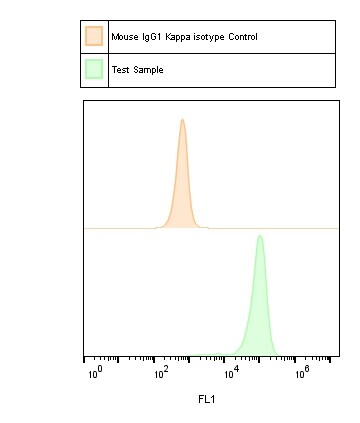
Flow Cytometry: Mouse IgG1 Kappa Isotype Control (P3.6.2.8.1) [NBP1-43319]
Flow Cytometry: Mouse IgG1 Kappa Isotype Control (P3.6.2.8.1) [NBP1-43319] - Analysis of Involucrin signal in human keratinocytes. Red: anti-IVL antibody. BLACK: Isotype control NBP1-43319APC at 1:100. Flow cytometry image submitted by a verified customer review.![Flow Cytometry: Mouse IgG1 Kappa Isotype Control (P3.6.2.8.1) [NBP1-43319]](https://resources.rndsystems.com/images/products/Mouse-IgG1-Kappa-Light-Chain-Isotype-Control-P3-6-2-8-1-Flow-Cytometry-NBP1-43319-img0001.jpg "Flow Cytometry: Mouse IgG1 Kappa Isotype Control (P3.6.2.8.1) [NBP1-43319]")
Flow Cytometry: Mouse IgG1 Kappa Isotype Control (P3.6.2.8.1) [NBP1-43319]
Flow Cytometry: Mouse IgG1 Kappa Isotype Control (P3.6.2.8.1) [NBP1-43319] - Analysis of Alexa Fluor (R) 647 conjugate of NBP1-43319. Mouse IgG1 isotype control was used as a negative control. Flow cytometry image submitted by a verified customer review.![Flow Cytometry: Mouse IgG1 Kappa Isotype Control (P3.6.2.8.1) [NBP1-43319]](https://resources.rndsystems.com/images/products/Mouse-IgG1-Kappa-Light-Chain-Isotype-Control-P3-6-2-8-1-Flow-Cytometry-NBP1-43319-img0002.jpg "Flow Cytometry: Mouse IgG1 Kappa Isotype Control (P3.6.2.8.1) [NBP1-43319]")
Flow Cytometry: Mouse IgG1 Kappa Isotype Control (P3.6.2.8.1) [NBP1-43319]
Flow Cytometry: Mouse IgG1 Kappa Isotype Control (P3.6.2.8.1) [NBP1-43319] - Analysis of Alexa Fluor (R) 700 conjugate of NBP1-43319. Human buffy coat stained with mouse IgG1 Kappa isotype control. Flow cytometry image submitted by a verified customer review.![Flow Cytometry: Mouse IgG1 Kappa Isotype Control (P3.6.2.8.1) [NBP1-43319]](https://resources.rndsystems.com/images/products/Mouse-IgG1-Kappa-Light-Chain-Isotype-Control-P3-6-2-8-1-Flow-Cytometry-NBP1-43319-img0003.jpg "Flow Cytometry: Mouse IgG1 Kappa Isotype Control (P3.6.2.8.1) [NBP1-43319]")
Flow Cytometry: Mouse IgG1 Kappa Isotype Control (P3.6.2.8.1) [NBP1-43319]
Flow Cytometry: Mouse IgG1 Kappa Isotype Control (P3.6.2.8.1) [NBP1-43319] - Flow cytometry image submitted by a verified customer review.Applications for Mouse IgG1 Kappa Isotype Control (P3.6.2.8.1)
Application
Recommended Usage
Flow Cytometry
1:10 - 1:1000
Immunocytochemistry/ Immunofluorescence
1:10 - 1:500
Immunohistochemistry
1:10 - 1:500
Immunoprecipitation
1:10 - 1:500
Western Blot
1:100 - 1:2000
Application Notes
Tested for Flow analysis on normal human peripheral blood cells and mouse spleen cells. Use the isotype control at the same concentration as the experimental antibody. Optimal dilution of this antibody should be experimentally determined.
Flow Cytometry Panel Builder
Bio-Techne Knows Flow Cytometry
Save time and reduce costly mistakes by quickly finding compatible reagents using the Panel Builder Tool.
Advanced Features
- Spectra Viewer - Custom analysis of spectra from multiple fluorochromes
- Spillover Popups - Visualize the spectra of individual fluorochromes
- Antigen Density Selector - Match fluorochrome brightness with antigen density

Formulation, Preparation, and Storage
Purification
Affinity purified
Formulation
PBS (pH 7.2)
Preservative
0.09% Sodium Azide
Concentration
0.5 mg/ml
Shipping
The product is shipped with polar packs. Upon receipt, store it immediately at the temperature recommended below.
Stability & Storage
Store at 4C. Do not freeze.
Product Documents for Mouse IgG1 Kappa Isotype Control (P3.6.2.8.1)
Certificate of Analysis
To download a Certificate of Analysis, please enter a lot or batch number in the search box below.
Product Specific Notices for Mouse IgG1 Kappa Isotype Control (P3.6.2.8.1)
This product is for research use only and is not approved for use in humans or in clinical diagnosis. Isotype Controls are guaranteed for 1 year from date of receipt.
Citations for Mouse IgG1 Kappa Isotype Control (P3.6.2.8.1)
 Powered by Bioz
Customer Reviews for Mouse IgG1 Kappa Isotype Control (P3.6.2.8.1) (1)
5 out of 5
1 Customer Rating
Have you used Mouse IgG1 Kappa Isotype Control (P3.6.2.8.1)?
Submit a review and receive an Amazon gift card!
$25/€18/£15/$25CAN/¥2500 Yen for a review with an image
$10/€7/£6/$10CAN/¥1110 Yen for a review without an image
Submit a review
Customer Images
Showing
1
-
1 of
1 review
Showing All
Filter By:
-
Verified Customer | Posted 07/19/2018

There are no reviews that match your criteria.
Protocols
Find general support by application which include: protocols, troubleshooting, illustrated assays, videos and webinars.
- 7-Amino Actinomycin D (7-AAD) Cell Viability Flow Cytometry Protocol
- Antigen Retrieval Protocol (PIER)
- Antigen Retrieval for Frozen Sections Protocol
- Appropriate Fixation of IHC/ICC Samples
- Cellular Response to Hypoxia Protocols
- Chromogenic IHC Staining of Formalin-Fixed Paraffin-Embedded (FFPE) Tissue Protocol
- Chromogenic Immunohistochemistry Staining of Frozen Tissue
- ClariTSA™ Fluorophore Kits
- Detection & Visualization of Antibody Binding
- Extracellular Membrane Flow Cytometry Protocol
- Flow Cytometry Protocol for Cell Surface Markers
- Flow Cytometry Protocol for Staining Membrane Associated Proteins
- Flow Cytometry Staining Protocols
- Flow Cytometry Troubleshooting Guide
- Fluorescent IHC Staining of Frozen Tissue Protocol
- Graphic Protocol for Heat-induced Epitope Retrieval
- Graphic Protocol for the Preparation and Fluorescent IHC Staining of Frozen Tissue Sections
- Graphic Protocol for the Preparation and Fluorescent IHC Staining of Paraffin-embedded Tissue Sections
- Graphic Protocol for the Preparation of Gelatin-coated Slides for Histological Tissue Sections
- ICC Cell Smear Protocol for Suspension Cells
- ICC Immunocytochemistry Protocol Videos
- ICC for Adherent Cells
- IHC Sample Preparation (Frozen sections vs Paraffin)
- Immunocytochemistry (ICC) Protocol
- Immunocytochemistry Troubleshooting
- Immunofluorescence of Organoids Embedded in Cultrex Basement Membrane Extract
- Immunofluorescent IHC Staining of Formalin-Fixed Paraffin-Embedded (FFPE) Tissue Protocol
- Immunohistochemistry (IHC) and Immunocytochemistry (ICC) Protocols
- Immunohistochemistry Frozen Troubleshooting
- Immunohistochemistry Paraffin Troubleshooting
- Immunoprecipitation Protocol
- Intracellular Flow Cytometry Protocol Using Alcohol (Methanol)
- Intracellular Flow Cytometry Protocol Using Detergents
- Intracellular Nuclear Staining Flow Cytometry Protocol Using Detergents
- Intracellular Staining Flow Cytometry Protocol Using Alcohol Permeabilization
- Intracellular Staining Flow Cytometry Protocol Using Detergents to Permeabilize Cells
- Preparing Samples for IHC/ICC Experiments
- Preventing Non-Specific Staining (Non-Specific Binding)
- Primary Antibody Selection & Optimization
- Propidium Iodide Cell Viability Flow Cytometry Protocol
- Protocol for Heat-Induced Epitope Retrieval (HIER)
- Protocol for Liperfluo
- Protocol for Making a 4% Formaldehyde Solution in PBS
- Protocol for VisUCyte™ HRP Polymer Detection Reagent
- Protocol for the Characterization of Human Th22 Cells
- Protocol for the Characterization of Human Th9 Cells
- Protocol for the Fluorescent ICC Staining of Cell Smears - Graphic
- Protocol for the Fluorescent ICC Staining of Cultured Cells on Coverslips - Graphic
- Protocol for the Preparation & Fixation of Cells on Coverslips
- Protocol for the Preparation and Chromogenic IHC Staining of Frozen Tissue Sections
- Protocol for the Preparation and Chromogenic IHC Staining of Frozen Tissue Sections - Graphic
- Protocol for the Preparation and Chromogenic IHC Staining of Paraffin-embedded Tissue Sections
- Protocol for the Preparation and Chromogenic IHC Staining of Paraffin-embedded Tissue Sections - Graphic
- Protocol for the Preparation and Fluorescent ICC Staining of Cells on Coverslips
- Protocol for the Preparation and Fluorescent ICC Staining of Non-adherent Cells
- Protocol for the Preparation and Fluorescent ICC Staining of Stem Cells on Coverslips
- Protocol for the Preparation and Fluorescent IHC Staining of Frozen Tissue Sections
- Protocol for the Preparation and Fluorescent IHC Staining of Paraffin-embedded Tissue Sections
- Protocol for the Preparation of Gelatin-coated Slides for Histological Tissue Sections
- Protocol for the Preparation of a Cell Smear for Non-adherent Cell ICC - Graphic
- Protocol: Annexin V and PI Staining by Flow Cytometry
- Protocol: Annexin V and PI Staining for Apoptosis by Flow Cytometry
- R&D Systems Quality Control Western Blot Protocol
- TUNEL and Active Caspase-3 Detection by IHC/ICC Protocol
- The Importance of IHC/ICC Controls
- Troubleshooting Guide: Fluorokine Flow Cytometry Kits
- Troubleshooting Guide: Immunohistochemistry
- Troubleshooting Guide: Western Blot Figures
- Western Blot Conditions
- Western Blot Protocol
- Western Blot Protocol for Cell Lysates
- Western Blot Troubleshooting
- Western Blot Troubleshooting Guide
- View all Protocols, Troubleshooting, Illustrated assays and Webinars
Loading...