IL-17E/IL-25 Antibody (68C1039.2) - BSA Free
Novus Biologicals | Catalog # NB100-56541
Key Product Details
Species Reactivity
Validated:
Cited:
Applications
Validated:
Cited:
Label
Antibody Source
Format
Product Specifications
Immunogen
Clonality
Host
Isotype
Theoretical MW
Disclaimer note: The observed molecular weight of the protein may vary from the listed predicted molecular weight due to post translational modifications, post translation cleavages, relative charges, and other experimental factors.
Scientific Data Images for IL-17E/IL-25 Antibody (68C1039.2) - BSA Free
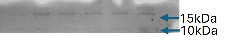
![Western Blot: IL-17E/IL-25 Antibody (68C1039.2)BSA Free [NB100-56541]](https://resources.rndsystems.com/images/products/IL-17E-IL-25-Antibody-68C1039-2-Western-Blot-NB100-56541-img0007.jpg "Western Blot: IL-17E/IL-25 Antibody (68C1039.2)BSA Free [NB100-56541]")
Western Blot: IL-17E/IL-25 Antibody (68C1039.2)BSA Free [NB100-56541]
Western Blot: IL-17E/IL-25 Antibody (68C1039.2) [NB100-56541] - Analysis of IL-17E using IL-17E monoclonal antibody. Mouse testis lysate probed with IL-17E antibody at 2 ug/mL.![Immunohistochemistry: IL-17E/IL-25 Antibody (68C1039.2) - BSA Free [NB100-56541]](https://resources.rndsystems.com/images/products/IL-17E-IL-25-Antibody-68C1039-2-BSA-Free-Immunohistochemistry-NB100-56541-img0008.jpg "Immunohistochemistry: IL-17E/IL-25 Antibody (68C1039.2) - BSA Free [NB100-56541]")
Immunohistochemistry: IL-17E/IL-25 Antibody (68C1039.2) - BSA Free [NB100-56541]
IL-17E-IL-25-Antibody-68C1039-2-BSA-Free-Immunohistochemistry-NB100-56541-img0008.jpg![Immunohistochemistry-Paraffin: IL-17E/IL-25 Antibody (68C1039.2) - BSA Free [NB100-56541]](https://resources.rndsystems.com/images/products/IL-17E-IL-25-Antibody-68C1039-2-Immunohistochemistry-Paraffin-NB100-56541-img0004.jpg "Immunohistochemistry-Paraffin: IL-17E/IL-25 Antibody (68C1039.2) - BSA Free [NB100-56541]")
Immunohistochemistry-Paraffin: IL-17E/IL-25 Antibody (68C1039.2) - BSA Free [NB100-56541]
Immunohistochemistry-Paraffin: IL-17E/IL-25 Antibody (68C1039.2) [NB100-56541] - Tissue section of mouse liver using IL17E/IL25 antibody clone 68C1039.2 at 1:150. The antibody generated a diffused cytoplasmic staining in the hepatocytes with a very strong signal in the sinusoidal endothelial cells.![Immunohistochemistry-Paraffin: IL-17E/IL-25 Antibody (68C1039.2) - BSA Free [NB100-56541]](https://resources.rndsystems.com/images/products/IL-17E-IL-25-Antibody-68C1039-2-Immunohistochemistry-Paraffin-NB100-56541-img0005.jpg "Immunohistochemistry-Paraffin: IL-17E/IL-25 Antibody (68C1039.2) - BSA Free [NB100-56541]")
Immunohistochemistry-Paraffin: IL-17E/IL-25 Antibody (68C1039.2) - BSA Free [NB100-56541]
Immunohistochemistry-Paraffin: IL-17E/IL-25 Antibody (68C1039.2) [NB100-56541] - Tissue section of mouse kidney using IL17E/IL25 antibody clone 68C1039.2 at 1:150. The antibody generated a diffused cytoplasmic staining in the tubular and ductal epithelia cells, and a strong positivity in the inter-tubular/ductal blood vessels.![Immunohistochemistry-Paraffin: IL-17E/IL-25 Antibody (68C1039.2) - BSA Free [NB100-56541]](https://resources.rndsystems.com/images/products/IL-17E-IL-25-Antibody-68C1039-2-Immunohistochemistry-NB100-56541-img0006.jpg "Immunohistochemistry-Paraffin: IL-17E/IL-25 Antibody (68C1039.2) - BSA Free [NB100-56541]")
Immunohistochemistry-Paraffin: IL-17E/IL-25 Antibody (68C1039.2) - BSA Free [NB100-56541]
Immunohistochemistry-Paraffin: IL-17E/IL-25 Antibody (68C1039.2) [NB100-56541] - Adrenal cortex, Human - BSA Free [NB100-56541] -")
Western Blot: IL-17E/IL-25 Antibody (68C1039.2) - BSA Free [NB100-56541] -
Western Blot: IL-17E/IL-25 Antibody (68C1039.2) - BSA Free [NB100-56541] - IL25 upregulated GLI1 by inhibiting p-AMPK. (A) HT-29 cells were treated with cycloheximide (CHX, 50 μg/ml) for the indicated time, & cell lysates were analyzed by Western blotting with the indicated antibodies. (B) Western blotting of p-AMPK & AMPK in the WT & IL25KO AOM/DSS-induced tumor tissue. (C) The expression levels of GLI1, p-AMPK, & AMPK were examined in HT-29 & SW620 cells treated with recombinant IL25 in a time-dependent manner by Western blotting. (D, E) Western blotting of GLI1, p-AMPK, & AMPK in SW620 cells treated with AMPK activator A769662 & Metformin following IL25 treatment. (F) GLI1 expression was detected by immunofluorescence staining in SW620 cells treated with AMPK activator Metformin following IL25 treatment. (G) SW620 cells were treated with 10 μM MG132 & then incubated with or without 50 ng/ml recombinant IL25 & 1 mM Metformin, then immunoprecipitated with GLI1 antibody. GLI1 ubiquitination was determined using an anti-ubiquitin antibody. IP, immunoprecipitation. (H) Sphere formation analysis of SW620 cells treated with AMPK activator Metformin following IL25 treatment. Representative images (left) & the mean numbers & sphere size (right) of spheres are shown. Data present as mean ± SEM; *p <0.05, **p <0.01, ***p <0.001. Image collected & cropped by CiteAb from the following publication (https://pubmed.ncbi.nlm.nih.gov/35359953), licensed under a CC-BY license. Not internally tested by Novus Biologicals. - BSA Free [NB100-56541] -")
Immunohistochemistry: IL-17E/IL-25 Antibody (68C1039.2) - BSA Free [NB100-56541] -
Immunohistochemistry: IL-17E/IL-25 Antibody (68C1039.2) - BSA Free [NB100-56541] - Overexpression of IL25 was found in CRC patients & predicts a poor prognosis. (A) Immunohistochemistry (IHC) staining of IL25 was performed in a tissue microarray consisting of 74 CRC tumor tissues & adjacent colon tissues (left). Statistical analysis of IL25 staining in adjacent specimens & CRC specimens (right). (B) Protein levels of IL25 were detected by Western blotting in normal intestinal cells (CCD841) & CRC cell lines (left). The right panel showed the quantitative analysis of the gray scan. The ImageJ software was used for gray scanning. (C) Representative images of IL25 IHC staining at different clinical stages (up). Correlation between IL25 expression & various clinical stages (down). (D) Overall survival curves of 49 CRC patients in correlation with intra-tumor IL25 IHC-scores. High IL25 expression was considered IHC-Score >6. The patients with CRC were divided into 2 groups according to the intra-tumor IL25 IHC-score: low group (n = 34), high group (n = 15). (E) Representative images of IL25 IHC staining from WT colon & AOM/DSS induced tumors on weeks 10 & 16 (down). Statistical analysis of IL25 staining in con colon, adjacent tissues, & AOM/DSS-induced CRC tissues (up). Data present as mean ± SEM; *p < 0.05, **p < 0.01, ***p < 0.001. Image collected & cropped by CiteAb from the following publication (https://pubmed.ncbi.nlm.nih.gov/35359953), licensed under a CC-BY license. Not internally tested by Novus Biologicals. - BSA Free [NB100-56541] -")
Western Blot: IL-17E/IL-25 Antibody (68C1039.2) - BSA Free [NB100-56541] -
Western Blot: IL-17E/IL-25 Antibody (68C1039.2) - BSA Free [NB100-56541] - IL25 upregulated GLI1 by inhibiting p-AMPK. (A) HT-29 cells were treated with cycloheximide (CHX, 50 μg/ml) for the indicated time, & cell lysates were analyzed by Western blotting with the indicated antibodies. (B) Western blotting of p-AMPK & AMPK in the WT & IL25KO AOM/DSS-induced tumor tissue. (C) The expression levels of GLI1, p-AMPK, & AMPK were examined in HT-29 & SW620 cells treated with recombinant IL25 in a time-dependent manner by Western blotting. (D, E) Western blotting of GLI1, p-AMPK, & AMPK in SW620 cells treated with AMPK activator A769662 & Metformin following IL25 treatment. (F) GLI1 expression was detected by immunofluorescence staining in SW620 cells treated with AMPK activator Metformin following IL25 treatment. (G) SW620 cells were treated with 10 μM MG132 & then incubated with or without 50 ng/ml recombinant IL25 & 1 mM Metformin, then immunoprecipitated with GLI1 antibody. GLI1 ubiquitination was determined using an anti-ubiquitin antibody. IP, immunoprecipitation. (H) Sphere formation analysis of SW620 cells treated with AMPK activator Metformin following IL25 treatment. Representative images (left) & the mean numbers & sphere size (right) of spheres are shown. Data present as mean ± SEM; *p <0.05, **p <0.01, ***p <0.001. Image collected & cropped by CiteAb from the following publication (https://pubmed.ncbi.nlm.nih.gov/35359953), licensed under a CC-BY license. Not internally tested by Novus Biologicals. - BSA Free [NB100-56541] -")
Western Blot: IL-17E/IL-25 Antibody (68C1039.2) - BSA Free [NB100-56541] -
Western Blot: IL-17E/IL-25 Antibody (68C1039.2) - BSA Free [NB100-56541] - Overexpression of IL25 was found in CRC patients & predicts a poor prognosis. (A) Immunohistochemistry (IHC) staining of IL25 was performed in a tissue microarray consisting of 74 CRC tumor tissues & adjacent colon tissues (left). Statistical analysis of IL25 staining in adjacent specimens & CRC specimens (right). (B) Protein levels of IL25 were detected by Western blotting in normal intestinal cells (CCD841) & CRC cell lines (left). The right panel showed the quantitative analysis of the gray scan. The ImageJ software was used for gray scanning. (C) Representative images of IL25 IHC staining at different clinical stages (up). Correlation between IL25 expression & various clinical stages (down). (D) Overall survival curves of 49 CRC patients in correlation with intra-tumor IL25 IHC-scores. High IL25 expression was considered IHC-Score >6. The patients with CRC were divided into 2 groups according to the intra-tumor IL25 IHC-score: low group (n = 34), high group (n = 15). (E) Representative images of IL25 IHC staining from WT colon & AOM/DSS induced tumors on weeks 10 & 16 (down). Statistical analysis of IL25 staining in con colon, adjacent tissues, & AOM/DSS-induced CRC tissues (up). Data present as mean ± SEM; *p < 0.05, **p < 0.01, ***p < 0.001. Image collected & cropped by CiteAb from the following publication (https://pubmed.ncbi.nlm.nih.gov/35359953), licensed under a CC-BY license. Not internally tested by Novus Biologicals. - BSA Free [NB100-56541] -")
Immunohistochemistry: IL-17E/IL-25 Antibody (68C1039.2) - BSA Free [NB100-56541] -
Immunohistochemistry: IL-17E/IL-25 Antibody (68C1039.2) - BSA Free [NB100-56541] - IL25 upregulated GLI1 by inhibiting p-AMPK. (A) HT-29 cells were treated with cycloheximide (CHX, 50 μg/ml) for the indicated time, & cell lysates were analyzed by Western blotting with the indicated antibodies. (B) Western blotting of p-AMPK & AMPK in the WT & IL25KO AOM/DSS-induced tumor tissue. (C) The expression levels of GLI1, p-AMPK, & AMPK were examined in HT-29 & SW620 cells treated with recombinant IL25 in a time-dependent manner by Western blotting. (D, E) Western blotting of GLI1, p-AMPK, & AMPK in SW620 cells treated with AMPK activator A769662 & Metformin following IL25 treatment. (F) GLI1 expression was detected by immunofluorescence staining in SW620 cells treated with AMPK activator Metformin following IL25 treatment. (G) SW620 cells were treated with 10 μM MG132 & then incubated with or without 50 ng/ml recombinant IL25 & 1 mM Metformin, then immunoprecipitated with GLI1 antibody. GLI1 ubiquitination was determined using an anti-ubiquitin antibody. IP, immunoprecipitation. (H) Sphere formation analysis of SW620 cells treated with AMPK activator Metformin following IL25 treatment. Representative images (left) & the mean numbers & sphere size (right) of spheres are shown. Data present as mean ± SEM; *p <0.05, **p <0.01, ***p <0.001. Image collected & cropped by CiteAb from the following publication (https://pubmed.ncbi.nlm.nih.gov/35359953), licensed under a CC-BY license. Not internally tested by Novus Biologicals. - BSA Free [NB100-56541] -")
Western Blot: IL-17E/IL-25 Antibody (68C1039.2) - BSA Free [NB100-56541] -
Western Blot: IL-17E/IL-25 Antibody (68C1039.2) - BSA Free [NB100-56541] - IL25 upregulated GLI1 by inhibiting p-AMPK. (A) HT-29 cells were treated with cycloheximide (CHX, 50 μg/ml) for the indicated time, & cell lysates were analyzed by Western blotting with the indicated antibodies. (B) Western blotting of p-AMPK & AMPK in the WT & IL25KO AOM/DSS-induced tumor tissue. (C) The expression levels of GLI1, p-AMPK, & AMPK were examined in HT-29 & SW620 cells treated with recombinant IL25 in a time-dependent manner by Western blotting. (D, E) Western blotting of GLI1, p-AMPK, & AMPK in SW620 cells treated with AMPK activator A769662 & Metformin following IL25 treatment. (F) GLI1 expression was detected by immunofluorescence staining in SW620 cells treated with AMPK activator Metformin following IL25 treatment. (G) SW620 cells were treated with 10 μM MG132 & then incubated with or without 50 ng/ml recombinant IL25 & 1 mM Metformin, then immunoprecipitated with GLI1 antibody. GLI1 ubiquitination was determined using an anti-ubiquitin antibody. IP, immunoprecipitation. (H) Sphere formation analysis of SW620 cells treated with AMPK activator Metformin following IL25 treatment. Representative images (left) & the mean numbers & sphere size (right) of spheres are shown. Data present as mean ± SEM; *p <0.05, **p <0.01, ***p <0.001. Image collected & cropped by CiteAb from the following publication (https://pubmed.ncbi.nlm.nih.gov/35359953), licensed under a CC-BY license. Not internally tested by Novus Biologicals. [IMGENEX: IMG-323A] [NB100-56541]")
Western Blot: Mouse Monoclonal IL-17E/IL-25 Antibody (68C1039.2) [IMGENEX: IMG-323A] [NB100-56541]
Mouse liver lysates probed with IL-17E/IL-25 Antibody at dilution 1:1000 in 1xTBST. Image from a verified customer review.Applications for IL-17E/IL-25 Antibody (68C1039.2) - BSA Free
Immunocytochemistry/ Immunofluorescence
Immunohistochemistry
Immunohistochemistry-Paraffin
Western Blot
Reviewed Applications
Read 1 review rated 4 using NB100-56541 in the following applications:
Formulation, Preparation, and Storage
Purification
Formulation
Format
Preservative
Concentration
Shipping
Stability & Storage
Background: IL-17E/IL-25
Long Name
Alternate Names
Gene Symbol
UniProt
Additional IL-17E/IL-25 Products
Product Documents for IL-17E/IL-25 Antibody (68C1039.2) - BSA Free
Certificate of Analysis
To download a Certificate of Analysis, please enter a lot or batch number in the search box below.
Product Specific Notices for IL-17E/IL-25 Antibody (68C1039.2) - BSA Free
This product is for research use only and is not approved for use in humans or in clinical diagnosis. Primary Antibodies are guaranteed for 1 year from date of receipt.
Related Research Areas
Citations for IL-17E/IL-25 Antibody (68C1039.2) - BSA Free
 Powered by Bioz
Customer Reviews for IL-17E/IL-25 Antibody (68C1039.2) - BSA Free (1)
Have you used IL-17E/IL-25 Antibody (68C1039.2) - BSA Free?
Submit a review and receive an Amazon gift card!
$25/€18/£15/$25CAN/¥2500 Yen for a review with an image
$10/€7/£6/$10CAN/¥1110 Yen for a review without an image
Submit a review
Customer Images
-
Application: Western BlotSample Tested: mouse liver lysateSpecies: MouseVerified Customer | Posted 10/21/2025Mouse liver lysates probed with IL-17E/IL-25 Antibody at dilution 1:1000 in 1xTBST.

There are no reviews that match your criteria.
Protocols
View specific protocols for IL-17E/IL-25 Antibody (68C1039.2) - BSA Free (NB100-56541):
Antigen Unmasking:
Bring slides to a boil in 10 mM sodium citrate buffer (pH 6.0) then maintain at a sub-boiling temperature for 10 minutes. Cool slides on bench-top for 30 minutes (keep slides in the sodium citrate buffer at all times).
Staining:
1. Wash sections in deionized water three times for 5 minutes each.
2. Wash sections in PBS for 5 minutes.
3. Block each section with 100-400 ul blocking solution (1% BSA in PBS) for 1 hour at room temperature.
4. Remove blocking solution and add 100-400 ul diluted primary antibody. Incubate overnight at 4 C.
5. Remove antibody solution and wash sections in wash buffer three times for 5 minutes each.
6. Add 100-400 ul HRP polymer conjugated secondary antibody. Incubate 30 minutes at room temperature.
7. Wash sections three times in wash buffer for 5 minutes each.
8. Add 100-400 ul DAB substrate to each section and monitor staining closely.
9. As soon as the sections develop, immerse slides in deionized water.
10. Counterstain sections in hematoxylin.
11. Wash sections in deionized water two times for 5 minutes each.
12. Dehydrate sections.
13. Mount coverslips.
Find general support by application which include: protocols, troubleshooting, illustrated assays, videos and webinars.
- Antigen Retrieval Protocol (PIER)
- Antigen Retrieval for Frozen Sections Protocol
- Appropriate Fixation of IHC/ICC Samples
- Cellular Response to Hypoxia Protocols
- Chromogenic IHC Staining of Formalin-Fixed Paraffin-Embedded (FFPE) Tissue Protocol
- Chromogenic Immunohistochemistry Staining of Frozen Tissue
- ClariTSA™ Fluorophore Kits
- Detection & Visualization of Antibody Binding
- Fluorescent IHC Staining of Frozen Tissue Protocol
- Graphic Protocol for Heat-induced Epitope Retrieval
- Graphic Protocol for the Preparation and Fluorescent IHC Staining of Frozen Tissue Sections
- Graphic Protocol for the Preparation and Fluorescent IHC Staining of Paraffin-embedded Tissue Sections
- Graphic Protocol for the Preparation of Gelatin-coated Slides for Histological Tissue Sections
- ICC Cell Smear Protocol for Suspension Cells
- ICC Immunocytochemistry Protocol Videos
- ICC for Adherent Cells
- IHC Sample Preparation (Frozen sections vs Paraffin)
- Immunocytochemistry (ICC) Protocol
- Immunocytochemistry Troubleshooting
- Immunofluorescence of Organoids Embedded in Cultrex Basement Membrane Extract
- Immunofluorescent IHC Staining of Formalin-Fixed Paraffin-Embedded (FFPE) Tissue Protocol
- Immunohistochemistry (IHC) and Immunocytochemistry (ICC) Protocols
- Immunohistochemistry Frozen Troubleshooting
- Immunohistochemistry Paraffin Troubleshooting
- Preparing Samples for IHC/ICC Experiments
- Preventing Non-Specific Staining (Non-Specific Binding)
- Primary Antibody Selection & Optimization
- Protocol for Heat-Induced Epitope Retrieval (HIER)
- Protocol for Making a 4% Formaldehyde Solution in PBS
- Protocol for VisUCyte™ HRP Polymer Detection Reagent
- Protocol for the Fluorescent ICC Staining of Cell Smears - Graphic
- Protocol for the Fluorescent ICC Staining of Cultured Cells on Coverslips - Graphic
- Protocol for the Preparation & Fixation of Cells on Coverslips
- Protocol for the Preparation and Chromogenic IHC Staining of Frozen Tissue Sections
- Protocol for the Preparation and Chromogenic IHC Staining of Frozen Tissue Sections - Graphic
- Protocol for the Preparation and Chromogenic IHC Staining of Paraffin-embedded Tissue Sections
- Protocol for the Preparation and Chromogenic IHC Staining of Paraffin-embedded Tissue Sections - Graphic
- Protocol for the Preparation and Fluorescent ICC Staining of Cells on Coverslips
- Protocol for the Preparation and Fluorescent ICC Staining of Non-adherent Cells
- Protocol for the Preparation and Fluorescent ICC Staining of Stem Cells on Coverslips
- Protocol for the Preparation and Fluorescent IHC Staining of Frozen Tissue Sections
- Protocol for the Preparation and Fluorescent IHC Staining of Paraffin-embedded Tissue Sections
- Protocol for the Preparation of Gelatin-coated Slides for Histological Tissue Sections
- Protocol for the Preparation of a Cell Smear for Non-adherent Cell ICC - Graphic
- R&D Systems Quality Control Western Blot Protocol
- TUNEL and Active Caspase-3 Detection by IHC/ICC Protocol
- The Importance of IHC/ICC Controls
- Troubleshooting Guide: Immunohistochemistry
- Troubleshooting Guide: Western Blot Figures
- Western Blot Conditions
- Western Blot Protocol
- Western Blot Protocol for Cell Lysates
- Western Blot Troubleshooting
- Western Blot Troubleshooting Guide
- View all Protocols, Troubleshooting, Illustrated assays and Webinars
FAQs for IL-17E/IL-25 Antibody (68C1039.2) - BSA Free
-
Q: Are the following monoclonals purified from ascites or from tissue culture supernatant? NB100-56541, NB100-56705, NB600-1298, NB100-56534, NB100-56505, NBP2-24873, NB600-1107, NBP2-24917, NB100-56524, NB100-56712
A: These antibodies are all purified from tissue culture supernatant.
-
Q: Would you be able to tell me if this Ab has neutralizing capabilities? Has it been tested?
A: This antibody has only been validated in Western blot and IHC on paraffin-embedded tissues. It has not been tested for its neutralizing capabilities.
-
Q: Are the following monoclonals purified from ascites or from tissue culture supernatant? NB100-56541, NB100-56705, NB600-1298, NB100-56534, NB100-56505, NBP2-24873, NB600-1107, NBP2-24917, NB100-56524, NB100-56712
A: These antibodies are all purified from tissue culture supernatant.
-
Q: Would you be able to tell me if this Ab has neutralizing capabilities? Has it been tested?
A: This antibody has only been validated in Western blot and IHC on paraffin-embedded tissues. It has not been tested for its neutralizing capabilities.
Associated Pathways


