Integrin beta 1/CD29 Antibody (12G10) - BSA Free
Novus Biologicals | Catalog # NB100-63255
![Immunocytochemistry/ Immunofluorescence: Integrin beta 1/CD29 Antibody (12G10) - BSA Free [NB100-63255]](https://resources.rndsystems.com/images/products/Integrin-beta-1-CD29-Antibody-12G10-Immunocytochemistry-Immunofluorescence-NB100-63255-img0005.jpg "Immunocytochemistry/ Immunofluorescence: Integrin beta 1/CD29 Antibody (12G10) - BSA Free [NB100-63255]")
Key Product Details
Validated by
Species Reactivity
Validated:
Cited:
Applications
Validated:
Cited:
Label
Antibody Source
Format
Product Specifications
Immunogen
Epitope
Reactivity Notes
Localization
Marker
Specificity
Clonality
Host
Isotype
Scientific Data Images for Integrin beta 1/CD29 Antibody (12G10) - BSA Free
![Flow Cytometry: Integrin beta 1/CD29 Antibody (12G10) - BSA Free [NB100-63255]](https://resources.rndsystems.com/images/products/Integrin-beta-1-CD29-Antibody-12G10-Flow-Cytometry-NB100-63255-img0003.jpg "Flow Cytometry: Integrin beta 1/CD29 Antibody (12G10) - BSA Free [NB100-63255]")
Flow Cytometry: Integrin beta 1/CD29 Antibody (12G10) - BSA Free [NB100-63255]
Flow Cytometry: Integrin beta 1/CD29 Antibody (12G10) [NB100-63255] - Flow Cytometry: Integrin beta 1/CD29 Antibody (12G10) [Alexa Fluor 488] [NB100-63255AF488] - A cell surface stain was performed on A549 cells with Integrin beta 1/CD29 (12G10) antibody NB100-63255AF488 (blue) and a matched isotype control NBP1-97005AF488 (orange). Cells were incubated in an antibody dilution of 5 ug/mL for 20 minutes at room temperature. Both antibodies were conjugated to Alexa Fluor 488. Image using the Alexa Fluor 488 form of this antibody.![Immunocytochemistry/ Immunofluorescence: Integrin beta 1/CD29 Antibody (12G10) - BSA Free [NB100-63255]](https://resources.rndsystems.com/images/products/Integrin-beta-1-CD29-Antibody-12G10-Immunocytochemistry-Immunofluorescence-NB100-63255-img0004.jpg "Immunocytochemistry/ Immunofluorescence: Integrin beta 1/CD29 Antibody (12G10) - BSA Free [NB100-63255]")
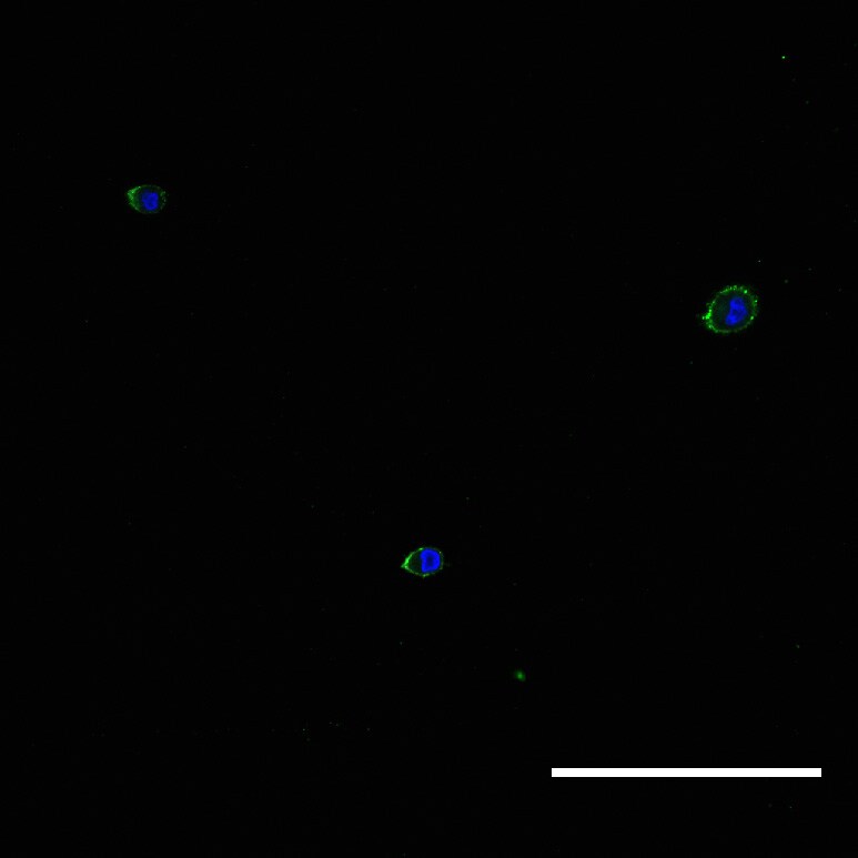
Immunocytochemistry/ Immunofluorescence: Integrin beta 1/CD29 Antibody (12G10) - BSA Free [NB100-63255]
Immunocytochemistry/Immunofluorescence: Integrin beta 1/CD29 Antibody (12G10) [NB100-63255] - Human salivary gland progenitor cells express integrin beta 1 by day 1 in three-dimensional hyaluronan-based hydrogels. Image from verified customer review.![Flow Cytometry: Integrin beta 1/CD29 Antibody (12G10) - BSA Free [NB100-63255]](https://resources.rndsystems.com/images/products/Integrin-beta-1-CD29-Antibody-12G10-Flow-Cytometry-NB100-63255-img0002.jpg "Flow Cytometry: Integrin beta 1/CD29 Antibody (12G10) - BSA Free [NB100-63255]")
Flow Cytometry: Integrin beta 1/CD29 Antibody (12G10) - BSA Free [NB100-63255]
Flow Cytometry: Integrin beta 1/CD29 Antibody (12G10) [NB100-63255] - Staining of human peripheral blood monocytes with MOUSE ANTI HUMAN CD29. - BSA Free [NB100-63255] -")
Immunocytochemistry/ Immunofluorescence: Integrin beta 1/CD29 Antibody (12G10) - BSA Free [NB100-63255] -
Quantification of knockdown of beta 1 integrin in hS/PC microstructures. hS/PC microstructures formed in HA-based hydrogels and were transfected with scrambled siRNA or ITGB1 siRNA. Immunocytochemistry was used to determine beta 1 integrin expression and spatial distribution of beta 1 integrin 46 h post-transfection. Image analysis software was used to quantify beta 1 integrin expression at microstructure surfaces from confocal micrographs and coverage indices were calculated for scrambled siRNA (A) and ITGB1 siRNA (B) groups where a 63% reduction in beta 1 integrin expression at microstructure surfaces was quantified (C). Image collected and cropped by CiteAb from the following open publication (https://pubmed.ncbi.nlm.nih.gov/31750298), licensed under a CC-BY license. Not internally tested by Novus Biologicals.Applications for Integrin beta 1/CD29 Antibody (12G10) - BSA Free
ELISA
Flow Cytometry
Immunohistochemistry
Immunohistochemistry-Frozen
Reviewed Applications
Read 1 review rated 5 using NB100-63255 in the following applications:
Flow Cytometry Panel Builder
Bio-Techne Knows Flow Cytometry
Save time and reduce costly mistakes by quickly finding compatible reagents using the Panel Builder Tool.
Advanced Features
- Spectra Viewer - Custom analysis of spectra from multiple fluorochromes
- Spillover Popups - Visualize the spectra of individual fluorochromes
- Antigen Density Selector - Match fluorochrome brightness with antigen density

Formulation, Preparation, and Storage
Purification
Formulation
Format
Preservative
Concentration
Shipping
Stability & Storage
Background: Integrin beta 1/CD29
Additional Integrin beta 1/CD29 Products
Product Documents for Integrin beta 1/CD29 Antibody (12G10) - BSA Free
Certificate of Analysis
To download a Certificate of Analysis, please enter a lot or batch number in the search box below.
Product Specific Notices for Integrin beta 1/CD29 Antibody (12G10) - BSA Free
This product is for research use only and is not approved for use in humans or in clinical diagnosis. Primary Antibodies are guaranteed for 1 year from date of receipt.
Citations for Integrin beta 1/CD29 Antibody (12G10) - BSA Free
 Powered by Bioz
Customer Reviews for Integrin beta 1/CD29 Antibody (12G10) - BSA Free (1)
Have you used Integrin beta 1/CD29 Antibody (12G10) - BSA Free?
Submit a review and receive an Amazon gift card!
$25/€18/£15/$25CAN/¥2500 Yen for a review with an image
$10/€7/£6/$10CAN/¥1110 Yen for a review without an image
Submit a review
Customer Images
-
Application: ImmunocytochemistrySample Tested: Human salivary gland primary progenitor cellsSpecies: HumanVerified Customer | Posted 11/29/2018Human salivary gland progenitor cells express integrin beta 1 by day 1 in three-dimensional hyaluronan-based hydrogels.Used dilution of 1:100

There are no reviews that match your criteria.
Protocols
Find general support by application which include: protocols, troubleshooting, illustrated assays, videos and webinars.
- 7-Amino Actinomycin D (7-AAD) Cell Viability Flow Cytometry Protocol
- Antigen Retrieval Protocol (PIER)
- Antigen Retrieval for Frozen Sections Protocol
- Appropriate Fixation of IHC/ICC Samples
- Cellular Response to Hypoxia Protocols
- Chromogenic IHC Staining of Formalin-Fixed Paraffin-Embedded (FFPE) Tissue Protocol
- Chromogenic Immunohistochemistry Staining of Frozen Tissue
- ClariTSA™ Fluorophore Kits
- Detection & Visualization of Antibody Binding
- ELISA Sample Preparation & Collection Guide
- ELISA Troubleshooting Guide
- Extracellular Membrane Flow Cytometry Protocol
- Flow Cytometry Protocol for Cell Surface Markers
- Flow Cytometry Protocol for Staining Membrane Associated Proteins
- Flow Cytometry Staining Protocols
- Flow Cytometry Troubleshooting Guide
- Fluorescent IHC Staining of Frozen Tissue Protocol
- Graphic Protocol for Heat-induced Epitope Retrieval
- Graphic Protocol for the Preparation and Fluorescent IHC Staining of Frozen Tissue Sections
- Graphic Protocol for the Preparation and Fluorescent IHC Staining of Paraffin-embedded Tissue Sections
- Graphic Protocol for the Preparation of Gelatin-coated Slides for Histological Tissue Sections
- How to Run an R&D Systems DuoSet ELISA
- How to Run an R&D Systems Quantikine ELISA
- How to Run an R&D Systems Quantikine™ QuicKit™ ELISA
- IHC Sample Preparation (Frozen sections vs Paraffin)
- Immunofluorescent IHC Staining of Formalin-Fixed Paraffin-Embedded (FFPE) Tissue Protocol
- Immunohistochemistry (IHC) and Immunocytochemistry (ICC) Protocols
- Immunohistochemistry Frozen Troubleshooting
- Immunohistochemistry Paraffin Troubleshooting
- Intracellular Flow Cytometry Protocol Using Alcohol (Methanol)
- Intracellular Flow Cytometry Protocol Using Detergents
- Intracellular Nuclear Staining Flow Cytometry Protocol Using Detergents
- Intracellular Staining Flow Cytometry Protocol Using Alcohol Permeabilization
- Intracellular Staining Flow Cytometry Protocol Using Detergents to Permeabilize Cells
- Preparing Samples for IHC/ICC Experiments
- Preventing Non-Specific Staining (Non-Specific Binding)
- Primary Antibody Selection & Optimization
- Propidium Iodide Cell Viability Flow Cytometry Protocol
- Protocol for Heat-Induced Epitope Retrieval (HIER)
- Protocol for Liperfluo
- Protocol for Making a 4% Formaldehyde Solution in PBS
- Protocol for VisUCyte™ HRP Polymer Detection Reagent
- Protocol for the Characterization of Human Th22 Cells
- Protocol for the Characterization of Human Th9 Cells
- Protocol for the Preparation & Fixation of Cells on Coverslips
- Protocol for the Preparation and Chromogenic IHC Staining of Frozen Tissue Sections
- Protocol for the Preparation and Chromogenic IHC Staining of Frozen Tissue Sections - Graphic
- Protocol for the Preparation and Chromogenic IHC Staining of Paraffin-embedded Tissue Sections
- Protocol for the Preparation and Chromogenic IHC Staining of Paraffin-embedded Tissue Sections - Graphic
- Protocol for the Preparation and Fluorescent IHC Staining of Frozen Tissue Sections
- Protocol for the Preparation and Fluorescent IHC Staining of Paraffin-embedded Tissue Sections
- Protocol for the Preparation of Gelatin-coated Slides for Histological Tissue Sections
- Protocol: Annexin V and PI Staining by Flow Cytometry
- Protocol: Annexin V and PI Staining for Apoptosis by Flow Cytometry
- Quantikine HS ELISA Kit Assay Principle, Alkaline Phosphatase
- Quantikine HS ELISA Kit Principle, Streptavidin-HRP Polymer
- Sandwich ELISA (Colorimetric) – Biotin/Streptavidin Detection Protocol
- Sandwich ELISA (Colorimetric) – Direct Detection Protocol
- TUNEL and Active Caspase-3 Detection by IHC/ICC Protocol
- The Importance of IHC/ICC Controls
- Troubleshooting Guide: ELISA
- Troubleshooting Guide: Fluorokine Flow Cytometry Kits
- Troubleshooting Guide: Immunohistochemistry
- View all Protocols, Troubleshooting, Illustrated assays and Webinars
FAQs for Integrin beta 1/CD29 Antibody (12G10) - BSA Free
-
Q: I'm looking for a good antibody for integrins that works in rats. I would need it for Western Blot, Immunocyto- as well as Immunohistochemistry. I'm especially interested in beta1.
A: I would recommend NB110-57123 as it has been validated using rat samples in WB, IHC-P, and FACS. Although we do not list ICC on the datasheet for this antibody I believe it will work in ICC because it works in Flow Cytometry and has been validated to stain tissue. We have several other Integrin targets. To search for what you are looking for I recommend typing Integrin into the search area at the top of our website, then review the list of suggested targets that poplulate below the search. Once you click on a particular Integrin you can narrow the results with our filter. I recommend filtering to primary antibodies, by target name, species of interest you can filter to rat. If you so choose you can also filter further by application, host, clonality, etc.
Associated Pathways


