KLF4 Antibody - BSA Free
Novus Biologicals | Catalog # NBP1-83940
![Immunohistochemistry-Paraffin: KLF4 Antibody [NBP1-83940]](https://resources.rndsystems.com/images/products/KLF4-Antibody-Immunohistochemistry-Paraffin-NBP1-83940-img0021.jpg "Immunohistochemistry-Paraffin: KLF4 Antibody [NBP1-83940]")
Loading...
Key Product Details
Validated by
Orthogonal Validation, Biological Validation
Species Reactivity
Validated:
Human
Cited:
Human
Applications
Validated:
Immunohistochemistry, Immunohistochemistry-Paraffin, Western Blot, Immunocytochemistry/ Immunofluorescence, Chromatin Immunoprecipitation-exo-Seq
Cited:
Immunohistochemistry-Paraffin, Western Blot, Immunocytochemistry/ Immunofluorescence, Immunoprecipitation, Chemotaxis, IF/IHC, Knockdown Validated
Label
Unconjugated
Antibody Source
Polyclonal Rabbit IgG
Format
BSA Free
Loading...
Product Specifications
Immunogen
This antibody was developed against Recombinant Protein corresponding to amino acids: ETEEFNDLLDLDFILSNSLTHPPESVAATVSSSASASSSSSPSSSGPASAPSTCSFTYPIRAGNDPGVAPGGTGGGLLYGRESAPPPTAPFNLADINDVSP
Reactivity Notes
Immunogen displays the following percentage of sequence identity for non-tested species: Mouse (89%), Rat (89%).
Clonality
Polyclonal
Host
Rabbit
Isotype
IgG
Theoretical MW
56 kDa.
Disclaimer note: The observed molecular weight of the protein may vary from the listed predicted molecular weight due to post translational modifications, post translation cleavages, relative charges, and other experimental factors.
Disclaimer note: The observed molecular weight of the protein may vary from the listed predicted molecular weight due to post translational modifications, post translation cleavages, relative charges, and other experimental factors.
Scientific Data Images for KLF4 Antibody - BSA Free
![Western Blot: KLF4 Antibody [NBP1-83940]](https://resources.rndsystems.com/images/products/KLF4-Antibody-Western-Blot-NBP1-83940-img0025.jpg "Western Blot: KLF4 Antibody [NBP1-83940]")
Western Blot: KLF4 Antibody [NBP1-83940]
KLF4-Antibody-Western-Blot-NBP1-83940-img0025.jpg![Western Blot: KLF4 Antibody [NBP1-83940]](https://resources.rndsystems.com/images/products/KLF4-Antibody-Western-Blot-NBP1-83940-img0022.jpg "Western Blot: KLF4 Antibody [NBP1-83940]")
Western Blot: KLF4 Antibody [NBP1-83940]
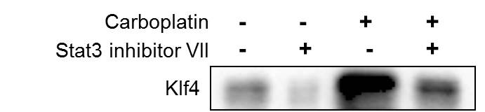
Western Blot: KLF4 Antibody [NBP1-83940] - Human breast cancer cell MCF-7 was treated with carboplatin, alone or in combination with Stat3 inhibitor, for 72 hours, and the expression of Klf4 was detected by Western blot. WB image submitted by a verified customer review.![Western Blot: KLF4 Antibody [NBP1-83940]](https://resources.rndsystems.com/images/products/KLF4-Antibody-Western-Blot-NBP1-83940-img0023.jpg "Western Blot: KLF4 Antibody [NBP1-83940]")
Western Blot: KLF4 Antibody [NBP1-83940]
Western Blot: KLF4 Antibody [NBP1-83940] - Analysis in control (vector only transfected HEK293T lysate) and KLF4 over-expression lysate (Co-expressed with a C-terminal myc-DDK tag (3.1 kDa) in mammalian HEK293T cells).![Western Blot: KLF4 Antibody [NBP1-83940]](https://resources.rndsystems.com/images/products/KLF4-Antibody-Western-Blot-NBP1-83940-img0024.jpg "Western Blot: KLF4 Antibody [NBP1-83940]")
![Immunohistochemistry-Paraffin: KLF4 Antibody [NBP1-83940]](https://resources.rndsystems.com/images/products/KLF4-Antibody-Immunohistochemistry-Paraffin-NBP1-83940-img0017.jpg "Immunohistochemistry-Paraffin: KLF4 Antibody [NBP1-83940]")
Immunohistochemistry-Paraffin: KLF4 Antibody [NBP1-83940]
Immunohistochemistry-Paraffin: KLF4 Antibody [NBP1-83940] - Staining of human colon shows moderate to strong nuclear positivity in glandular cells.![Immunohistochemistry-Paraffin: KLF4 Antibody [NBP1-83940]](https://resources.rndsystems.com/images/products/KLF4-Antibody-Immunohistochemistry-Paraffin-NBP1-83940-img0018.jpg "Immunohistochemistry-Paraffin: KLF4 Antibody [NBP1-83940]")
Immunohistochemistry-Paraffin: KLF4 Antibody [NBP1-83940]
Immunohistochemistry-Paraffin: KLF4 Antibody [NBP1-83940] - Staining of human pancreas shows no positivity in exocrine glandular cells as expected.![Immunohistochemistry-Paraffin: KLF4 Antibody [NBP1-83940]](https://resources.rndsystems.com/images/products/KLF4-Antibody-Immunohistochemistry-Paraffin-NBP1-83940-img0019.jpg "Immunohistochemistry-Paraffin: KLF4 Antibody [NBP1-83940]")
Immunohistochemistry-Paraffin: KLF4 Antibody [NBP1-83940]
Immunohistochemistry-Paraffin: KLF4 Antibody [NBP1-83940] - Staining of human skin shows moderate to strong nuclear positivity in keratinocytes.![Immunohistochemistry-Paraffin: KLF4 Antibody [NBP1-83940]](https://resources.rndsystems.com/images/products/KLF4-Antibody-Immunohistochemistry-Paraffin-NBP1-83940-img0020.jpg "Immunohistochemistry-Paraffin: KLF4 Antibody [NBP1-83940]")
Immunohistochemistry-Paraffin: KLF4 Antibody [NBP1-83940]
Immunohistochemistry-Paraffin: KLF4 Antibody [NBP1-83940] - Staining of human testis shows moderate to strong nuclear positivity in a subset of cells in seminiferous ducts.
Western Blot: KLF4 Antibody [NBP1-83940] -
Western Blot: KLF4 Antibody [NBP1-83940] - HIFs are required for hypoxia-induced expression of pluripotency factorsA-C. Breast cancer cell lines were exposed to 20% or 1% O2 for 24 h & NANOG (A), KLF4 (B), & SOX2 (C) mRNA levels were determined by RT-qPCR, relative to 18S rRNA, & normalized to the mean value for MDA-MB-231 cells (MDA231) at 20% O2 (mean ± SEM; n = 3). *P < 0.05, **P < 0.01, ***P < 0.001 vs. same cell line at 20% O2 by Student's t test. D & E. HCC-1954 (D) & MCF-7 (E) subclones, which were stably transfected with an expression vector encoding a non-targeting control (NTC) shRNA, or vector encoding shRNA targeting HIF-1 alpha (sh1 alpha ) or HIF-2 alpha (sh2 alpha ), or vectors encoding shRNAs targeting both HIF-1 alpha & HIF-2 alpha (DKD), were exposed to 20% or 1% O2 for 24 h & RT-qPCR was performed to determine NANOG (D) or KLF4 (E) mRNA levels relative to 18S rRNA. The results were normalized to NTC at 20% O2 (mean ± SEM; n = 3). *P < 0.05, **P < 0.01, ***P < 0.001 vs. NTC at 20% O2; #P < 0.05, ##P < 0.01, ###P < 0.001 vs. NTC at 1% O2 by ANOVA. F. ZR75.1 cells treated with vehicle or digoxin (200 nM) were exposed to 20% or 1% O2 for 24 h & SOX2 mRNA was measured (mean ± SEM; n = 3). *P < 0.05, **P < 0.01 vs. NTC at 20% O2; ###P < 0.001 vs. NTC at 1% O2 by ANOVA. G & H. NTC & DKD subclones of HCC-1954 (G) & MCF-7 (H) were exposed to 20% or 1% O2 for 48 h, whole cell lysates were prepared, & immunoblot assays were performed to analyze HIF-1 alpha, HIF-2 alpha, NANOG & KLF4 protein expression. Actin was also analyzed as a loading control. I. ZR75.1 cells were treated with vehicle or digoxin (200 nM), exposed to 20% or 1% O2 for 48 h, & HIF-1 alpha, NANOG & SOX2 immunoblot assays were performed. Image collected & cropped by CiteAb from the following publication (https://www.oncotarget.com/lookup/doi/10.18632/oncotarget.11743), licensed under a CC-BY license. Not internally tested by Novus Biologicals.![KLF4 Antibody - BSA Free Western Blot: KLF4 Antibody - BSA Free [NBP1-83940]](https://resources.rndsystems.com/images/products/nbp1-83940_rabbit-polyclonal-klf4-antibody-8420251719569.jpg "Western Blot: KLF4 Antibody - BSA Free [NBP1-83940]")
![KLF4 Antibody - BSA Free Chromatin Immunoprecipitation-exo-Seq: KLF4 Antibody - BSA Free [NBP1-83940]](https://resources.rndsystems.com/images/products/nbp1-83940_rabbit-polyclonal-klf4-antibody-25620259121918.jpg "Chromatin Immunoprecipitation-exo-Seq: KLF4 Antibody - BSA Free [NBP1-83940]")
Chromatin Immunoprecipitation-exo-Seq: KLF4 Antibody - BSA Free [NBP1-83940]
ChIP-Exo-Seq composite graph for Anti-KLF4 (NBP1-83940) tested in NCCIT cells. Strand-specific reads (blue: forward, red: reverse) and IgG controls (black: forward, grey: reverse) are plotted against the distance from a composite set of reference binding sites. The antibody exhibits robust target enrichment compared to a non-specific IgG control and precisely reveals its structural organization around the binding site. Data generated by Prof. B. F. Pugh´s Lab at Cornell University.![KLF4 Antibody - BSA Free Immunocytochemistry/ Immunofluorescence: KLF4 Antibody [NBP1-83940]](https://resources.rndsystems.com/images/products/nbp1-83940_-immunocytochemistry-immunofluorescence-639174076497912393.jpg "Immunocytochemistry/ Immunofluorescence: KLF4 Antibody [NBP1-83940]")
Immunocytochemistry/ Immunofluorescence: KLF4 Antibody [NBP1-83940]
Staining of human cell line A-431 shows localization to nucleoplasm.Applications for KLF4 Antibody - BSA Free
Application
Recommended Usage
Chromatin Immunoprecipitation-exo-Seq
1-10ug per reaction
Immunocytochemistry/ Immunofluorescence
0.25-2 ug/ml
Immunohistochemistry
1:1000 - 1:2500
Immunohistochemistry-Paraffin
1:1000 - 1:2500
Western Blot
0.04-0.4 ug/ml
Application Notes
For IHC-Paraffin, HIER pH 6 retrieval is recommended. ICC/IF, fixation/permeabilization: PFA/Triton X-100.
Reviewed Applications
Read 2 reviews rated 4 using NBP1-83940 in the following applications:
Formulation, Preparation, and Storage
Purification
Affinity purified
Formulation
PBS (pH 7.2) and 40% Glycerol
Format
BSA Free
Preservative
0.02% Sodium Azide
Concentration
Concentrations vary lot to lot. See vial label for concentration. If unlisted please contact technical services.
Shipping
The product is shipped with polar packs. Upon receipt, store it immediately at the temperature recommended below.
Stability & Storage
Store at 4C short term. Aliquot and store at -20C long term. Avoid freeze-thaw cycles.
Background: KLF4
Long Name
Kruppel-Like Factor 4
Alternate Names
EZF
Entrez Gene IDs
9314 (Human)
Gene Symbol
KLF4
UniProt
Additional KLF4 Products
Product Documents for KLF4 Antibody - BSA Free
Certificate of Analysis
To download a Certificate of Analysis, please enter a lot or batch number in the search box below.
Product Specific Notices for KLF4 Antibody - BSA Free
This product is for research use only and is not approved for use in humans or in clinical diagnosis. Primary Antibodies are guaranteed for 1 year from date of receipt.
Citations for KLF4 Antibody - BSA Free
 Powered by Bioz
Customer Reviews for KLF4 Antibody - BSA Free (2)
4 out of 5
2 Customer Ratings
Have you used KLF4 Antibody - BSA Free?
Submit a review and receive an Amazon gift card!
$25/€18/£15/$25CAN/¥2500 Yen for a review with an image
$10/€7/£6/$10CAN/¥1110 Yen for a review without an image
Submit a review
Customer Images
Showing
1
-
2 of
2 reviews
Showing All
Filter By:
-
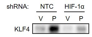
Application: Western BlotSample Tested: Breast cancer cellsSpecies: HumanVerified Customer | Posted 05/18/2020MDA-MB-231 subclones transfected with NTC or HIF-1 alpha shRNA vector were treated with vehicle (V) or paclitaxel (P) and immunoblot assay was performed.

-
Application: Western BlotSample Tested: breast cancer MCF7 cellsSpecies: HumanVerified Customer | Posted 09/21/2018Human breast cancer cell MCF-7 was treated with carboplatin, alone or in combination with Stat3 inhibitor, for 72 hours, and the expression of Klf4 was detected by western blot.
There are no reviews that match your criteria.
Protocols
Find general support by application which include: protocols, troubleshooting, illustrated assays, videos and webinars.
- Antigen Retrieval Protocol (PIER)
- Antigen Retrieval for Frozen Sections Protocol
- Appropriate Fixation of IHC/ICC Samples
- Cellular Response to Hypoxia Protocols
- Chromogenic IHC Staining of Formalin-Fixed Paraffin-Embedded (FFPE) Tissue Protocol
- Chromogenic Immunohistochemistry Staining of Frozen Tissue
- ClariTSA™ Fluorophore Kits
- Detection & Visualization of Antibody Binding
- Fluorescent IHC Staining of Frozen Tissue Protocol
- Graphic Protocol for Heat-induced Epitope Retrieval
- Graphic Protocol for the Preparation and Fluorescent IHC Staining of Frozen Tissue Sections
- Graphic Protocol for the Preparation and Fluorescent IHC Staining of Paraffin-embedded Tissue Sections
- Graphic Protocol for the Preparation of Gelatin-coated Slides for Histological Tissue Sections
- ICC Cell Smear Protocol for Suspension Cells
- ICC Immunocytochemistry Protocol Videos
- ICC for Adherent Cells
- IHC Sample Preparation (Frozen sections vs Paraffin)
- Immunocytochemistry (ICC) Protocol
- Immunocytochemistry Troubleshooting
- Immunofluorescence of Organoids Embedded in Cultrex Basement Membrane Extract
- Immunofluorescent IHC Staining of Formalin-Fixed Paraffin-Embedded (FFPE) Tissue Protocol
- Immunohistochemistry (IHC) and Immunocytochemistry (ICC) Protocols
- Immunohistochemistry Frozen Troubleshooting
- Immunohistochemistry Paraffin Troubleshooting
- Preparing Samples for IHC/ICC Experiments
- Preventing Non-Specific Staining (Non-Specific Binding)
- Primary Antibody Selection & Optimization
- Protocol for Heat-Induced Epitope Retrieval (HIER)
- Protocol for Making a 4% Formaldehyde Solution in PBS
- Protocol for VisUCyte™ HRP Polymer Detection Reagent
- Protocol for the Fluorescent ICC Staining of Cell Smears - Graphic
- Protocol for the Fluorescent ICC Staining of Cultured Cells on Coverslips - Graphic
- Protocol for the Preparation & Fixation of Cells on Coverslips
- Protocol for the Preparation and Chromogenic IHC Staining of Frozen Tissue Sections
- Protocol for the Preparation and Chromogenic IHC Staining of Frozen Tissue Sections - Graphic
- Protocol for the Preparation and Chromogenic IHC Staining of Paraffin-embedded Tissue Sections
- Protocol for the Preparation and Chromogenic IHC Staining of Paraffin-embedded Tissue Sections - Graphic
- Protocol for the Preparation and Fluorescent ICC Staining of Cells on Coverslips
- Protocol for the Preparation and Fluorescent ICC Staining of Non-adherent Cells
- Protocol for the Preparation and Fluorescent ICC Staining of Stem Cells on Coverslips
- Protocol for the Preparation and Fluorescent IHC Staining of Frozen Tissue Sections
- Protocol for the Preparation and Fluorescent IHC Staining of Paraffin-embedded Tissue Sections
- Protocol for the Preparation of Gelatin-coated Slides for Histological Tissue Sections
- Protocol for the Preparation of a Cell Smear for Non-adherent Cell ICC - Graphic
- R&D Systems Quality Control Western Blot Protocol
- TUNEL and Active Caspase-3 Detection by IHC/ICC Protocol
- The Importance of IHC/ICC Controls
- Troubleshooting Guide: Immunohistochemistry
- Troubleshooting Guide: Western Blot Figures
- Western Blot Conditions
- Western Blot Protocol
- Western Blot Protocol for Cell Lysates
- Western Blot Troubleshooting
- Western Blot Troubleshooting Guide
- View all Protocols, Troubleshooting, Illustrated assays and Webinars

