Klotho Antibody - BSA Free
Novus Biologicals | Catalog # NBP1-76511
![Western Blot: Klotho AntibodyBSA Free [NBP1-76511]](https://resources.rndsystems.com/images/products/Klotho-Antibody---BSA-Free-Western-Blot-NBP1-76511-img0010.jpg "Western Blot: Klotho AntibodyBSA Free [NBP1-76511]")
Key Product Details
Validated by
Species Reactivity
Validated:
Cited:
Applications
Validated:
Cited:
Label
Antibody Source
Format
Product Specifications
Immunogen
Specificity
Clonality
Host
Isotype
Theoretical MW
Disclaimer note: The observed molecular weight of the protein may vary from the listed predicted molecular weight due to post translational modifications, post translation cleavages, relative charges, and other experimental factors.
Scientific Data Images for Klotho Antibody - BSA Free
Western Blot: Klotho AntibodyBSA Free [NBP1-76511]
Western Blot: Klotho Antibody - BSA Free [NBP1-76511] - Western Blot Validation with Recombinant Protein. Loading: 30 ng of human KLOTHO recombinant protein per lane. Antibodies: KLOTHO NBP1-76511 (Lane 1: 1 ug/mL, Lane 2: 2 ug/mL and Lane 3: 4 ug/mL), 1h incubation at RT in 5% NFDM/TBST.Secondary: Goat anti-rabbit IgG HRP conjugate at 1:10000 dilution.
Immunohistochemistry: Klotho Antibody - BSA Free [NBP1-76511] -
Immunohistochemistry: Klotho Antibody - BSA Free [NBP1-76511] - Figure 6 Immunohistochemistry Validation of Klotho in Rat Heart TissueImmunohistochemical analysis of paraffin-embedded rat heart tissue using anti-Klotho antibody at 2.5 u/ml. Tissue was fixed with formaldehyde and blocked with 10% serum for 1 h at RT; antigen retrieval was by heat mediation with a citrate buffer (pH6). Samples were incubated with primary antibody overnight at 4;C. A goat anti-rabbit IgG H&L (HRP) at 1/250 was used as secondary. Counter stained with Hematoxylin.
![Immunohistochemistry-Paraffin: Klotho Antibody - BSA Free [NBP1-76511]](https://resources.rndsystems.com/images/products/Klotho-Antibody---BSA-Free-Immunohistochemistry-Paraffin-NBP1-76511-img0003.jpg "Immunohistochemistry-Paraffin: Klotho Antibody - BSA Free [NBP1-76511]")
Immunohistochemistry-Paraffin: Klotho Antibody - BSA Free [NBP1-76511]
Immunohistochemistry-Paraffin: Klotho Antibody - BSA Free [NBP1-76511] - Mouse heart tissue diluted at 2.5 ug/ml.
Immunocytochemistry/ Immunofluorescence: Klotho Antibody - BSA Free [NBP1-76511] -
Immunocytochemistry/ Immunofluorescence: Klotho Antibody - BSA Free [NBP1-76511] - Figure 5 Immunofluorescence Validation of Klotho in Mouse Heart TissueImmunofluorescent analysis of 4% paraformaldehyde-fixed mouse heart tissue labeling Klotho with at 20 u/mL, followed by goat anti-rabbit IgG secondary antibody at 1/500 dilution (red).![Western Blot: Klotho AntibodyBSA Free [NBP1-76511]](https://resources.rndsystems.com/images/products/Klotho-Antibody---BSA-Free-Western-Blot-NBP1-76511-img0007.jpg "Western Blot: Klotho AntibodyBSA Free [NBP1-76511]")
Western Blot: Klotho AntibodyBSA Free [NBP1-76511]
Western Blot: Klotho Antibody - BSA Free [NBP1-76511] - Validation in Rat Kidney Tissue LysateLoading: 15ug of lysates per lane.Antibodies: KLOTHO 6107 (1ug/mL), 1h incubation at RT in 5% NFDM/TBST.Secondary: Goat anti-rabbit IgG HRP conjugate at 1:10000 dilution.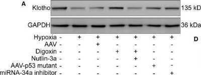
![Immunohistochemistry: Klotho Antibody - BSA Free [NBP1-76511]](https://resources.rndsystems.com/images/products/Klotho-Antibody---BSA-Free-Immunohistochemistry-NBP1-76511-img0008.jpg "Immunohistochemistry: Klotho Antibody - BSA Free [NBP1-76511]")
Immunohistochemistry: Klotho Antibody - BSA Free [NBP1-76511]
Klotho-Antibody---BSA-Free-Immunohistochemistry-NBP1-76511-img0008.jpg![Western Blot: Klotho AntibodyBSA Free [NBP1-76511]](https://resources.rndsystems.com/images/products/Klotho-Antibody---BSA-Free-Western-Blot-NBP1-76511-img0009.jpg "Western Blot: Klotho AntibodyBSA Free [NBP1-76511]")
Western Blot: Klotho AntibodyBSA Free [NBP1-76511]
Western Blot: Klotho Antibody - BSA Free [NBP1-76511] - Independent Antibody Validation (IAV) via Protein Expression Profile in Human and Mouse Cell Lines. Loading: 15 ug of lysates per lane. Antibodies: KLOTHO NBP1-76511 (2 ug/mL), KLOTHO, competitor antibody (4 ug/mL) and beta-actin (1 ug/mL), 1h incubation at RT in 5% NFDM/TBST. Secondary: Goat anti-rabbit IgG HRP conjugate at 1:10000 dilution.
Western Blot: Klotho Antibody - BSA Free [NBP1-76511] -
Western Blot: Klotho Antibody - BSA Free [NBP1-76511] - Blockade of the HIF‐1 alpha /p53/miRNA‐34a/Klotho axis decreases the leakage & area of mouse laser‐induced CNV. The mice were divided into the following groups: normal, CNV 7 d, CNV 7 d + 0.1% DMSO, CNV 7 d + digoxin (oral; 2 mg/kg for 7 d), CNV 7 d + AAV‐p53 mutant (intravitreal injection; approximately 3 μL, 3 × 10¹⁰ viral particles/mL), CNV 7 d + miRNA‐34a inhibitor (intravitreal injection; 1 μg) & CNV 7 d + AAV‐Klotho full‐length plasmid (intravitreal injection, 2 μL, 5 × 10¹⁰ viral particles/mL). A, Western blot was performed to measure HIF‐1 alpha, p‐p53 (S15), p53 (S20), p‐p53 (S46), p53 & Klotho protein levels. B, The relative protein levels of HIF‐1 alpha /GAPDH (B), p‐p53 (S15)/p53 (C), p‐p53 (S20)/p53 (D), p‐p53 (S46)/p53 (E), p53/GAPDH (F) & Klotho/GAPDH (G) were analysed. ***P < .001, CNV 7‐d group vs normal group. ##P < .01, compared with the CNV 7‐d group. NS, CNV 7 d + KL OE group vs CNV 7‐d group. H, RT‐PCR was performed to measure the expression of miRNA‐34a. **P < .01, CNV 7‐d group vs normal group. ##P < .01, compared with the CNV 7‐d group. NS, CNV 7 d + KL OE group vs CNV 7‐d group. I, FFA was performed to measure the leakage of CNV. J, The leakage of CNV was analysed. K, ICGA was performed to measure the area of CNV. L, The area of CNV was analysed. **P < .01, ***P < .001, compared with the CNV 7‐d group Image collected & cropped by CiteAb from the following publication (https://pubmed.ncbi.nlm.nih.gov/33438362), licensed under a CC-BY license. Not internally tested by Novus Biologicals.
Western Blot: Klotho Antibody - BSA Free [NBP1-76511] -
Western Blot: Klotho Antibody - BSA Free [NBP1-76511] - microrna‐34a inhibits the expression of Klotho. ARPE‐19 cells were divided into the following groups: normal, hypoxia, hypoxia + AAV vector, hypoxia + digoxin, hypoxia + digoxin + nutlin‐3a, hypoxia + AAV‐p53 mutant infection & hypoxia + miRNA‐34a inhibitor. A, Western blot assay of Klotho in ARPE‐19 cells was performed. B, The relative protein level of Klotho compared with the GAPDH level was analysed. ***P < .001, hypoxia group vs normal group. ###P < .001, ##P < .01, compared with the hypoxia group. %%P < .01, hypoxia + digoxin + nutlin‐3a group vs hypoxia + digoxin group. C, Schematic of the Klotho 3' UTR with the WT or mutated putative‐binding site of miRNA‐34a inserted into a luciferase (Luc) reporter. D, qRT‐PCR analysis of Klotho mRNA expression in HEK293T cells transfected with miRNA‐34a mimic for 24 h. **P < .01, miRNA‐34a mimic group vs control group. E, Western blot of Klotho in HEK293T cells transfected with a miRNA‐34a mimic for 24 h was performed. F, Immunoblotted Klotho was quantified & normalized to GAPDH. ***P < .001, miRNA‐34a mimic group vs control group. G Relative luciferase activity was detected in HEK293T cells co‐transfected with plasmids containing firefly luciferase & wild‐type (WT) or mutant (Mut) Klotho 3′‐UTR & miRNA‐34a mimic for 24 h. The luciferase activity values were normalized to Renilla reniformis luciferase (TK‐RL) activity. **P < .01, Luc‐Klotho 3′ UTR WT group vs vector group. Statistically non‐significant (NS), Luc‐Klotho 3′ UTR mutant group vs vector group Image collected & cropped by CiteAb from the following publication (https://pubmed.ncbi.nlm.nih.gov/33438362), licensed under a CC-BY license. Not internally tested by Novus Biologicals.
Western Blot: Klotho Antibody - BSA Free [NBP1-76511] -
Western Blot: Klotho Antibody - BSA Free [NBP1-76511] - High phytate-induced renal phosphate wasting is independent of FGF23.(A) The renal expression of downregulated genes associated with Ca2+ & phosphate homeostasis in rats fed control or phytate-supplemented diets (n = 8 per group). (B) Results of immunoblot analyses & protein levels of renal aKlotho, NHERF1, & NaPi-2a after 2 weeks for rats fed control, HP- LCa2+, & HP-HCa2+ diets. (C) The levels of renal proteins were quantified using ImageJ software (n = 6 per group). (D, E) Time course analysis of serum levels of intact FGF23 (D) & C-terminal FGF23 (E) in rats fed control, HP-LCa2+, & HP-HCa2+ diets. (F) Representative immunohistochemical staining of EDTA-decalcified femur sections for FGF23 from rats fed control, HP-LCa2+, & HP-HCa2+ diets. All data are presented as the mean ± SD of each group (n = 3–8 per group). *, p<0.05; **, p<0.01; ***, p<0.001, compared with controls. Image collected & cropped by CiteAb from the following publication (https://pubmed.ncbi.nlm.nih.gov/32271147), licensed under a CC-BY license. Not internally tested by Novus Biologicals.
Immunohistochemistry-Paraffin: Klotho Antibody - BSA Free [NBP1-76511] -
WD feeding induced changes in FGF-23 and Klotho expression are restored by DPP-4 inhibition. a FGF-23 staining; b Endothelial FGF-23; c Adventitia FGF-23; d Endothelial klotho staining; e Endothelial klotho staining f Adventitia klotho staining; Average gray intensities in the different cohorts. Values are mean +/- SE. CDC control diet control, CDL control diet linagliptin, WDC western diet control and WDL western diet linagliptin. Post-hoc comparisons within a time point; *p < 0 0.05 CDC vs WDC; †p < 0.05 WDC vs WDL; #p < 0.05 CDC vs CDL. Scale bars represent 50 mμ Image collected and cropped by CiteAb from the following open publication (https://pubmed.ncbi.nlm.nih.gov/27391040), licensed under a CC-BY license. Not internally tested by Novus Biologicals.Applications for Klotho Antibody - BSA Free
ELISA
Immunocytochemistry/ Immunofluorescence
Immunohistochemistry
Immunohistochemistry-Paraffin
Western Blot
Formulation, Preparation, and Storage
Purification
Formulation
Format
Preservative
Concentration
Shipping
Stability & Storage
Background: Klotho
Additional Klotho Products
Product Documents for Klotho Antibody - BSA Free
Certificate of Analysis
To download a Certificate of Analysis, please enter a lot or batch number in the search box below.
Product Specific Notices for Klotho Antibody - BSA Free
This product is for research use only and is not approved for use in humans or in clinical diagnosis. Primary Antibodies are guaranteed for 1 year from date of receipt.
Related Research Areas
Citations for Klotho Antibody - BSA Free
 Powered by Bioz
Customer Reviews for Klotho Antibody - BSA Free
There are currently no reviews for this product. Be the first to review Klotho Antibody - BSA Free and earn rewards!
Have you used Klotho Antibody - BSA Free?
Submit a review and receive an Amazon gift card!
$25/€18/£15/$25CAN/¥2500 Yen for a review with an image
$10/€7/£6/$10CAN/¥1110 Yen for a review without an image
Submit a review
Protocols
Find general support by application which include: protocols, troubleshooting, illustrated assays, videos and webinars.
- Antigen Retrieval Protocol (PIER)
- Antigen Retrieval for Frozen Sections Protocol
- Appropriate Fixation of IHC/ICC Samples
- Cellular Response to Hypoxia Protocols
- Chromogenic IHC Staining of Formalin-Fixed Paraffin-Embedded (FFPE) Tissue Protocol
- Chromogenic Immunohistochemistry Staining of Frozen Tissue
- ClariTSA™ Fluorophore Kits
- Detection & Visualization of Antibody Binding
- ELISA Sample Preparation & Collection Guide
- ELISA Troubleshooting Guide
- Fluorescent IHC Staining of Frozen Tissue Protocol
- Graphic Protocol for Heat-induced Epitope Retrieval
- Graphic Protocol for the Preparation and Fluorescent IHC Staining of Frozen Tissue Sections
- Graphic Protocol for the Preparation and Fluorescent IHC Staining of Paraffin-embedded Tissue Sections
- Graphic Protocol for the Preparation of Gelatin-coated Slides for Histological Tissue Sections
- How to Run an R&D Systems DuoSet ELISA
- How to Run an R&D Systems Quantikine ELISA
- How to Run an R&D Systems Quantikine™ QuicKit™ ELISA
- ICC Cell Smear Protocol for Suspension Cells
- ICC Immunocytochemistry Protocol Videos
- ICC for Adherent Cells
- IHC Sample Preparation (Frozen sections vs Paraffin)
- Immunocytochemistry (ICC) Protocol
- Immunocytochemistry Troubleshooting
- Immunofluorescence of Organoids Embedded in Cultrex Basement Membrane Extract
- Immunofluorescent IHC Staining of Formalin-Fixed Paraffin-Embedded (FFPE) Tissue Protocol
- Immunohistochemistry (IHC) and Immunocytochemistry (ICC) Protocols
- Immunohistochemistry Frozen Troubleshooting
- Immunohistochemistry Paraffin Troubleshooting
- Preparing Samples for IHC/ICC Experiments
- Preventing Non-Specific Staining (Non-Specific Binding)
- Primary Antibody Selection & Optimization
- Protocol for Heat-Induced Epitope Retrieval (HIER)
- Protocol for Making a 4% Formaldehyde Solution in PBS
- Protocol for VisUCyte™ HRP Polymer Detection Reagent
- Protocol for the Fluorescent ICC Staining of Cell Smears - Graphic
- Protocol for the Fluorescent ICC Staining of Cultured Cells on Coverslips - Graphic
- Protocol for the Preparation & Fixation of Cells on Coverslips
- Protocol for the Preparation and Chromogenic IHC Staining of Frozen Tissue Sections
- Protocol for the Preparation and Chromogenic IHC Staining of Frozen Tissue Sections - Graphic
- Protocol for the Preparation and Chromogenic IHC Staining of Paraffin-embedded Tissue Sections
- Protocol for the Preparation and Chromogenic IHC Staining of Paraffin-embedded Tissue Sections - Graphic
- Protocol for the Preparation and Fluorescent ICC Staining of Cells on Coverslips
- Protocol for the Preparation and Fluorescent ICC Staining of Non-adherent Cells
- Protocol for the Preparation and Fluorescent ICC Staining of Stem Cells on Coverslips
- Protocol for the Preparation and Fluorescent IHC Staining of Frozen Tissue Sections
- Protocol for the Preparation and Fluorescent IHC Staining of Paraffin-embedded Tissue Sections
- Protocol for the Preparation of Gelatin-coated Slides for Histological Tissue Sections
- Protocol for the Preparation of a Cell Smear for Non-adherent Cell ICC - Graphic
- Quantikine HS ELISA Kit Assay Principle, Alkaline Phosphatase
- Quantikine HS ELISA Kit Principle, Streptavidin-HRP Polymer
- R&D Systems Quality Control Western Blot Protocol
- Sandwich ELISA (Colorimetric) – Biotin/Streptavidin Detection Protocol
- Sandwich ELISA (Colorimetric) – Direct Detection Protocol
- TUNEL and Active Caspase-3 Detection by IHC/ICC Protocol
- The Importance of IHC/ICC Controls
- Troubleshooting Guide: ELISA
- Troubleshooting Guide: Immunohistochemistry
- Troubleshooting Guide: Western Blot Figures
- Western Blot Conditions
- Western Blot Protocol
- Western Blot Protocol for Cell Lysates
- Western Blot Troubleshooting
- Western Blot Troubleshooting Guide
- View all Protocols, Troubleshooting, Illustrated assays and Webinars
FAQs for Klotho Antibody - BSA Free
-
Q: I study physiology (PhD) and I need a klotho kit.
A:
A list of our klotho products can be found here. I am not entirely sure what you are looking for as I don't have your complete message for some reason. Can you please clarify what you are looking for?
-
Q: This data sheet contains figures including a western blot and fluorescence images of rat or mouse heart tissue. Can you give me the information about the source of these figures?
A: The images supplied on the datasheet for NBP1-76511 were generated from our lab's testing of this antibody.
-
Q: I study physiology (PhD) and I need a klotho kit.
A:
A list of our klotho products can be found here. I am not entirely sure what you are looking for as I don't have your complete message for some reason. Can you please clarify what you are looking for?
-
Q: This data sheet contains figures including a western blot and fluorescence images of rat or mouse heart tissue. Can you give me the information about the source of these figures?
A: The images supplied on the datasheet for NBP1-76511 were generated from our lab's testing of this antibody.