![Knockout Validated: LC3 Antibody Pack [NB910-40435]](https://resources.rndsystems.com/images/products/LC3-Antibody-Pack-Knockout-Validated-NB910-40435-img0013.jpg "Western Blot: LC3 Antibody Pack [NB910-40435]")
Loading...
Key Product Details
Species
Human, Mouse, Rat, Porcine, Alligator, Amphibian, Avian, Bacteria, Bovine, Canine, Chicken, Chinese Hamster, Fish, Golden Syrian Hamster, Guinea Pig, Hamster, Invertebrate, Monkey, Plant, Primate, Rabbit, Zebrafish
Applications
Chromatin Immunoprecipitation, ELISA, Flow Cytometry, Functional Assay, Immunocytochemistry/ Immunofluorescence, Immunohistochemistry, Immunoprecipitation, Knockout Validated, Mycoplasma, SDS-PAGE, Simple Western, Western Blot
Product Summary for LC3 Antibody Pack
This pack contains 1 vial each of: NB100-2220 (0.1 mL) and NB100-2331 (0.1 mL).
Loading...
Product Specifications
Application Notes
Antibodies in this pack are validated for the following applications:
NB100-2331: Chromatin Immunoprecipitation, ELISA, Flow Cytometry, Immunoblotting, Immunocytochemistry/Immunofluorescence, Immunohistochemistry, Immunohistochemistry Whole-Mount, Immunohistochemistry-Frozen, Immunohistochemistry-Paraffin, Immunoprecipitation, Simple Western, Southern Blot, Western Blot
NB100-2220: Chromatin Immunoprecipitation (ChIP), ELISA, Flow Cytometry, Immunoblotting, Immunocytochemistry/Immunofluorescence, Immunohistochemistry, Immunohistochemistry-Frozen, Immunohistochemistry-Paraffin, Immunoprecipitation, Knockdown Validated, Knockout Validated, Proximity Ligation Assay, SDS-Page, Simple Western, Western Blot
Reactivity Notes
Antibodies in this pack are validated for use in the following species:
NB100-2331: Amphibian, Canine, Fish, Human, Mouse, Plant, Rat, Zebrafish
NB100-2220: Alligator, Avian, Bacteria, Bovine, Canine, Chicken, Chinese Hamster, Golden Syrian Hamster, Guinea Pig, Hamster, Human, Invertebrate, Monkey, Mouse, Porcine, Primate, Rabbit, Rat, Zebrafish
Fish reactivity reported in scientific literature (PMID:32818499).
Marker
Autophagosome Marker
Scientific Data Images for LC3 Antibody Pack
Western Blot: LC3 Antibody Pack [NB910-40435]
Western Blot: LC3 Antibody Pack [NB910-40435] - Lysates of HeLa parental cell line and LC3B knockout HeLa cell line (KO) untreated (-) or treated (+) with 50 uM Chloroquine for 18 hours. PVDF (Polyvinylidene difluoride) membrane was probed with 0.5 ug/mL of Rabbit Anti-LC3B Polyclonal Antibody (Catalog # NB100-2220) followed by HRP-conjugated Anti-Rabbit IgG Secondary Antibody (Catalog# HAF008). A specific band was detected for LC3B at a molecular weight of approximately 15 kDa (as indicated) in the parental HeLa cell line, but is not detectable in the knockout HeLa cell line. GAPDH is shown as a loading control. This experiment was conducted under reducing conditions. LC3B Antibody [NB100-2220]![Immunohistochemistry: LC3 Antibody Pack [NB910-40435]](https://resources.rndsystems.com/images/products/LC3-Antibody-Pack-Immunohistochemistry-NB910-40435-img0009.jpg "Immunohistochemistry: LC3 Antibody Pack [NB910-40435]")
Immunohistochemistry: LC3 Antibody Pack [NB910-40435]
Immunohistochemistry: LC3 Antibody Pack [NB910-40435] - Rapamycin increases autophagy in brains of PDAPP mice. Representative epifluorescent (c200x) image of hippocampal CA1 in control- and rapamycin-fed transgenic PDAPP mice stained with an anti-LC3 antibody. An increase in LC3-immunoreactive puncta was observed in CA1 projections of transgenic PDAPP mice following rapamycin administration. Image collected and cropped by CiteAb from the following publication (//dx.plos.org/10.1371/journal.pone.0009979) licensed under a CC-BY license. LC3A Antibody [NB100-2331]![Knockout Validated: LC3 Antibody Pack [NB910-40435]](https://resources.rndsystems.com/images/products/LC3-Antibody-Pack-Knockout-Validated-NB910-40435-img0014.jpg "Immunocytochemistry/ Immunofluorescence: LC3 Antibody Pack [NB910-40435]")
Immunocytochemistry/ Immunofluorescence: LC3 Antibody Pack [NB910-40435]
Immunocytochemistry/ Immunofluorescence: LC3 Antibody Pack [NB910-40435] - LC3B was detected in immersion fixed Cloroquine treated HeLa cells (left) but was not detected in LC3B knockout HeLa cells (right) using rabbit anti-human LC3B polyclonal antibody (Catalog #NB100-2220) at 0.3 ug/mL for 3 hours at room temperature. Cells were stained using the NorthernLights(TM) 557-conjugated anti-Rabbit IgG Secondary Antibody (red; Catalog # NL004) and counterstained with DAPI (blue). Specific staining was localized to cytoplasm.![Western Blot: LC3 Antibody Pack [NB910-40435]](https://resources.rndsystems.com/images/products/LC3-Antibody-Pack-Western-Blot-NB910-40435-img0011.jpg "Western Blot: LC3 Antibody Pack [NB910-40435]")
Western Blot: LC3 Antibody Pack [NB910-40435]
Western Blot: LC3 Antibody Pack [NB910-40435] - High autophagosome concentration is consumed during early immortalized human mesenchymal stem cell differentiation. Immortalized human mesenchymal stem cells were differentiated under osteogenic conditions (see Materials and methods) and assayed for changes in LC3I and LC3II during a 72-hour window. Cells were differentiated under standard conditions (top) or with addition of 5 uM rapamycin (middle) or 5 nM bafilomycin (bottom) for the first 3 hours of differentiation to modulate autophagy. Immunoblots were performed for LC3 at the indicated time points to assess autophagosome degradation via relative changes in LC3II (lower band; 17 kDa). Studies were repeated three times with similar trends seen consistently. Image collected and cropped by CiteAb from the following publication (//stemcellres.com/content/5/6/140), licensed under a CC-BY license. LC3A Antibody [NB100-2331]![Immunohistochemistry: LC3 Antibody Pack [NB910-40435]](https://resources.rndsystems.com/images/products/LC3-Antibody-Pack-Immunohistochemistry-NB910-40435-img0015.jpg "Immunohistochemistry: LC3 Antibody Pack [NB910-40435]")
Immunohistochemistry: LC3 Antibody Pack [NB910-40435]
Immunohistochemistry: LC3 Antibody Pack [NB910-40435] - Localization of LC3 by immunohistochemical method. The immunohistochemical staining shows that LC3, CRF, and HIF-1alpha have an overlapping localization in villous and extravillous trophoblast. d: decidua; v: villi; DeVe: decidual vessel. Original magnification 40x. Image collected and cropped by CiteAb from the following publication (//www.hindawi.com/journals/bmri/2013/689768/) licensed under a CC-BY license. LC3B Antibody [NB100-2220]![Immunohistochemistry: LC3 Antibody Pack [NB910-40435]](https://resources.rndsystems.com/images/products/LC3-Antibody-Pack-Immunohistochemistry-NB910-40435-img0008.jpg "Immunohistochemistry: LC3 Antibody Pack [NB910-40435]")
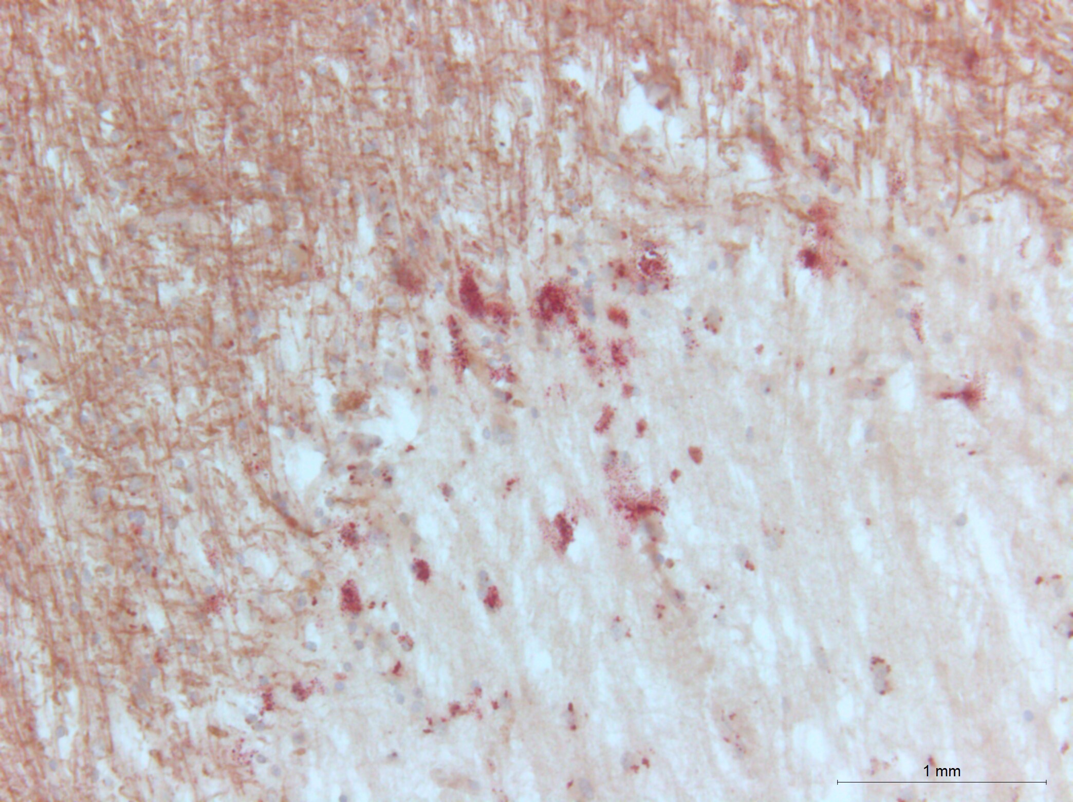
Immunohistochemistry: LC3 Antibody Pack [NB910-40435]
Immunohistochemistry: LC3 Antibody Pack [NB910-40435] - Brain, Cerebral Cortex, Cell Processes in Gray Matter 40x.![Immunohistochemistry: LC3 Antibody Pack [NB910-40435]](https://resources.rndsystems.com/images/products/LC3-Antibody-Pack-Immunohistochemistry-NB910-40435-img0012.jpg "Immunohistochemistry: LC3 Antibody Pack [NB910-40435]")
Immunohistochemistry: LC3 Antibody Pack [NB910-40435]
Immunohistochemistry: LC3 Antibody Pack [NB910-40435] - Analysis in PFA fixed NIH/3T3 cells using anti-LC3A antibody. Image from verified customer review. LC3A Antibody [NB100-2331]
Immunocytochemistry/ Immunofluorescence: LC3 Antibody Pack [NB910-40435] -
Immunocytochemistry/ Immunofluorescence: LC3 Antibody Pack [NB910-40435] - The autophagy level was increased in degenerated mouse SGNs. (a-c) Western blot results revealed that the levels of the autophagy-related proteins LC3 & BECN1 were increased in the degenerated SGNs on the 5th, 15th & 30th day after ototoxic drug administration & were significantly different from those in the normal mice. *, P < 0.05. (d) Immunofluorescence staining of LC3 puncta (red) also demonstrated that the LC3 level in the degenerated SGNs (green) was significantly increased on the 30th day after drug administration. Con, normal mice without drug treatment; 30D, 30 days after drug administration. Images of immunofluorescence staining were taken from the middle turn of cochlea. Scale bar: 10 µm. Image collected & cropped by CiteAb from the following publication (https://pubmed.ncbi.nlm.nih.gov/30706760), licensed under a CC-BY license. Not internally tested by Novus Biologicals.
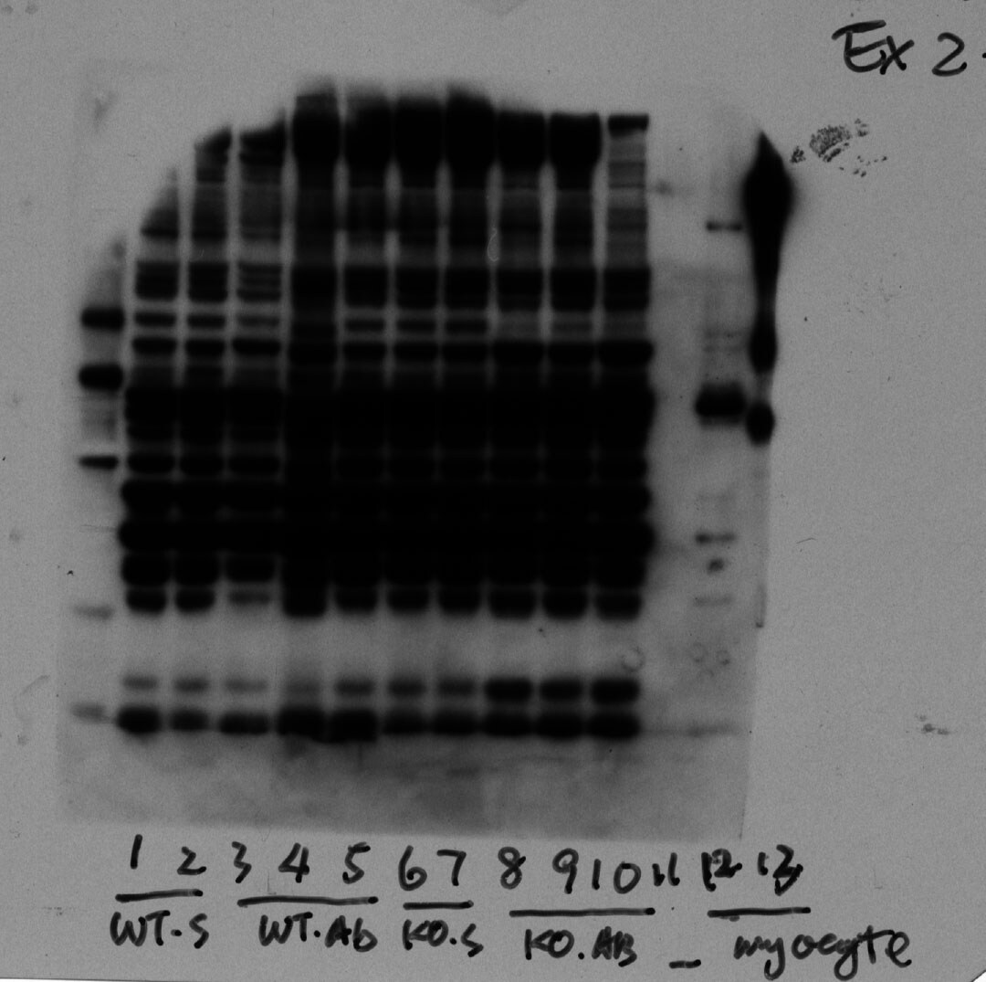
Western Blot: LC3 Antibody Pack [NB910-40435] -
Western Blot: LC3 Antibody Pack [NB910-40435] - The autophagy level was increased in degenerated mouse SGNs. (a-c) Western blot results revealed that the levels of the autophagy-related proteins LC3 & BECN1 were increased in the degenerated SGNs on the 5th, 15th & 30th day after ototoxic drug administration & were significantly different from those in the normal mice. *, P < 0.05. (d) Immunofluorescence staining of LC3 puncta (red) also demonstrated that the LC3 level in the degenerated SGNs (green) was significantly increased on the 30th day after drug administration. Con, normal mice without drug treatment; 30D, 30 days after drug administration. Images of immunofluorescence staining were taken from the middle turn of cochlea. Scale bar: 10 µm. Image collected & cropped by CiteAb from the following publication (https://pubmed.ncbi.nlm.nih.gov/30706760), licensed under a CC-BY license. Not internally tested by Novus Biologicals.
Western Blot: LC3 Antibody Pack [NB910-40435] -
Western Blot: LC3 Antibody Pack [NB910-40435] - CCI-779 significantly rescued the impaired autophagy-lysosomal pathway in degenerated SGNs of mice. (a) The levels of Ctsb, Ctsd, & Lamp1 & of the autophagic genes Becn1 & Lc3b were significantly higher, & Sqstm1 was lower in the experimental group than in the negative control group, which was determined by quantitative real-time PCR. (b & c) The LAMP1 & CTSD levels determined by western blotting were consistent with the quantitative real-time PCR results. (d & e) Compared with those in the negative control group, the LC3 & BECN1 levels in the experimental groups were significantly increased, as determined by western blot assays. (f & g) The western blot results revealed that the levels of the autophagic cargo receptor SQSTM1 & ubiquitinated proteins were decreased significantly in the experimental group compared with those in the negative control groups. *, the difference between the experimental group & the blank control group was significant (P < 0.05); #, the difference between the experimental group & the negative control group was significant (P < 0.05); CCI-779, experimental group; 30D, negative control group; Con, blank control group. Image collected & cropped by CiteAb from the following publication (https://pubmed.ncbi.nlm.nih.gov/30706760), licensed under a CC-BY license. Not internally tested by Novus Biologicals.Kit Contents for LC3 Antibody Pack
Formulation, Preparation, and Storage
Concentration
Concentration of individual antibodies may be found on the vial label. If unlisted please contact technical services.
Shipping
The product is shipped with polar packs. Upon receipt, store it immediately at the temperature recommended below.
Storage
Store at 4C short term. Aliquot and store at -20C long term. Avoid freeze-thaw cycles.
Background: LC3
Autophagic flux is supported by autophagy-related proteins (Atgs) initially identified in yeast (6,7). The core autophagy machinery is comprised of 17 Atg proteins that play specific roles in autophagosome formation. Among these Atg proteins, Atg8 is not only involved in autophagosome formation but also functions in cargo selection. In mammals, several Atg8 homologues have been identified including microtubule-associated protein 1 light chain 3 alpha, beta and gamma - LC3A, LC3B, and LC3C (8) respectively, as well as GABA type A receptor-associated protein (GABARAP), GABARAP-Like1, and GABARAP-Like2 (9). LC3 (predicted molecular weight 14kD) is ubiquitously expressed and undergoes posttranslational processing after synthesis. First, the cysteine protease Atg4 cleaves a carboxy terminal sequence to generate the cytosolic form LC3-I. Next, E1-like (Atg7) and E2-like (Atg3) enzymes conjugate phosphatidylethanolamine to the newly exposed carboxyterminal glycine, generating LC3-II. Finally, the Atg12-Atg5-Atg16L1 complex participates in LC3 lipidation and autophagosome formation (10). LC3B-I to LC3B-II conversion correlates with autophagosome number and is considered the best marker to monitor autophagy.
References
1. Yu, L., Chen, Y., & Tooze, S. A. (2018). Autophagy pathway: Cellular and molecular mechanisms. Autophagy. https://doi.org/10.1080/15548627.2017.1378838
2. Forrester, A., De Leonibus, C., Grumati, P., Fasana, E., Piemontese, M., Staiano, L.,... Settembre, C. (2019). A selective ER-phagy exerts procollagen quality control via a Calnexin-FAM 134B complex. The EMBO Journal. https://doi.org/10.15252/embj.201899847
3. He, X., Zhu, Y., Zhang, Y., Geng, Y., Gong, J., Geng, J.,... Zhong, H. (2019). RNF34 functions in immunity and selective mitophagy by targeting MAVS for autophagic degradation. The EMBO Journal. https://doi.org/10.15252/embj.2018100978
4. Mathai, B., Meijer, A., & Simonsen, A. (2017). Studying Autophagy in Zebrafish. Cells. https://doi.org/10.3390/cells6030021
5. Losier, T. T., Akuma, M., McKee-Muir, O. C., LeBlond, N. D., Suk, Y., Alsaadi, R. M.,... Russell, R. C. (2019). AMPK Promotes Xenophagy through Priming of Autophagic Kinases upon Detection of Bacterial Outer Membrane Vesicles. Cell Reports. https://doi.org/10.1016/j.celrep.2019.01.062
6. Nakatogawa, H., Suzuki, K., Kamada, Y., & Ohsumi, Y. (2009). Dynamics and diversity in autophagy mechanisms: Lessons from yeast. Nature Reviews Molecular Cell Biology. https://doi.org/10.1038/nrm2708
7. Tsukada, M., & Ohsumi, Y. (1993). Isolation and characterization of autophagy-defective mutants of Saccharomyces cerevisiae. FEBS Letters. https://doi.org/10.1016/0014-5793(93)80398-E
8. Wild, P., McEwan, D. G., & Dikic, I. (2014). The LC3 interactome at a glance. Journal of Cell Science. https://doi.org/10.1242/jcs.140426
9. Igloi, G. L. (2001). Cloning, expression patterns, and chromosome localization of three human and two mouse homologues of GABAA receptor-associated protein. Genomics. https://doi.org/10.1006/geno.2001.6555
10. Glick, D., Barth, S., & Macleod, K. F. (2010). Autophagy: Cellular and molecular mechanisms. Journal of Pathology. https://doi.org/10.1002/path.2697
Alternate Names
ATG8F, LC3B, MAP1A/1BLC3, map1lc3b, MAP1LC3B-a, microtubule associated protein 1 light chain 3 beta
Gene Symbol
MAP1LC3B
Additional LC3 Products
Product Documents for LC3 Antibody Pack
Certificate of Analysis
To download a Certificate of Analysis, please enter a lot or batch number in the search box below.
Product Specific Notices for LC3 Antibody Pack
This product is for research use only and is not approved for use in humans or in clinical diagnosis. Antibody Packs are guaranteed for 1 year from date of receipt.
Citations for LC3 Antibody Pack
 Powered by Bioz
Customer Reviews for LC3 Antibody Pack (3)
5 out of 5
3 Customer Ratings
Have you used LC3 Antibody Pack?
Submit a review and receive an Amazon gift card!
$25/€18/£15/$25CAN/¥2500 Yen for a review with an image
$10/€7/£6/$10CAN/¥1110 Yen for a review without an image
Submit a review
Customer Images
Showing
1
-
3 of
3 reviews
Showing All
Filter By:
-
Application: ImmunocytochemistrySample Tested: Human cellSpecies: MouseVerified Customer | Posted 02/12/2019
-
Application: Western BlotSample Tested:Species: MouseVerified Customer | Posted 07/11/2014LC3 expression in mouse heart 4 days after sTAC orperation

-
Application: ImmunohistochemistrySample Tested: Human BrainSpecies: MouseVerified Customer | Posted 04/29/2014LC3 positive cells in Brown and Myelin in Red
There are no reviews that match your criteria.
Protocols
Find general support by application which include: protocols, troubleshooting, illustrated assays, videos and webinars.
- 7-Amino Actinomycin D (7-AAD) Cell Viability Flow Cytometry Protocol
- Antigen Retrieval Protocol (PIER)
- Antigen Retrieval for Frozen Sections Protocol
- Appropriate Fixation of IHC/ICC Samples
- Cellular Response to Hypoxia Protocols
- ChIP Protocol Video
- Chromatin Immunoprecipitation (ChIP) Protocol
- Chromatin Immunoprecipitation Protocol
- Chromogenic IHC Staining of Formalin-Fixed Paraffin-Embedded (FFPE) Tissue Protocol
- Chromogenic Immunohistochemistry Staining of Frozen Tissue
- ClariTSA™ Fluorophore Kits
- Detection & Visualization of Antibody Binding
- ELISA Sample Preparation & Collection Guide
- ELISA Troubleshooting Guide
- Extracellular Membrane Flow Cytometry Protocol
- Flow Cytometry Protocol for Cell Surface Markers
- Flow Cytometry Protocol for Staining Membrane Associated Proteins
- Flow Cytometry Staining Protocols
- Flow Cytometry Troubleshooting Guide
- Fluorescent IHC Staining of Frozen Tissue Protocol
- Graphic Protocol for Heat-induced Epitope Retrieval
- Graphic Protocol for the Preparation and Fluorescent IHC Staining of Frozen Tissue Sections
- Graphic Protocol for the Preparation and Fluorescent IHC Staining of Paraffin-embedded Tissue Sections
- Graphic Protocol for the Preparation of Gelatin-coated Slides for Histological Tissue Sections
- How to Run an R&D Systems DuoSet ELISA
- How to Run an R&D Systems Quantikine ELISA
- How to Run an R&D Systems Quantikine™ QuicKit™ ELISA
- ICC Cell Smear Protocol for Suspension Cells
- ICC Immunocytochemistry Protocol Videos
- ICC for Adherent Cells
- IHC Sample Preparation (Frozen sections vs Paraffin)
- Immunocytochemistry (ICC) Protocol
- Immunocytochemistry Troubleshooting
- Immunofluorescence of Organoids Embedded in Cultrex Basement Membrane Extract
- Immunofluorescent IHC Staining of Formalin-Fixed Paraffin-Embedded (FFPE) Tissue Protocol
- Immunohistochemistry (IHC) and Immunocytochemistry (ICC) Protocols
- Immunohistochemistry Frozen Troubleshooting
- Immunohistochemistry Paraffin Troubleshooting
- Immunoprecipitation Protocol
- Intracellular Flow Cytometry Protocol Using Alcohol (Methanol)
- Intracellular Flow Cytometry Protocol Using Detergents
- Intracellular Nuclear Staining Flow Cytometry Protocol Using Detergents
- Intracellular Staining Flow Cytometry Protocol Using Alcohol Permeabilization
- Intracellular Staining Flow Cytometry Protocol Using Detergents to Permeabilize Cells
- Preparing Samples for IHC/ICC Experiments
- Preventing Non-Specific Staining (Non-Specific Binding)
- Primary Antibody Selection & Optimization
- Propidium Iodide Cell Viability Flow Cytometry Protocol
- Protocol for Heat-Induced Epitope Retrieval (HIER)
- Protocol for Liperfluo
- Protocol for Making a 4% Formaldehyde Solution in PBS
- Protocol for VisUCyte™ HRP Polymer Detection Reagent
- Protocol for the Characterization of Human Th22 Cells
- Protocol for the Characterization of Human Th9 Cells
- Protocol for the Fluorescent ICC Staining of Cell Smears - Graphic
- Protocol for the Fluorescent ICC Staining of Cultured Cells on Coverslips - Graphic
- Protocol for the Preparation & Fixation of Cells on Coverslips
- Protocol for the Preparation and Chromogenic IHC Staining of Frozen Tissue Sections
- Protocol for the Preparation and Chromogenic IHC Staining of Frozen Tissue Sections - Graphic
- Protocol for the Preparation and Chromogenic IHC Staining of Paraffin-embedded Tissue Sections
- Protocol for the Preparation and Chromogenic IHC Staining of Paraffin-embedded Tissue Sections - Graphic
- Protocol for the Preparation and Fluorescent ICC Staining of Cells on Coverslips
- Protocol for the Preparation and Fluorescent ICC Staining of Non-adherent Cells
- Protocol for the Preparation and Fluorescent ICC Staining of Stem Cells on Coverslips
- Protocol for the Preparation and Fluorescent IHC Staining of Frozen Tissue Sections
- Protocol for the Preparation and Fluorescent IHC Staining of Paraffin-embedded Tissue Sections
- Protocol for the Preparation of Gelatin-coated Slides for Histological Tissue Sections
- Protocol for the Preparation of a Cell Smear for Non-adherent Cell ICC - Graphic
- Protocol: Annexin V and PI Staining by Flow Cytometry
- Protocol: Annexin V and PI Staining for Apoptosis by Flow Cytometry
- Quantikine HS ELISA Kit Assay Principle, Alkaline Phosphatase
- Quantikine HS ELISA Kit Principle, Streptavidin-HRP Polymer
- R&D Systems Quality Control Western Blot Protocol
- Sandwich ELISA (Colorimetric) – Biotin/Streptavidin Detection Protocol
- Sandwich ELISA (Colorimetric) – Direct Detection Protocol
- TUNEL and Active Caspase-3 Detection by IHC/ICC Protocol
- The Importance of IHC/ICC Controls
- Troubleshooting Guide: ELISA
- Troubleshooting Guide: Fluorokine Flow Cytometry Kits
- Troubleshooting Guide: Immunohistochemistry
- Troubleshooting Guide: Western Blot Figures
- Western Blot Conditions
- Western Blot Protocol
- Western Blot Protocol for Cell Lysates
- Western Blot Troubleshooting
- Western Blot Troubleshooting Guide
- View all Protocols, Troubleshooting, Illustrated assays and Webinars
FAQs for LC3 Antibody Pack
Showing
1
-
3 of
3 FAQs
Showing All
-
Q: Can you recommend a positive control (like a recombinant LC3 purified protein) for LC3B antibody NB100-2220? I am using the antibody on a western blot of mouse tissue.
-
Q: Do you have a suggestion regarding the starting point for this antibody's dilution in Western blot?
A: We recommend starting with 2.0ug/ml when using this antibody in Western blot.
-
Q: May we ask if it is possible to perform IF to stain LC3-I and LC3-II separately with two different fluorescent colors?
A:
Yes, it is possible to perform IF stain for LC3-I and LC3-II separately with two different fluorescent colors! You will have to use two different primary antibodies and in order to avoid any potential background/cross reactivity issues, I would suggest that you employ conjugated primary antibodies for the testing. 1. Our LC3I antibody (NBP1-78964) has been designed to specifically detect the cytosolic form of the LC3 protein which is actually LC3 I (Note: LC3-II binds to the autophagic membranes). 2. There is not even a single antibody to our knowledge that would exclusively detect the LC3 II form, and you would have to detect LC3II/ autophagic membranes form with an antibody which detects LC3 I/LC3II together. Therefore you may opt second antibody from one of the followings: LC3 Antibody (NB100-2220), LC3 Antibody (NB100-2331), LC3 Antibody (NBP1-19167). All of these mentioned catalog #s come with different options for their conjugated forms and you may select appropriate conjugated forms for performing the IF staining using our explained criteria.
-
Q: Can you recommend a positive control (like a recombinant LC3 purified protein) for LC3B antibody NB100-2220? I am using the antibody on a western blot of mouse tissue.
-
Q: Do you have a suggestion regarding the starting point for this antibody's dilution in Western blot?
A: We recommend starting with 2.0ug/ml when using this antibody in Western blot.
-
Q: May we ask if it is possible to perform IF to stain LC3-I and LC3-II separately with two different fluorescent colors?
A:
Yes, it is possible to perform IF stain for LC3-I and LC3-II separately with two different fluorescent colors! You will have to use two different primary antibodies and in order to avoid any potential background/cross reactivity issues, I would suggest that you employ conjugated primary antibodies for the testing. 1. Our LC3I antibody (NBP1-78964) has been designed to specifically detect the cytosolic form of the LC3 protein which is actually LC3 I (Note: LC3-II binds to the autophagic membranes). 2. There is not even a single antibody to our knowledge that would exclusively detect the LC3 II form, and you would have to detect LC3II/ autophagic membranes form with an antibody which detects LC3 I/LC3II together. Therefore you may opt second antibody from one of the followings: LC3 Antibody (NB100-2220), LC3 Antibody (NB100-2331), LC3 Antibody (NBP1-19167). All of these mentioned catalog #s come with different options for their conjugated forms and you may select appropriate conjugated forms for performing the IF staining using our explained criteria.
Loading...