LDLR Antibody (C7) - BSA Free
Novus Biologicals | Catalog # NBP1-78159
Key Product Details
Species Reactivity
Validated:
Human, Rat, Porcine, Bovine
Cited:
Human, Mouse, Porcine, Bovine
Applications
Validated:
Immunohistochemistry, Immunohistochemistry-Paraffin, Immunohistochemistry-Frozen, Western Blot, ELISA, Flow Cytometry, Immunocytochemistry/ Immunofluorescence, Immunoprecipitation, In vitro assay, Radioimmunoassay, Proximity Ligation Assay, Electron Microscopy
Cited:
Immunohistochemistry-Frozen, Western Blot, ELISA, Flow Cytometry, Immunocytochemistry/ Immunofluorescence, Immunoprecipitation, In vitro assay, Radioimmunoassay, Electron Microscopy
Label
Unconjugated
Antibody Source
Monoclonal Mouse IgG2B Clone # C7
Format
BSA Free
Loading...
Product Specifications
Immunogen
Partially purified bovine LDL receptor [Swiss-Prot# P01131].
Localization
Cell membrane/cell surface, endomembrane system, endosomes, lysosome, clathrin-coated pit, and Golgi apparatus
Clonality
Monoclonal
Host
Mouse
Isotype
IgG2B
Scientific Data Images for LDLR Antibody (C7) - BSA Free
![Western Blot: LDLR Antibody (C7) [NBP1-78159]](https://resources.rndsystems.com/images/products/LDL-R-Antibody-C7-Western-Blot-NBP1-78159-img0008.jpg "Western Blot: LDLR Antibody (C7) [NBP1-78159]")
Western Blot: LDLR Antibody (C7) [NBP1-78159]
Western Blot: LDL R Antibody (C7) [NBP1-78159] - Total protein from human HeLa and HepG2 cells was separated on a 7.5% gel by SDS-PAGE, transferred to PVDF membrane and blocked in 5% non-fat milk in TBST. The membrane was probed with 0.5 ug/mL anti-LDL receptor in 1% non-fat milk in TBST and detected with an anti-mouse HRP secondary antibody using chemiluminescence.![Immunocytochemistry/ Immunofluorescence: LDLR Antibody (C7) [NBP1-78159]](https://resources.rndsystems.com/images/products/LDLR-Antibody-C7-Immunocytochemistry-Immunofluorescence-NBP1-78159-img0010.jpg "Immunocytochemistry/ Immunofluorescence: LDLR Antibody (C7) [NBP1-78159]")
Immunocytochemistry/ Immunofluorescence: LDLR Antibody (C7) [NBP1-78159]
LDLR-Antibody-C7-Immunocytochemistry-Immunofluorescence-NBP1-78159-img0010.jpg![Immunohistochemistry: LDLR Antibody (C7) [NBP1-78159]](https://resources.rndsystems.com/images/products/LDL-R-Antibody-C7-Immunohistochemistry-NBP1-78159-img0006.jpg "Immunohistochemistry: LDLR Antibody (C7) [NBP1-78159]")
Immunohistochemistry: LDLR Antibody (C7) [NBP1-78159]
Immunohistochemistry: LDL R Antibody (C7) [NBP1-78159] - Analysis of an FFPE tissue section of human liver using LDL Receptor antibody (clone C7) with HRP-DAB based detection and hematoxylin counterstaining.![Immunocytochemistry/ Immunofluorescence: LDLR Antibody (C7) [NBP1-78159]](https://resources.rndsystems.com/images/products/LDL-R-Antibody-C7-Immunocytochemistry-Immunofluorescence-NBP1-78159-img0005.jpg "Immunocytochemistry/ Immunofluorescence: LDLR Antibody (C7) [NBP1-78159]")
Immunocytochemistry/ Immunofluorescence: LDLR Antibody (C7) [NBP1-78159]
Immunocytochemistry/Immunofluorescence: LDL R Antibody (C7) [NBP1-78159] - Antibody was tested in HepG2 cells with FITC (green). Nuclei were counterstained with DAPI (blue).![Immunocytochemistry/ Immunofluorescence: LDLR Antibody (C7) [NBP1-78159]](https://resources.rndsystems.com/images/products/LDL-R-Antibody-C7-Immunocytochemistry-Immunofluorescence-NBP1-78159-img0007.jpg "Immunocytochemistry/ Immunofluorescence: LDLR Antibody (C7) [NBP1-78159]")
Immunocytochemistry/ Immunofluorescence: LDLR Antibody (C7) [NBP1-78159]
Immunocytochemistry/Immunofluorescence: LDL R Antibody (C7) [NBP1-78159] - Image from a customer review on porcine intestinal epithelial cells IPEC-J2. - BSA Free [NBP1-78159] -")
Immunocytochemistry/ Immunofluorescence: LDLR Antibody (C7) - BSA Free [NBP1-78159] -
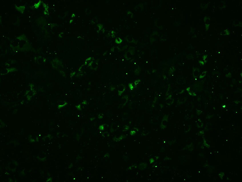
(A) Flowchart of the experiments used for testing the feasibility of the new system for enrichment of cells genome-edited at a single target locus. Four days after transfection with pCGsap1/LDLR, pgRNA#3, and pEGFP-N1, cells were treated with IB4SAP for a short period and cultured in normal medium for more than 10 days. The emerging colonies were propagated for molecular biological and cytochemical analyses; (B) cytochemical staining of clones LA-2 and -6 with AF594-IB4. Untransfected MPEFs were stained as positive control. Phase, photographs taken under light microscopy; AF594-IB4, photographs taken under UV illumination to detect AF594-IB4–derived red fluorescence. Bar = 30 μm; (C) electrophoretic pattern of polymerase chain reaction (PCR) products derived from the amplification of LA-1 to -7. Arrows indicate the size of PCR products for each gene (LDLR and GGTA1). M, 100 bp-ladder markers; (D) direct sequencing of PCR products from LA-2 (left panel) and LA-6 (right panel) using primers specific to LDLR or GGTA1 gene. Arrows above ideograms indicate the sites showing indels. In the bottom of each panel, sequencing results of inserts sub-cloned into TA cloning vector are shown. The numbers of clones examined are shown in parentheses. The translation initiation codon ATG is shown by boxes or in red. PAM is indicated by underlines. The deleted portion in the clones is shown by dotted lines; (E) immunocytochemistry of LA-6 and intact MPEFs (used as positive control) using anti-LDLR antibody. The parental MPEFs reactive to the second antibody (fluorescein-labeled anti-mouse IgG) alone are designated as negative control. All cells were counterstained with DAPI upon reaction with the second antibody. Bar = 30 μm. Image collected and cropped by CiteAb from the following open publication (https://pubmed.ncbi.nlm.nih.gov/29207527), licensed under a CC-BY license. Not internally tested by Novus Biologicals.Applications for LDLR Antibody (C7) - BSA Free
Application
Recommended Usage
Electron Microscopy
reported in scientific literature (PMID 11839845)
Flow Cytometry
reported in scientific literature (PMID 10906332)
Immunocytochemistry/ Immunofluorescence
1:100
Immunohistochemistry
1:200
Immunohistochemistry-Frozen
reported in scientific literature (PMID 11839845)
Immunohistochemistry-Paraffin
1:200
Immunoprecipitation
1:10 - 1:500
In vitro assay
reported in scientific literature (PMID 9642270)
Proximity Ligation Assay
reported in scientific literature (PMID 3263645)
Western Blot
1:1000
Application Notes
In WB assay, specific bands can be seen around 160 kDa (mature form) and 120 kDa (precursor) molecular weight positions.
Reviewed Applications
Read 1 review rated 4 using NBP1-78159 in the following applications:
Flow Cytometry Panel Builder
Bio-Techne Knows Flow Cytometry
Save time and reduce costly mistakes by quickly finding compatible reagents using the Panel Builder Tool.
Advanced Features
- Spectra Viewer - Custom analysis of spectra from multiple fluorochromes
- Spillover Popups - Visualize the spectra of individual fluorochromes
- Antigen Density Selector - Match fluorochrome brightness with antigen density

Formulation, Preparation, and Storage
Purification
Protein G purified
Formulation
Tris-Glycine, 0.15M NaCl
Format
BSA Free
Preservative
0.05% Sodium Azide
Concentration
1 mg/ml
Shipping
The product is shipped with polar packs. Upon receipt, store it immediately at the temperature recommended below.
Stability & Storage
Store at 4C short term. Aliquot and store at -20C long term. Avoid freeze-thaw cycles.
Background: LDLR
Long Name
Low Density Lipoprotein Receptor
Alternate Names
LDL R
Gene Symbol
LDLR
UniProt
Additional LDLR Products
Product Documents for LDLR Antibody (C7) - BSA Free
Certificate of Analysis
To download a Certificate of Analysis, please enter a lot or batch number in the search box below.
Product Specific Notices for LDLR Antibody (C7) - BSA Free
This product is for research use only and is not approved for use in humans or in clinical diagnosis. Primary Antibodies are guaranteed for 1 year from date of receipt.
Related Research Areas
Citations for LDLR Antibody (C7) - BSA Free
 Powered by Bioz
Customer Reviews for LDLR Antibody (C7) - BSA Free (1)
4 out of 5
1 Customer Rating
Have you used LDLR Antibody (C7) - BSA Free?
Submit a review and receive an Amazon gift card!
$25/€18/£15/$25CAN/¥2500 Yen for a review with an image
$10/€7/£6/$10CAN/¥1110 Yen for a review without an image
Submit a review
Customer Images
Showing
1
-
1 of
1 review
Showing All
Filter By:
-
Application: ImmunocytochemistrySample Tested: A porcine intestinal epithelial cell line, named as IPEC-J2Species: OtherVerified Customer | Posted 12/03/2013

There are no reviews that match your criteria.
Protocols
View specific protocols for LDLR Antibody (C7) - BSA Free (NBP1-78159):
Immunocytochemistry Protocol
Culture cells to appropriate density in 35 mm culture dishes or 6-well plates.
1. Remove culture medium and add 10% formalin to the dish. Fix at room temperature for 30 minutes.
2. Remove the formalin and add ice cold methanol. Incubate for 5-10 minutes.
3. Remove methanol and add washing solution (i.e. PBS). Be sure to not let the specimen dry out. Wash three times for 10 minutes.
4. To block nonspecific antibody binding incubate in 10% normal goat serum from 1 hour to overnight at room temperature.
5. Add primary antibody at appropriate dilution and incubate at room temperature from 2 hours to overnight at room temperature.
6. Remove primary antibody and replace with washing solution. Wash three times for 10 minutes.
7. Add secondary antibody at appropriate dilution. Incubate for 1 hour at room temperature.
8. Remove antibody and replace with wash solution, then wash for 10 minutes. Add Hoechst 33258 to wash solution at 1:25,0000 and incubate for 10 minutes. Wash a third time for 10 minutes.
9. Cells can be viewed directly after washing. The plates can also be stored in PBS containing Azide covered in Parafilm (TM). Cells can also be cover-slipped using Fluoromount, with appropriate sealing.
*The above information is only intended as a guide. The researcher should determine what protocol best meets their needs. Please follow safe laboratory procedures.
Immunohistochemistry-Paraffin Embedded Sections
Antigen Unmasking:
Bring slides to a boil in 10 mM sodium citrate buffer (pH 6.0) then maintain at a sub-boiling temperature for 10 minutes. Cool slides on bench-top for 30 minutes.
Staining:
1. Wash sections in deionized water three times for 5 minutes each.
2. Wash sections in wash buffer for 5 minutes.
3. Block each section with 100-400 ul blocking solution for 1 hour at room temperature.
4. Remove blocking solution and add 100-400 ul diluted primary antibody. Incubate overnight at 4C.
5. Remove antibody solution and wash sections in wash buffer three times for 5 minutes each.
6. Add 100-400 ul biotinylated diluted secondary antibody. Incubate 30 minutes at room temperature.
7. Remove secondary antibody solution and wash sections three times with wash buffer for 5 minutes each.
8. Add 100-400 ul Streptavidin-HRP reagent to each section and incubate for 30 minutes at room temperature.
9. Wash sections three times in wash buffer for 5 minutes each.
10. Add 100-400 ul DAB substrate to each section and monitor staining closely.
11. As soon as the sections develop, immerse slides in deionized water.
12. Counterstain sections in hematoxylin.
13. Wash sections in deionized water two times for 5 minutes each.
14. Dehydrate sections.
15. Mount coverslips.
*The above information is only intended as a guide. The researcher should determine what protocol best meets their needs. Please follow safe laboratory procedures.
Find general support by application which include: protocols, troubleshooting, illustrated assays, videos and webinars.
- 7-Amino Actinomycin D (7-AAD) Cell Viability Flow Cytometry Protocol
- Antigen Retrieval Protocol (PIER)
- Antigen Retrieval for Frozen Sections Protocol
- Appropriate Fixation of IHC/ICC Samples
- Cellular Response to Hypoxia Protocols
- Chromogenic IHC Staining of Formalin-Fixed Paraffin-Embedded (FFPE) Tissue Protocol
- Chromogenic Immunohistochemistry Staining of Frozen Tissue
- ClariTSA™ Fluorophore Kits
- Detection & Visualization of Antibody Binding
- ELISA Sample Preparation & Collection Guide
- ELISA Troubleshooting Guide
- Extracellular Membrane Flow Cytometry Protocol
- Flow Cytometry Protocol for Cell Surface Markers
- Flow Cytometry Protocol for Staining Membrane Associated Proteins
- Flow Cytometry Staining Protocols
- Flow Cytometry Troubleshooting Guide
- Fluorescent IHC Staining of Frozen Tissue Protocol
- Graphic Protocol for Heat-induced Epitope Retrieval
- Graphic Protocol for the Preparation and Fluorescent IHC Staining of Frozen Tissue Sections
- Graphic Protocol for the Preparation and Fluorescent IHC Staining of Paraffin-embedded Tissue Sections
- Graphic Protocol for the Preparation of Gelatin-coated Slides for Histological Tissue Sections
- How to Run an R&D Systems DuoSet ELISA
- How to Run an R&D Systems Quantikine ELISA
- How to Run an R&D Systems Quantikine™ QuicKit™ ELISA
- ICC Cell Smear Protocol for Suspension Cells
- ICC Immunocytochemistry Protocol Videos
- ICC for Adherent Cells
- IHC Sample Preparation (Frozen sections vs Paraffin)
- Immunocytochemistry (ICC) Protocol
- Immunocytochemistry Troubleshooting
- Immunofluorescence of Organoids Embedded in Cultrex Basement Membrane Extract
- Immunofluorescent IHC Staining of Formalin-Fixed Paraffin-Embedded (FFPE) Tissue Protocol
- Immunohistochemistry (IHC) and Immunocytochemistry (ICC) Protocols
- Immunohistochemistry Frozen Troubleshooting
- Immunohistochemistry Paraffin Troubleshooting
- Immunoprecipitation Protocol
- Intracellular Flow Cytometry Protocol Using Alcohol (Methanol)
- Intracellular Flow Cytometry Protocol Using Detergents
- Intracellular Nuclear Staining Flow Cytometry Protocol Using Detergents
- Intracellular Staining Flow Cytometry Protocol Using Alcohol Permeabilization
- Intracellular Staining Flow Cytometry Protocol Using Detergents to Permeabilize Cells
- Preparing Samples for IHC/ICC Experiments
- Preventing Non-Specific Staining (Non-Specific Binding)
- Primary Antibody Selection & Optimization
- Propidium Iodide Cell Viability Flow Cytometry Protocol
- Protocol for Heat-Induced Epitope Retrieval (HIER)
- Protocol for Liperfluo
- Protocol for Making a 4% Formaldehyde Solution in PBS
- Protocol for VisUCyte™ HRP Polymer Detection Reagent
- Protocol for the Characterization of Human Th22 Cells
- Protocol for the Characterization of Human Th9 Cells
- Protocol for the Fluorescent ICC Staining of Cell Smears - Graphic
- Protocol for the Fluorescent ICC Staining of Cultured Cells on Coverslips - Graphic
- Protocol for the Preparation & Fixation of Cells on Coverslips
- Protocol for the Preparation and Chromogenic IHC Staining of Frozen Tissue Sections
- Protocol for the Preparation and Chromogenic IHC Staining of Frozen Tissue Sections - Graphic
- Protocol for the Preparation and Chromogenic IHC Staining of Paraffin-embedded Tissue Sections
- Protocol for the Preparation and Chromogenic IHC Staining of Paraffin-embedded Tissue Sections - Graphic
- Protocol for the Preparation and Fluorescent ICC Staining of Cells on Coverslips
- Protocol for the Preparation and Fluorescent ICC Staining of Non-adherent Cells
- Protocol for the Preparation and Fluorescent ICC Staining of Stem Cells on Coverslips
- Protocol for the Preparation and Fluorescent IHC Staining of Frozen Tissue Sections
- Protocol for the Preparation and Fluorescent IHC Staining of Paraffin-embedded Tissue Sections
- Protocol for the Preparation of Gelatin-coated Slides for Histological Tissue Sections
- Protocol for the Preparation of a Cell Smear for Non-adherent Cell ICC - Graphic
- Protocol: Annexin V and PI Staining by Flow Cytometry
- Protocol: Annexin V and PI Staining for Apoptosis by Flow Cytometry
- Quantikine HS ELISA Kit Assay Principle, Alkaline Phosphatase
- Quantikine HS ELISA Kit Principle, Streptavidin-HRP Polymer
- R&D Systems Quality Control Western Blot Protocol
- Sandwich ELISA (Colorimetric) – Biotin/Streptavidin Detection Protocol
- Sandwich ELISA (Colorimetric) – Direct Detection Protocol
- TUNEL and Active Caspase-3 Detection by IHC/ICC Protocol
- The Importance of IHC/ICC Controls
- Troubleshooting Guide: ELISA
- Troubleshooting Guide: Fluorokine Flow Cytometry Kits
- Troubleshooting Guide: Immunohistochemistry
- Troubleshooting Guide: Western Blot Figures
- Western Blot Conditions
- Western Blot Protocol
- Western Blot Protocol for Cell Lysates
- Western Blot Troubleshooting
- Western Blot Troubleshooting Guide
- View all Protocols, Troubleshooting, Illustrated assays and Webinars
Loading...
Associated Pathways
