Lgr5/GPR49 Antibody - BSA Free
Novus Biologicals | Catalog # NBP1-28904
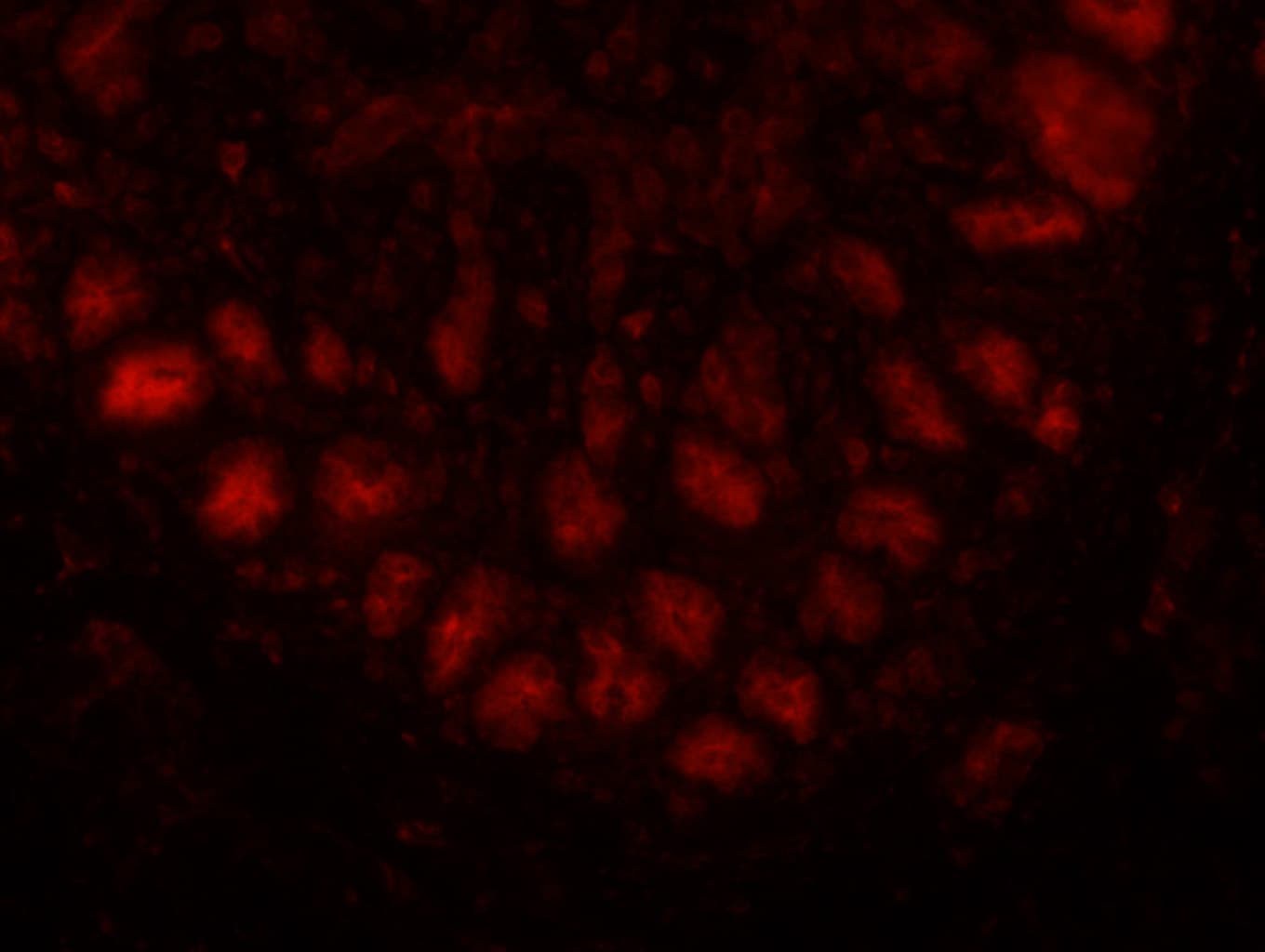
![Immunocytochemistry/ Immunofluorescence: Lgr5/GPR49 Antibody - BSA Free [NBP1-28904]](https://resources.rndsystems.com/images/products/Lgr5-GPR49-Antibody-Immunocytochemistry-Immunofluorescence-NBP1-28904-img0009.jpg "Immunocytochemistry/ Immunofluorescence: Lgr5/GPR49 Antibody - BSA Free [NBP1-28904]")
Key Product Details
Species Reactivity
Validated:
Cited:
Applications
Validated:
Cited:
Label
Antibody Source
Format
Product Specifications
Immunogen
Reactivity Notes
Localization
Clonality
Host
Isotype
Scientific Data Images for Lgr5/GPR49 Antibody - BSA Free
Immunocytochemistry/ Immunofluorescence: Lgr5/GPR49 Antibody - BSA Free [NBP1-28904]
Immunocytochemistry/Immunofluorescence: Lgr5/GPR49 Antibody [NBP1-28904] - Hek293 cells were fixed in 4% paraformaldehyde for 10 minutes and permeabilized in 0.05% Triton X-100 in PBS for 5 minutes. The cells were incubated with anti-Lgr5/GPR49 Antibody NBP1-28904 at 2 ug/ml overnight at 4C and detected with an anti-rabbit Dylight 488 (Green) at a 1:1000 dilution for 60 minutes. Nuclei were counterstained with DAPI (Blue). Cells were imaged using a 100X objective and digitally deconvolved.![Immunohistochemistry: Lgr5/GPR49 Antibody - BSA Free [NBP1-28904]](https://resources.rndsystems.com/images/products/Lgr5-GPR49-Antibody-Immunohistochemistry-NBP1-28904-img0011.jpg "Immunohistochemistry: Lgr5/GPR49 Antibody - BSA Free [NBP1-28904]")
Immunohistochemistry: Lgr5/GPR49 Antibody - BSA Free [NBP1-28904]
Lgr5-GPR49-Antibody-Immunohistochemistry-NBP1-28904-img0011.jpg![Immunohistochemistry: Lgr5/GPR49 Antibody - BSA Free [NBP1-28904]](https://resources.rndsystems.com/images/products/Lgr5-GPR49-Antibody-Immunohistochemistry-NBP1-28904-img0005.jpg "Immunohistochemistry: Lgr5/GPR49 Antibody - BSA Free [NBP1-28904]")
Immunohistochemistry: Lgr5/GPR49 Antibody - BSA Free [NBP1-28904]
Immunohistochemistry: Lgr5/GPR49 Antibody [NBP1-28904] - Analysis of human small intestine.
Immunohistochemistry-Paraffin: Lgr5/GPR49 Antibody - BSA Free [NBP1-28904] -
Immunohistochemistry-Paraffin: Lgr5/GPR49 Antibody - BSA Free [NBP1-28904] - Wnt-related genes show selective expression in the ZG of human adrenal. A, Heat map of 37 genes (shown on the vertical axis), which are 5-fold or greater differentially expressed in ZG & ZF. Genes previously associated with the Wnt signaling pathway are highlighted in red. On the horizontal axis, each pair of ZG & ZF from the 20 adrenals are anonymized & numbered. Probe set & detailed microarray information are available from the National Center for Biotechnology Information Gene Expression Omnibus under accession number GSE64957. B, IHC localization of LGR5 & downstream Wnt signaling proteins in the ZG: from the canonical beta -catenin pathway, beta -catenin & LEF1, & from the noncanonical AP1/JUN pathway, c-JUN & p-c-JUN. IHC was performed on formalin-fixed, paraffin-embedded human adrenal sections (4 μm) using a chromogen-based detection system (3,3'-Diaminobenzidine), which results in a positive brown staining. Interestingly, beta -catenin staining was mostly membranous or cytoplasmic (rather than nuclear staining as highlighted by the arrows), indicating limited canonical Wnt activation. Pictures are representative of six normal adrenal sections that were used in our microarray study; four from primary hyperaldosteronism patients & two from pheochromocytoma patients. Scale bar, 500 μm (low power) & 100 μm (high power). C, capsule. Image collected & cropped by CiteAb from the following publication (https://academic.oup.com/jcem/article-lookup/doi/10.1210/jc.2015-1734), licensed under a CC-BY license. Not internally tested by Novus Biologicals.
Western Blot: Lgr5/GPR49 Antibody - BSA Free [NBP1-28904] -
AKAP8L controls the chemoresistance in vivo.A 1 × 10⁶ AKAP8L-overexpressing or AKAP8L-knockdown BGC-823/Oxa were implanted in nude mice (tumor development following treatment with Oxa once a week), tumors were dissected and representative images of the tumors are shown. B, C Tumor growth curves for orthotopic models. D Representative images of immunofluorescence staining of AKAP8L, Ki-67 and quantification analysis of AKAP8L, Ki-67 positive cells in xenograft tumors. Scale bar indicates 50 μm. E, F qPCR and Western blot analysis of the expression of AKAP8L, Lgr5, CD133, CD44, Oct4, and Sox2 in xenograft tumors. G Representative immunohistochemical staining of AKAP8L and CD133 in the indicated tumor tissues. Scale bar indicates 20 μm. H Immunofluorescence staining of TUNEL was performed to evaluate the apoptotic cells. Scale bar indicates 50 μm. I Western blot analysis of cleaved-caspase 3 and cleaved-PAPR. The values indicate the mean +/- SD of three independent experiments. *P < 0.05, **P < 0.01, ***P < 0.001. Image collected and cropped by CiteAb from the following open publication (https://pubmed.ncbi.nlm.nih.gov/36522343), licensed under a CC-BY license. Not internally tested by Novus Biologicals.
Western Blot: Lgr5/GPR49 Antibody - BSA Free [NBP1-28904] -
The expression levels of CSC markers in HCC cells are decreased by JIB-04. (A) The mRNA expression of cancer stem cell marker genes was analyzed using qRT-PCR in PLC/PRF/5, Huh7, and HepG2 cultures after treatment with DMSO (control), 6 μM JIB-04, or 4 μM TSA (positive control) for 24 h. All data are normalized to GAPDH and plotted relative to the expression level in control cells. Data are represented as mean +/- SEM derived from triplicate measurements (n = 3); * p < 0.05, ** p < 0.01, and *** p < 0.001. (B) (Top panels) Protein levels of CD133, CD44, LGR5, and EpCAM in HCC cells after treatment with 6 μM JIB-04 for 48 h were confirmed with Western blot analysis. GAPDH was used as a loading control. (Bottom panels) Quantification based on densitometry of Western blotting data from top panels in (B). All data are normalized to GAPDH. Data are represented as mean +/- SEM of triplicate measurements; * p < 0.05 and ** p < 0.01. Image collected and cropped by CiteAb from the following open publication (https://pubmed.ncbi.nlm.nih.gov/35887001), licensed under a CC-BY license. Not internally tested by Novus Biologicals.
Western Blot: Lgr5/GPR49 Antibody - BSA Free [NBP1-28904] -
SCD1 mediates the effects of AKAP8L on GC cell stemness and chemoresistance.SCD1 siRNA was transiently transfected to AKAP8L overexpressing cells. SCD1 inhibitor (CAY10566) was added into AKAP8L overexpressing cells. A Representative images of spheroids in the SCD1 inhibitor, AKAP8L, AKAP8L/SCD1 inhibitor and the control groups. B, C qPCR and Western blot assays showed the stemness-related gene expression induced by AKAP8L/CAY10566 or AKAP8L/si-SCD1. D Cell viability was measured by MTT in four groups after the treatments of Oxa. E Representative images of flow cytometry showed the percentage of apoptotic cells in four groups after the treatments of Oxa. F Western blot analysis of cleaved-caspase 3 and cleaved-PAPR induced by AKAP8L/CAY10566 or AKAP8L/si-SCD1. Scale bar indicates 50 μm. The values indicate the mean +/- SD of three independent experiments. *P < 0.05, **P < 0.01, ***P < 0.001. Image collected and cropped by CiteAb from the following open publication (https://pubmed.ncbi.nlm.nih.gov/36522343), licensed under a CC-BY license. Not internally tested by Novus Biologicals.
Western Blot: Lgr5/GPR49 Antibody - BSA Free [NBP1-28904] -
AKAP8L enhances the stemness in GC cells.A Western blot analysis showed ectopic expression of AKAP8L in GC cells transfected with AKAP8L (AKAP8L) or control lentivirus (Con), and AKAP8L silencing in cells treated with scrambled shRNA (shCon) or shRNA against AKAP8L (shAKAP8L-1, shAKAP8L-2). B Representative images and quantification analysis of spheroids in AKAP8L overexpression or the control groups, and AKAP8L knockdown or the control groups. Scale bar indicates 50 μm. C In soft agar colony formation assay, representative images and numbers of spheroids formed by AKAP8L-overexpressing and AKAP8L-knockdown BGC-823/Oxa and MKN-45/Oxa. Scale bar indicates 50 μm. D, E qPCR and Western blot assays showed the expression of stem cell markers induced by AKAP8L or AKAP8L shRNAs. The values indicate the mean +/- standard deviation (SD) of three independent experiments. *P < 0.05, **P < 0.01,***P < 0.001. Image collected and cropped by CiteAb from the following open publication (https://pubmed.ncbi.nlm.nih.gov/36522343), licensed under a CC-BY license. Not internally tested by Novus Biologicals.Applications for Lgr5/GPR49 Antibody - BSA Free
Immunocytochemistry/ Immunofluorescence
Immunohistochemistry
Immunohistochemistry-Paraffin
Western Blot
Reviewed Applications
Read 1 review rated 4 using NBP1-28904 in the following applications:
Formulation, Preparation, and Storage
Purification
Formulation
Format
Preservative
Concentration
Shipping
Stability & Storage
Background: Lgr5/GPR49
Long Name
Alternate Names
Entrez Gene IDs
Gene Symbol
UniProt
Additional Lgr5/GPR49 Products
Product Documents for Lgr5/GPR49 Antibody - BSA Free
Certificate of Analysis
To download a Certificate of Analysis, please enter a lot or batch number in the search box below.
Product Specific Notices for Lgr5/GPR49 Antibody - BSA Free
This product is for research use only and is not approved for use in humans or in clinical diagnosis. Primary Antibodies are guaranteed for 1 year from date of receipt.
Related Research Areas
Citations for Lgr5/GPR49 Antibody - BSA Free
 Powered by Bioz
Customer Reviews for Lgr5/GPR49 Antibody - BSA Free (1)
Have you used Lgr5/GPR49 Antibody - BSA Free?
Submit a review and receive an Amazon gift card!
$25/€18/£15/$25CAN/¥2500 Yen for a review with an image
$10/€7/£6/$10CAN/¥1110 Yen for a review without an image
Submit a review
Customer Images
-
Application: Immunohistochemistry-FrozenSample Tested: Pig small intestineSpecies: OtherVerified Customer | Posted 01/14/2014

There are no reviews that match your criteria.
Protocols
View specific protocols for Lgr5/GPR49 Antibody - BSA Free (NBP1-28904):
Culture cells to appropriate density in 35 mm culture dishes or 6-well plates.
1. Remove culture medium and wash the cells briefly in PBS. Add 10% formalin to the dish and fix at room temperature for 10 minutes.
2. Remove the formalin and wash the cells in PBS.
3. Permeablize the cells with 0.1% Triton X100 or other suitable detergent for 10 min.
4. Remove the permeablization buffer and wash three times for 10 minutes each in PBS. Be sure to not let the specimen dry out.
5. To block nonspecific antibody binding, incubate in 10% normal goat serum from 1 hour to overnight at room temperature.
6. Add primary antibody at appropriate dilution and incubate overnight at 4C.
7. Remove primary antibody and replace with PBS. Wash three times for 10 minutes each.
8. Add secondary antibody at appropriate dilution. Incubate for 1 hour at room temperature.
9. Remove secondary antibody and replace with PBS. Wash three times for 10 minutes each.
10. Counter stain DNA with DAPi if required.
Antigen Unmasking: Bring slides to a boil in 10 mM sodium citrate buffer (pH 6.0) then maintain at a sub-boiling temperature for 10 minutes. Cool slides on bench-top for 30 minutes.
Immunostaining:
1. After the antigen retrieval, wash sections in deionized water three times for 5 minutes each.
2. Wash sections in wash buffer for 5 minutes.
3. Block each section with 100-400 ul blocking solution for 1 hour at room temperature.
4. Remove blocking solution and add 100-400 ul diluted primary antibody. Incubate overnight at 4 C.
5. Remove antibody solution and wash sections in wash buffer three times for 5 minutes each.
6. Add 100-400 ul biotinylated diluted secondary antibody. Incubate 30 minutes at room temperature.
7. Remove secondary antibody solution and wash sections three times with wash buffer for 5 minutes each.
8. Add 100-400 ul Streptavidin-HRP reagent to each section and incubate for 30 minutes at room temperature.
9. Wash sections three times in wash buffer for 5 minutes each.
10. Add 100-400 ul DAB substrate to each section and monitor staining closely.
11. As soon as the sections develop, immerse slides in deionized water.
12. Counterstain sections in hematoxylin.
13. Wash sections in deionized water two times for 5 minutes each.
14. Dehydrate sections.
15. Mount coverslips.
1. Perform SDS-PAGE on samples to be analyzed, loading 10-25 ug of total protein per lane.
2. Transfer proteins to PVDF membrane according to the instructions provided by the manufacturer of the membrane and transfer apparatus.
3. Stain the membrane with Ponceau S (or similar product) to assess transfer success, and mark molecular weight standards where appropriate.
4. Rinse the blot TBS -0.05% Tween 20 (TBST).
5. Block the membrane in 5% Non-fat milk in TBST (blocking buffer) for at least 1 hour.
6. Wash the membrane in TBST three times for 10 minutes each.
7. Dilute primary antibody in blocking buffer and incubate overnight at 4C with gentle rocking.
8. Wash the membrane in TBST three times for 10 minutes each.
9. Incubate the membrane in diluted HRP conjugated secondary antibody in blocking buffer (as per manufacturer's instructions) for 1 hour at room temperature.
10. Wash the blot in TBST three times for 10 minutes each (this step can be repeated as required to reduce background).
11. Apply the detection reagent of choice in accordance with the manufacturer's instructions.
Find general support by application which include: protocols, troubleshooting, illustrated assays, videos and webinars.
- Antigen Retrieval Protocol (PIER)
- Antigen Retrieval for Frozen Sections Protocol
- Appropriate Fixation of IHC/ICC Samples
- Cellular Response to Hypoxia Protocols
- Chromogenic IHC Staining of Formalin-Fixed Paraffin-Embedded (FFPE) Tissue Protocol
- Chromogenic Immunohistochemistry Staining of Frozen Tissue
- ClariTSA™ Fluorophore Kits
- Detection & Visualization of Antibody Binding
- Fluorescent IHC Staining of Frozen Tissue Protocol
- Graphic Protocol for Heat-induced Epitope Retrieval
- Graphic Protocol for the Preparation and Fluorescent IHC Staining of Frozen Tissue Sections
- Graphic Protocol for the Preparation and Fluorescent IHC Staining of Paraffin-embedded Tissue Sections
- Graphic Protocol for the Preparation of Gelatin-coated Slides for Histological Tissue Sections
- ICC Cell Smear Protocol for Suspension Cells
- ICC Immunocytochemistry Protocol Videos
- ICC for Adherent Cells
- IHC Sample Preparation (Frozen sections vs Paraffin)
- Immunocytochemistry (ICC) Protocol
- Immunocytochemistry Troubleshooting
- Immunofluorescence of Organoids Embedded in Cultrex Basement Membrane Extract
- Immunofluorescent IHC Staining of Formalin-Fixed Paraffin-Embedded (FFPE) Tissue Protocol
- Immunohistochemistry (IHC) and Immunocytochemistry (ICC) Protocols
- Immunohistochemistry Frozen Troubleshooting
- Immunohistochemistry Paraffin Troubleshooting
- Preparing Samples for IHC/ICC Experiments
- Preventing Non-Specific Staining (Non-Specific Binding)
- Primary Antibody Selection & Optimization
- Protocol for Heat-Induced Epitope Retrieval (HIER)
- Protocol for Making a 4% Formaldehyde Solution in PBS
- Protocol for VisUCyte™ HRP Polymer Detection Reagent
- Protocol for the Fluorescent ICC Staining of Cell Smears - Graphic
- Protocol for the Fluorescent ICC Staining of Cultured Cells on Coverslips - Graphic
- Protocol for the Preparation & Fixation of Cells on Coverslips
- Protocol for the Preparation and Chromogenic IHC Staining of Frozen Tissue Sections
- Protocol for the Preparation and Chromogenic IHC Staining of Frozen Tissue Sections - Graphic
- Protocol for the Preparation and Chromogenic IHC Staining of Paraffin-embedded Tissue Sections
- Protocol for the Preparation and Chromogenic IHC Staining of Paraffin-embedded Tissue Sections - Graphic
- Protocol for the Preparation and Fluorescent ICC Staining of Cells on Coverslips
- Protocol for the Preparation and Fluorescent ICC Staining of Non-adherent Cells
- Protocol for the Preparation and Fluorescent ICC Staining of Stem Cells on Coverslips
- Protocol for the Preparation and Fluorescent IHC Staining of Frozen Tissue Sections
- Protocol for the Preparation and Fluorescent IHC Staining of Paraffin-embedded Tissue Sections
- Protocol for the Preparation of Gelatin-coated Slides for Histological Tissue Sections
- Protocol for the Preparation of a Cell Smear for Non-adherent Cell ICC - Graphic
- R&D Systems Quality Control Western Blot Protocol
- TUNEL and Active Caspase-3 Detection by IHC/ICC Protocol
- The Importance of IHC/ICC Controls
- Troubleshooting Guide: Immunohistochemistry
- Troubleshooting Guide: Western Blot Figures
- Western Blot Conditions
- Western Blot Protocol
- Western Blot Protocol for Cell Lysates
- Western Blot Troubleshooting
- Western Blot Troubleshooting Guide
- View all Protocols, Troubleshooting, Illustrated assays and Webinars
FAQs for Lgr5/GPR49 Antibody - BSA Free
-
Q: Is it possible to confirm that the actual immunogen is taken from within the actual cytoplasmic domain (aa 660-682)?
A: Yes, I can confirm that the immunogen falls within the cytoplasmic domain of this protein.
-
Q: Please inform us this item NBP1-28904 is stain cytoplasmic or stain Nuclus?
A: NBP1-28904 detects Lgr5/GPR49 protein which localize to Cell membrane and the Golgi apparatus. Therefore, this antibody should generate a staining pattern with more signal in membranes and in cytoplasm as patches around the nucleus of the cells. More information on this protein may be found at Uniprot site
-
Q: This morning I have a customer who has purchased NBP1-28904 and would like to test it in flow cytometry. She is new to flow and is wondering if you might have a recommended protocol. Also, I see that the immunogen is within aa 650-700 which appears to span both helical and cytoplasmic domains. Do you know if the exact immunogen is fully within one domain or the other? Is permeabilization required? Any information you have is much appreciated.
A:
Unfortunately I cannot disclose anything more about the immunogen sequence; however, our lab does recommend permeabilization for using this antibody. This is our general flow cytometry protocol. Note that if she tests this antibody in flow, she would be eligible for our Innovators Reward Program.
-
Q: Is it possible to confirm that the actual immunogen is taken from within the actual cytoplasmic domain (aa 660-682)?
A: Yes, I can confirm that the immunogen falls within the cytoplasmic domain of this protein.
-
Q: Please inform us this item NBP1-28904 is stain cytoplasmic or stain Nuclus?
A: NBP1-28904 detects Lgr5/GPR49 protein which localize to Cell membrane and the Golgi apparatus. Therefore, this antibody should generate a staining pattern with more signal in membranes and in cytoplasm as patches around the nucleus of the cells. More information on this protein may be found at Uniprot site
-
Q: This morning I have a customer who has purchased NBP1-28904 and would like to test it in flow cytometry. She is new to flow and is wondering if you might have a recommended protocol. Also, I see that the immunogen is within aa 650-700 which appears to span both helical and cytoplasmic domains. Do you know if the exact immunogen is fully within one domain or the other? Is permeabilization required? Any information you have is much appreciated.
A:
Unfortunately I cannot disclose anything more about the immunogen sequence; however, our lab does recommend permeabilization for using this antibody. This is our general flow cytometry protocol. Note that if she tests this antibody in flow, she would be eligible for our Innovators Reward Program.


