MBP Antibody (12) - BSA Free
Novus Biologicals | Catalog # NB600-717
![Western Blot: MBP Antibody (12) [NB600-717]](https://resources.rndsystems.com/images/products/MBP-Antibody-12-Western-Blot-NB600-717-img0002.jpg "Western Blot: MBP Antibody (12) [NB600-717]")
Loading...
Key Product Details
Validated by
Biological Validation
Species Reactivity
Validated:
Bovine
Cited:
Human, Mouse, Rat, Bovine, Canine, Feline, Guinea Pig, Primate
Applications
Validated:
Immunohistochemistry, Immunohistochemistry-Paraffin, Immunohistochemistry-Frozen, Western Blot, ELISA, Immunocytochemistry/ Immunofluorescence, Radioimmunodiffusion, Radioimmunoassay
Cited:
Immunohistochemistry, Immunohistochemistry-Paraffin, Immunohistochemistry-Frozen, Western Blot, Immunocytochemistry/ Immunofluorescence, IF/IHC
Label
Unconjugated
Antibody Source
Monoclonal Rat IgG2A Clone # 12
Format
BSA Free
Loading...
Product Specifications
Immunogen
Bovine MBP
Epitope
Amino acids 82-87.
Reactivity Notes
Predicted cross-reactivities: Mouse, Rabbit, Porcine, Mammals, Human, Guinea Pig, Chicken, Rat, Sheep
Localization
Cytoplasmic side of myelin
Marker
Oligodendrocyte Marker, Myelin Marker
Specificity
NB600-717 reacts with myelin basic protein from a wide range of species. The antibody reacts weakly with peptides ending in the Phe 91 where the 91-92 Phe-Phe bond is broken. Synthetic peptide 82-99 reacts very well, as does intact MBP. Further epitope analysis indicates binding to a region defined by amino acids 82-87 (DENPVV). Clone 12 has been reported as being suitable for use in Western blotting.
Clonality
Monoclonal
Host
Rat
Isotype
IgG2A
Scientific Data Images for MBP Antibody (12) - BSA Free
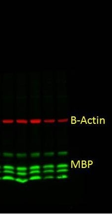
Western Blot: MBP Antibody (12) [NB600-717]
Western Blot: MBP Antibody (12) [NB600-717] - Mouse Brain Tissue lysate probed with Rat anti MBP.![Immunocytochemistry/ Immunofluorescence: MBP Antibody (12) [NB600-717]](https://resources.rndsystems.com/images/products/MBP-Antibody-12-Immunocytochemistry-Immunofluorescence-NB600-717-img0005.jpg "Immunocytochemistry/ Immunofluorescence: MBP Antibody (12) [NB600-717]")
![Immunohistochemistry-Frozen: MBP Antibody (12) [NB600-717]](https://resources.rndsystems.com/images/products/MBP-Antibody-12-Immunohistochemistry-Frozen-NB600-717-img0004.jpg "Immunohistochemistry-Frozen: MBP Antibody (12) [NB600-717]")
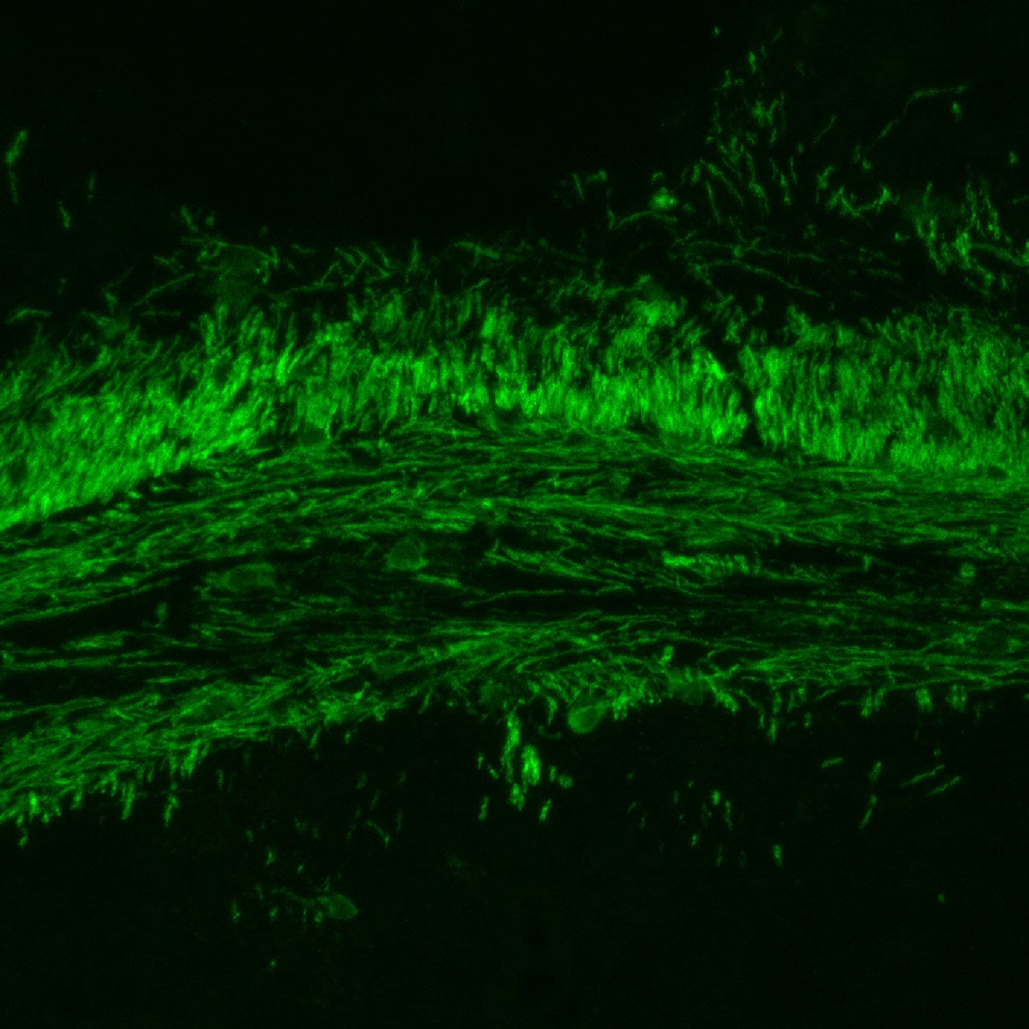
Immunohistochemistry-Frozen: MBP Antibody (12) [NB600-717]
Immunohistochemistry-Frozen: MBP Antibody (12) [NB600-717] - Staining of MBP in mouse brain corpus callosum. Image from verified customer review.![Immunohistochemistry-Frozen: MBP Antibody (12) [NB600-717]](https://resources.rndsystems.com/images/products/MBP-Antibody-12-Immunohistochemistry-Frozen-NB600-717-img0003.jpg "Immunohistochemistry-Frozen: MBP Antibody (12) [NB600-717]")
Immunohistochemistry-Frozen: MBP Antibody (12) [NB600-717]
Immunohistochemistry-Frozen: MBP Antibody (12) [NB600-717] - Staining of MBP in mouse spinal cord ventral white matter. Image from verified customer review. [NB600-717] -")
Immunocytochemistry/ Immunofluorescence: MBP Antibody (12) [NB600-717] -
Immunocytochemistry/ Immunofluorescence: MBP Antibody (12) [NB600-717] - Microglia processes appear to preferentially contact TBI-induced proximal axonal swellings. Representative 3D reconstructions of MBP+ myelinated axons (red) or APP+ axonal swellings (green) & Iba-1+ microglia (white) in sham-injured (a) or central fluid percussion injured (b) thalami. c Bar graph depicting the average number of Iba-1+ microglial processes contacting either MBP+ myelinated fibers in the sham animals or APP+ axonal swellings in injured animals. Graph depicts the mean ± standard error of the mean. *p < 0.05. Scale bar: 5 μm Image collected & cropped by CiteAb from the following publication (https://jneuroinflammation.biomedcentral.com/articles/10.1186/s12974-01…), licensed under a CC-BY license. Not internally tested by Novus Biologicals. - BSA Free [NB600-717] -")
Western Blot: MBP Antibody (12) - BSA Free [NB600-717] -
Western blots based on protein quantification with the PDB assay. (A,B) Ponceau S-stained dot blot. Undiluted BSA standards and BSA standards diluted 1:1 in 2x SDS lysis buffer were spotted in duplicate onto a membrane (fixed concentration, variable volumes). A quantity of 1 uL of sciatic nerve (SN) and brain samples lysed in 2x SDS LB or RIPA buffer was also applied onto the same membrane for quantification with the PDB assay. (C) The nerve and brain lysates containing 50, 25 or 15 ug of total proteins (based on the PDB assay) were loaded for SDS-PAGE and Western blot. After protein transfer to a nitrocellulose membrane, the membrane was also stained with Ponceau S. Image collected and cropped by CiteAb from the following open publication (https://pubmed.ncbi.nlm.nih.gov/35049578), licensed under a CC-BY license. Not internally tested by Novus Biologicals. - BSA Free [NB600-717] -")
Western Blot: MBP Antibody (12) - BSA Free [NB600-717] -
Liraglutide enhances the normal physiological function of SCs via insulin-Akt signaling. (a) Western blot analysis of Ser473-phosphorylated Akt confirmed that Liraglutide and insulin reverse the glucolipotoxicity-induced insulin signaling blockade. (b) MTT assays showed that the protective effect of Liraglutide and insulin were inhibited by co-treatment with 20 μM LY294002. (c) mRNA levels of neurotrophic factors, including CNTF, NGF, NT-3, and BDNF, were measured by qPCR. Liraglutide and insulin significantly elevated the mRNA levels of neurotrophic factors suppressed by glucolipotoxicity. However, LY294002 counteracted the effects of Liraglutide and insulin. (d) Western blots demonstrated that Liraglutide and insulin show efficacy in improving SC synthesis of essential myelin components and decrease the expression of the demyelination marker RAGE. Similarly, LY294002 blocked the effects of Liraglutide and insulin in promoting myelination in RSC96 SCs. All values are presented as the mean +/- SEM. Significant difference was determined using multiple comparisons of Dunnett’s posthoc test for * p < 0.05 and ** p < 0.01. N.S., not significant. Image collected and cropped by CiteAb from the following open publication (https://pubmed.ncbi.nlm.nih.gov/36291547), licensed under a CC-BY license. Not internally tested by Novus Biologicals.Applications for MBP Antibody (12) - BSA Free
Application
Recommended Usage
ELISA
1:100-1:2000
Immunocytochemistry/ Immunofluorescence
1:10-1:500
Immunohistochemistry
1:10-1:500
Immunohistochemistry-Frozen
1:10-1:500
Immunohistochemistry-Paraffin
1:10-1:500
Western Blot
1:100-1:2000
Reviewed Applications
Read 3 reviews rated 4.7 using NB600-717 in the following applications:
Formulation, Preparation, and Storage
Purification
Tissue culture supernatant
Formulation
0.1 M Tris
Format
BSA Free
Preservative
0.1% Sodium Azide
Concentration
This product is unpurified. The exact concentration of antibody is not quantifiable.
Shipping
The product is shipped with polar packs. Upon receipt, store it immediately at the temperature recommended below.
Stability & Storage
Store at 4C short term. Aliquot and store at -20C long term. Avoid freeze-thaw cycles.
Background: MBP
Long Name
Myelin Basic Protein
Alternate Names
Hmbpr, mld, shi
Gene Symbol
MBP
UniProt
Additional MBP Products
Product Documents for MBP Antibody (12) - BSA Free
Certificate of Analysis
To download a Certificate of Analysis, please enter a lot or batch number in the search box below.
Product Specific Notices for MBP Antibody (12) - BSA Free
This product is for research use only and is not approved for use in humans or in clinical diagnosis. Primary Antibodies are guaranteed for 1 year from date of receipt.
Related Research Areas
Citations for MBP Antibody (12) - BSA Free
 Powered by Bioz
Customer Reviews for MBP Antibody (12) - BSA Free (3)
4.7 out of 5
3 Customer Ratings
Have you used MBP Antibody (12) - BSA Free?
Submit a review and receive an Amazon gift card!
$25/€18/£15/$25CAN/¥2500 Yen for a review with an image
$10/€7/£6/$10CAN/¥1110 Yen for a review without an image
Submit a review
Customer Images

Showing
1
-
3 of
3 reviews
Showing All
Filter By:
-
Application: Western BlotSample Tested: brain and spinal cordSpecies: MouseVerified Customer | Posted 04/13/2019MBP Western blot in adult mice brain tissue

-
Application: Immunohistochemistry-FrozenSample Tested: brain and spinal cordSpecies: MouseVerified Customer | Posted 04/13/2019MBP stain in brain corpus callosum
-
Application: Immunohistochemistry-FrozenSample Tested: brain and spinal cordSpecies: FelineVerified Customer | Posted 12/08/2017NB600-717 was incubated at 1:100 dilution for overnight at 4C degree and Alexa488 conjugated antibody was used. Image was captured with epifluorescent microscope.
There are no reviews that match your criteria.
Protocols
Find general support by application which include: protocols, troubleshooting, illustrated assays, videos and webinars.
- Antigen Retrieval Protocol (PIER)
- Antigen Retrieval for Frozen Sections Protocol
- Appropriate Fixation of IHC/ICC Samples
- Cellular Response to Hypoxia Protocols
- Chromogenic IHC Staining of Formalin-Fixed Paraffin-Embedded (FFPE) Tissue Protocol
- Chromogenic Immunohistochemistry Staining of Frozen Tissue
- ClariTSA™ Fluorophore Kits
- Detection & Visualization of Antibody Binding
- ELISA Sample Preparation & Collection Guide
- ELISA Troubleshooting Guide
- Fluorescent IHC Staining of Frozen Tissue Protocol
- Graphic Protocol for Heat-induced Epitope Retrieval
- Graphic Protocol for the Preparation and Fluorescent IHC Staining of Frozen Tissue Sections
- Graphic Protocol for the Preparation and Fluorescent IHC Staining of Paraffin-embedded Tissue Sections
- Graphic Protocol for the Preparation of Gelatin-coated Slides for Histological Tissue Sections
- How to Run an R&D Systems DuoSet ELISA
- How to Run an R&D Systems Quantikine ELISA
- How to Run an R&D Systems Quantikine™ QuicKit™ ELISA
- ICC Cell Smear Protocol for Suspension Cells
- ICC Immunocytochemistry Protocol Videos
- ICC for Adherent Cells
- IHC Sample Preparation (Frozen sections vs Paraffin)
- Immunocytochemistry (ICC) Protocol
- Immunocytochemistry Troubleshooting
- Immunofluorescence of Organoids Embedded in Cultrex Basement Membrane Extract
- Immunofluorescent IHC Staining of Formalin-Fixed Paraffin-Embedded (FFPE) Tissue Protocol
- Immunohistochemistry (IHC) and Immunocytochemistry (ICC) Protocols
- Immunohistochemistry Frozen Troubleshooting
- Immunohistochemistry Paraffin Troubleshooting
- Preparing Samples for IHC/ICC Experiments
- Preventing Non-Specific Staining (Non-Specific Binding)
- Primary Antibody Selection & Optimization
- Protocol for Heat-Induced Epitope Retrieval (HIER)
- Protocol for Making a 4% Formaldehyde Solution in PBS
- Protocol for VisUCyte™ HRP Polymer Detection Reagent
- Protocol for the Fluorescent ICC Staining of Cell Smears - Graphic
- Protocol for the Fluorescent ICC Staining of Cultured Cells on Coverslips - Graphic
- Protocol for the Preparation & Fixation of Cells on Coverslips
- Protocol for the Preparation and Chromogenic IHC Staining of Frozen Tissue Sections
- Protocol for the Preparation and Chromogenic IHC Staining of Frozen Tissue Sections - Graphic
- Protocol for the Preparation and Chromogenic IHC Staining of Paraffin-embedded Tissue Sections
- Protocol for the Preparation and Chromogenic IHC Staining of Paraffin-embedded Tissue Sections - Graphic
- Protocol for the Preparation and Fluorescent ICC Staining of Cells on Coverslips
- Protocol for the Preparation and Fluorescent ICC Staining of Non-adherent Cells
- Protocol for the Preparation and Fluorescent ICC Staining of Stem Cells on Coverslips
- Protocol for the Preparation and Fluorescent IHC Staining of Frozen Tissue Sections
- Protocol for the Preparation and Fluorescent IHC Staining of Paraffin-embedded Tissue Sections
- Protocol for the Preparation of Gelatin-coated Slides for Histological Tissue Sections
- Protocol for the Preparation of a Cell Smear for Non-adherent Cell ICC - Graphic
- Quantikine HS ELISA Kit Assay Principle, Alkaline Phosphatase
- Quantikine HS ELISA Kit Principle, Streptavidin-HRP Polymer
- R&D Systems Quality Control Western Blot Protocol
- Sandwich ELISA (Colorimetric) – Biotin/Streptavidin Detection Protocol
- Sandwich ELISA (Colorimetric) – Direct Detection Protocol
- TUNEL and Active Caspase-3 Detection by IHC/ICC Protocol
- The Importance of IHC/ICC Controls
- Troubleshooting Guide: ELISA
- Troubleshooting Guide: Immunohistochemistry
- Troubleshooting Guide: Western Blot Figures
- Western Blot Conditions
- Western Blot Protocol
- Western Blot Protocol for Cell Lysates
- Western Blot Troubleshooting
- Western Blot Troubleshooting Guide
- View all Protocols, Troubleshooting, Illustrated assays and Webinars
Loading...