AHR (Aryl-hydrocarbon receptor; also bHLHE76) is a 100-105 kDa member of the bHLH/PAS transcription factor family. It is widely expressed and serves many functions. First, it binds multiple xenobiotic chemicals in the cytoplasm. This induces dimerization with ARNT, translocation to the nucleus, and activation of P450 genes such as CYP1A1 and UGT1A6. Second, it appears to block cell cycle progression, possibly via a downregulation of CDK proteins. And third, it blocks apoptosis by interacting with E2F1, thus silencing Tap73 and Apaf1 genes. Mouse AHR precursor is 848 amino acids (aa) in length. It contains a nine aa prosegment, plus an 839 aa mature molecule that contains a DNA binding motif (aa 12-39), a bHLH region (aa 40-80), two PAS domains (aa 116-336) and one PAC segment that stabilizes the PAS domains (aa 342-383). There are multiple alleles for mouse AHR. One 95-97 kDa allele shows a premature truncation after Ser805, while a second 112 kDa allele shows a 41 aa substitution for aa 843-848. Over aa 706-805, mouse AHR shares 87% and 63% aa identity with rat and human AHR, respectively.

Key Product Details
Validated by
Biological Validation
Species Reactivity
Validated:
Mouse
Cited:
Human, Mouse
Applications
Validated:
Western Blot, Immunocytochemistry
Cited:
Western Blot, Immunocytochemistry
Label
Unconjugated
Antibody Source
Polyclonal Sheep IgG
Loading...
Product Specifications
Immunogen
E. coli-derived recombinant mouse AHR
Asn706-Ser805 (Thr758Ala)
Accession # P30561
Asn706-Ser805 (Thr758Ala)
Accession # P30561
Specificity
Detects mouse AHR in Western blots.
Clonality
Polyclonal
Host
Sheep
Isotype
IgG
Scientific Data Images for Mouse AHR Antibody
Detection of Mouse AHR by Western Blot.
Western blot shows lysates of C2C12 mouse myoblast cell line, mouse lung tissue, Hepa 1-6 mouse hepatoma cell line, Neuro-2A mouse neuroblastoma cell line (negative control), NIH-3T3 mouse embryonic fibroblast cell line (negative control), and mouse cerebellum tissue (negative control). PVDF membrane was probed with 1 µg/mL of Sheep Anti-Mouse AHR Antigen Affinity-purified Polyclonal Antibody (Catalog # AF6697) followed by HRP-conjugated Anti-Sheep IgG Secondary Antibody (Catalog # HAF016). A specific band was detected for AHR at approximately 110 kDa (as indicated). GAPDH(Catalog # AF5718) is shown as a loading control. This experiment was conducted under reducing conditions and using Immunoblot Buffer Group 1.
AHR in C2C12 Mouse Cell Line.
AHR was detected in immersion fixed C2C12 mouse myoblast cell line using Sheep Anti-Mouse AHR Antigen Affinity-purified Polyclonal Antibody (Catalog # AF6697) at 10 µg/mL for 3 hours at room temperature. Cells were stained using the NorthernLights™ 557-conjugated Anti-Sheep IgG Secondary Antibody (red, upper panel; Catalog # NL010) and counterstained with DAPI (blue, lower panel). Specific staining was localized to nuclei and cytoplasm. View our protocol for Fluorescent ICC Staining of Cells on Coverslips.
Detection of Mouse AHR by Western Blot
HIF1 alpha and AhR mediate butyrate induction of IL-22 in CD4+ T cells.a WT CD4+ T cells were activated with anti-CD3/CD28 mAbs under Th1 conditions ± butyrate (0.5 mM) for 2 days (n = 3 biologically independent samples per group). RNA sequencing was performed. Hif1 alpha, Ahr, and Prdm1 were shown in heatmap. b–f CD4+ T cells were activated with anti-CD3/CD28 mAbs ± butyrate (0.5 mM) under Th1 conditions (n = 3/group). Hif1a (b) and Ahr (c) were analyzed by qRT-PCR. HIF1 alpha (d) and AhR (e) protein was analyzed by western blot on day 2. HIF1 alpha activity was measured using HIF1 alpha Transcription Factor Assay Kit (f). g Raw 264.7 cells were transduced with XRE/AhR Luciferase Reporter Gene Lentivirus, and treated ± butyrate (0.5 mM) 3 days post transduction. AhR activity was assessed by luciferase. h–j Cbir1 Tg CD4+ T cells were activated with APCs and Cbir1 peptide under Th1 conditions with butyrate (0.5 mM) ± YC-1 (5 µM) or/and CH-223191 (3 µM) for 60 h (n = 3/group). IL-22 mRNA (h) and protein (i) were measured by qRT-PCR and ELISA. j IL-22 was measured by flow cytometry on day 5. k WT and HIF1 alpha −/− CD4+ T cells were activated with anti-CD3/CD28 mAbs ± butyrate (0.5 mM) for 5 days (n = 3/group). IL-22 was assessed by flow cytometry. l, m CD4+ T cells were activated with anti-CD3/CD28 mAbs under Th1 conditions with or without butyrate (0.5 mM), AR420626 (5 µM), or TSA (10 nM) for 60 h (n = 3/group). Hif1a (l) and Ahr (m) were measured by qRT-PCR. One representative of three independent experiments was shown (b–m). Data were expressed as mean ± SD. Statistical significance was tested by two-tailed unpaired Student t-test (b–g) or two-tailed one-way ANOVA (h–m). b **p = 0.0033 (24 h), ***p = 0.0002 (36 h), **p = 0.0032 (48 h), *p = 0.0310 (60 h); c *p = 0.0338 (24 h), **p = 0.0054 (36 h), ***p = 0.0003 (48 h), ***p = 0.0007 (60 h); d *p = 0.0178; e *p = 0.0325; f *p = 0.0273; g *p = 0.0435; h ****p < 0.0001; i ****p < 0.0001, ***p = 0.0002 (butyrate + YC-1 vs butyrate), ***p = 0.0006; jApplications for Mouse AHR Antibody
Application
Recommended Usage
Immunocytochemistry
5-15 µg/mL
Sample: Immersion fixed C2C12 mouse myoblast cell line
Sample: Immersion fixed C2C12 mouse myoblast cell line
Western Blot
1 µg/mL
Sample: C2C12 mouse myoblast cell line, mouse lung tissue, and Hepa 1‑6 mouse hepatoma cell line
Sample: C2C12 mouse myoblast cell line, mouse lung tissue, and Hepa 1‑6 mouse hepatoma cell line
Reviewed Applications
Read 1 review rated 5 using AF6697 in the following applications:
Formulation, Preparation, and Storage
Purification
Antigen Affinity-purified
Reconstitution
Sterile PBS to a final concentration of 0.2 mg/mL. For liquid material, refer to CoA for concentration.
Loading...
Formulation
Lyophilized from a 0.2 μm filtered solution in PBS with Trehalose. *Small pack size (SP) is supplied either lyophilized or as a 0.2 µm filtered solution in PBS.
Shipping
Lyophilized product is shipped at ambient temperature. Liquid small pack size (-SP) is shipped with polar packs. Upon receipt, store immediately at the temperature recommended below.
Stability & Storage
Use a manual defrost freezer and avoid repeated freeze-thaw cycles.
- 12 months from date of receipt, -20 to -70 °C as supplied.
- 1 month, 2 to 8 °C under sterile conditions after reconstitution.
- 6 months, -20 to -70 °C under sterile conditions after reconstitution.
Calculators
Background: AHR
Long Name
Aryl Hydrocarbon Receptor
Alternate Names
BHLHE76
Gene Symbol
AHR
UniProt
Additional AHR Products
Product Documents for Mouse AHR Antibody
Certificate of Analysis
To download a Certificate of Analysis, please enter a lot or batch number in the search box below.
Note: Certificate of Analysis not available for kit components.
Product Specific Notices for Mouse AHR Antibody
For research use only
Citations for Mouse AHR Antibody
 Powered by Bioz
Customer Reviews for Mouse AHR Antibody (1)
5 out of 5
1 Customer Rating
Have you used Mouse AHR Antibody?
Submit a review and receive an Amazon gift card!
$25/€18/£15/$25CAN/¥2500 Yen for a review with an image
$10/€7/£6/$10CAN/¥1110 Yen for a review without an image
Submit a review
Customer Images
Showing
1
-
1 of
1 review
Showing All
Filter By:
-
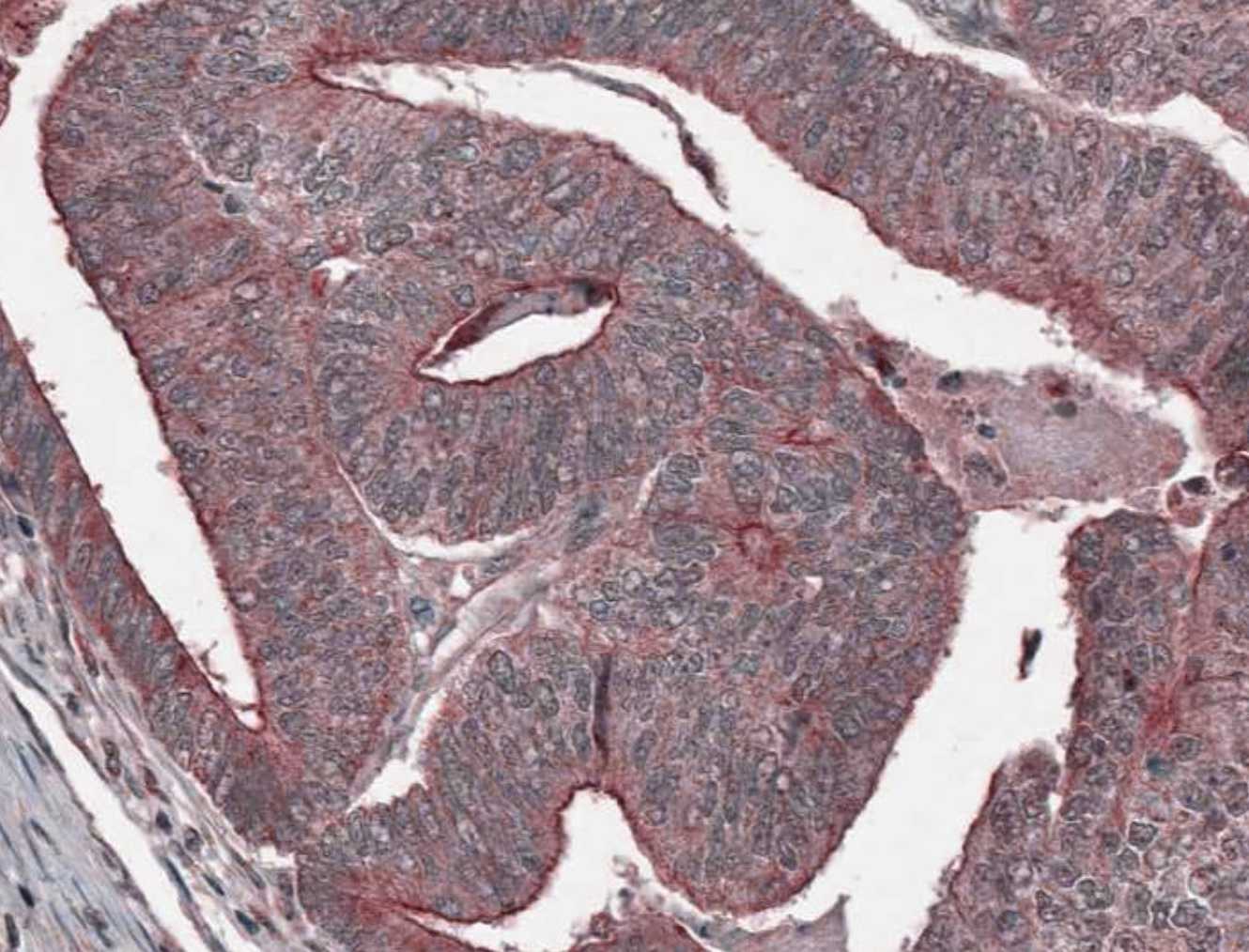
Application: ImmunohistochemistrySample Tested: Colon cancer tissueSpecies: MouseVerified Customer | Posted 10/20/2021

There are no reviews that match your criteria.
Protocols
Find general support by application which include: protocols, troubleshooting, illustrated assays, videos and webinars.
- Appropriate Fixation of IHC/ICC Samples
- Cellular Response to Hypoxia Protocols
- ClariTSA™ Fluorophore Kits
- Detection & Visualization of Antibody Binding
- ICC Cell Smear Protocol for Suspension Cells
- ICC Immunocytochemistry Protocol Videos
- ICC for Adherent Cells
- Immunocytochemistry (ICC) Protocol
- Immunocytochemistry Troubleshooting
- Immunofluorescence of Organoids Embedded in Cultrex Basement Membrane Extract
- Immunohistochemistry (IHC) and Immunocytochemistry (ICC) Protocols
- Preparing Samples for IHC/ICC Experiments
- Preventing Non-Specific Staining (Non-Specific Binding)
- Primary Antibody Selection & Optimization
- Protocol for VisUCyte™ HRP Polymer Detection Reagent
- Protocol for the Fluorescent ICC Staining of Cell Smears - Graphic
- Protocol for the Fluorescent ICC Staining of Cultured Cells on Coverslips - Graphic
- Protocol for the Preparation and Fluorescent ICC Staining of Cells on Coverslips
- Protocol for the Preparation and Fluorescent ICC Staining of Non-adherent Cells
- Protocol for the Preparation and Fluorescent ICC Staining of Stem Cells on Coverslips
- Protocol for the Preparation of a Cell Smear for Non-adherent Cell ICC - Graphic
- R&D Systems Quality Control Western Blot Protocol
- TUNEL and Active Caspase-3 Detection by IHC/ICC Protocol
- The Importance of IHC/ICC Controls
- Troubleshooting Guide: Western Blot Figures
- Western Blot Conditions
- Western Blot Protocol
- Western Blot Protocol for Cell Lysates
- Western Blot Troubleshooting
- Western Blot Troubleshooting Guide
- View all Protocols, Troubleshooting, Illustrated assays and Webinars
Loading...
Associated Pathways
