Coagulation Factors VII and VIIa refer to the pro and active forms of the same protease, respectively (1). Factor VII is synthesized in the liver and circulates in the plasma where it binds to tissue factor (TF), an integral membrane protein found in a variety of cell types. Upon binding of TF, factor VII is rapidly converted into VIIa. The resulting 1:1 complex of VIIa and TF initiates the coagulation pathway and has also important coagulation-independent functions such as angiogenesis (2). The cleavage and activation of Coagulation Factors VII, IX and X by VIIa:TF is phospholipid-dependent whereas the cleavage of small peptide substrates is not (1). The deduced amino acid sequence of mouse factor VII predicts a signal peptide (residues 1 to 24), propeptide (residues 25 to 41), and the mature chain that can be further processed into the light chain (residues 42 to 193) and the heavy chain (residues 194 to 446). The amino acid sequence of mouse Factor VII is 89%, 71%, 65% and 55% identical to that of rat, human/dog, chimpanzee and chicken.
Mouse Coagulation Factor VII Antibody (406707)
R&D Systems | Catalog # MAB33051

Key Product Details
Species Reactivity
Mouse
Applications
Immunohistochemistry, Western Blot
Label
Unconjugated
Antibody Source
Monoclonal Rat IgG2A Clone # 406707
Loading...
Product Specifications
Immunogen
Chinese hamster ovary cell line CHO-derived recombinant human mouse Coagulation Factor VII
Ala42-Leu446
Accession # P70375
Ala42-Leu446
Accession # P70375
Specificity
Detects mouse Coagulation Factor VII in direct ELISAs and Western blots. In direct ELISAs and Western blots, no cross-reactivity with recombinant human (rh) Factor X, rhFactor XI, rhPROC, rhThrombin, rhKallikrein 1, 2, 3, 4, 5, 6, 7, 8, 9, 10, 11, 12, 13, 14, 15, or Kallikrein B1 is observed.
Clonality
Monoclonal
Host
Rat
Isotype
IgG2A
Scientific Data Images for Mouse Coagulation Factor VII Antibody (406707)
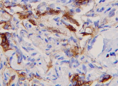
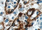
Coagulation Factor VII in Mouse Liver.
Coagulation Factor VII was detected in perfusion fixed frozen sections of mouse liver using Rat Anti-Mouse Coagulation Factor VII Monoclonal Antibody (Catalog # MAB33051) at 15 µg/mL overnight at 4 °C. Tissue was stained using the Anti-Rat HRP-DAB Cell & Tissue Staining Kit (brown; Catalog # CTS017) and counterstained with hematoxylin (blue). Lower panel shows a lack of labeling if primary antibodies are omitted and tissue is stained only with secondary antibody followed by incubation with detection reagents. View our protocol for Chromogenic IHC Staining of Frozen Tissue Sections.Applications for Mouse Coagulation Factor VII Antibody (406707)
Application
Recommended Usage
Immunohistochemistry
8-25 µg/mL
Sample: Perfusion fixed frozen sections of mouse liver
Sample: Perfusion fixed frozen sections of mouse liver
Western Blot
Optimal dilutions of this antibody should be experimentally determined.
Reviewed Applications
Read 2 reviews rated 5 using MAB33051 in the following applications:
Formulation, Preparation, and Storage
Purification
Protein A or G purified from hybridoma culture supernatant
Reconstitution
Reconstitute at 0.5 mg/mL in sterile PBS. For liquid material, refer to CoA for concentration.
Loading...
Formulation
Lyophilized from a 0.2 μm filtered solution in PBS with Trehalose. *Small pack size (SP) is supplied either lyophilized or as a 0.2 µm filtered solution in PBS.
Shipping
Lyophilized product is shipped at ambient temperature. Liquid small pack size (-SP) is shipped with polar packs. Upon receipt, store immediately at the temperature recommended below.
Stability & Storage
Use a manual defrost freezer and avoid repeated freeze-thaw cycles.
- 12 months from date of receipt, -20 to -70 °C as supplied.
- 1 month, 2 to 8 °C under sterile conditions after reconstitution.
- 6 months, -20 to -70 °C under sterile conditions after reconstitution.
Calculators
Background: Coagulation Factor VII
References
- Morrissey, J.H. (2004) in Handbook of Proteolytic Enzymes, Barrett, A.J. et al. (eds. ), Academic Press, San Diego, p. 1659.
- Versteeg, H.H. et al. (2003) Carcinogenesis 24:1009.
Long Name
Coagulation Factor VII (Serum Prothrombin Conversion Accelerator)
Alternate Names
F7
Gene Symbol
F7
UniProt
Additional Coagulation Factor VII Products
Product Documents for Mouse Coagulation Factor VII Antibody (406707)
Certificate of Analysis
To download a Certificate of Analysis, please enter a lot or batch number in the search box below.
Note: Certificate of Analysis not available for kit components.
Product Specific Notices for Mouse Coagulation Factor VII Antibody (406707)
For research use only
Related Research Areas
Customer Reviews for Mouse Coagulation Factor VII Antibody (406707) (2)
5 out of 5
2 Customer Ratings
Have you used Mouse Coagulation Factor VII Antibody (406707)?
Submit a review and receive an Amazon gift card!
$25/€18/£15/$25CAN/¥2500 Yen for a review with an image
$10/€7/£6/$10CAN/¥1110 Yen for a review without an image
Submit a review
Customer Images
Showing
1
-
2 of
2 reviews
Showing All
Filter By:
-
Application: ImmunohistochemistrySample Tested: Liver tissueSpecies: MouseVerified Customer | Posted 08/04/2022

-
Application: ImmunohistochemistrySample Tested: Liver tissueSpecies: MouseVerified Customer | Posted 01/24/2022
There are no reviews that match your criteria.
Protocols
Find general support by application which include: protocols, troubleshooting, illustrated assays, videos and webinars.
- Antigen Retrieval Protocol (PIER)
- Antigen Retrieval for Frozen Sections Protocol
- Appropriate Fixation of IHC/ICC Samples
- Cellular Response to Hypoxia Protocols
- Chromogenic IHC Staining of Formalin-Fixed Paraffin-Embedded (FFPE) Tissue Protocol
- Chromogenic Immunohistochemistry Staining of Frozen Tissue
- ClariTSA™ Fluorophore Kits
- Detection & Visualization of Antibody Binding
- Fluorescent IHC Staining of Frozen Tissue Protocol
- Graphic Protocol for Heat-induced Epitope Retrieval
- Graphic Protocol for the Preparation and Fluorescent IHC Staining of Frozen Tissue Sections
- Graphic Protocol for the Preparation and Fluorescent IHC Staining of Paraffin-embedded Tissue Sections
- Graphic Protocol for the Preparation of Gelatin-coated Slides for Histological Tissue Sections
- IHC Sample Preparation (Frozen sections vs Paraffin)
- Immunofluorescent IHC Staining of Formalin-Fixed Paraffin-Embedded (FFPE) Tissue Protocol
- Immunohistochemistry (IHC) and Immunocytochemistry (ICC) Protocols
- Immunohistochemistry Frozen Troubleshooting
- Immunohistochemistry Paraffin Troubleshooting
- Preparing Samples for IHC/ICC Experiments
- Preventing Non-Specific Staining (Non-Specific Binding)
- Primary Antibody Selection & Optimization
- Protocol for Heat-Induced Epitope Retrieval (HIER)
- Protocol for Making a 4% Formaldehyde Solution in PBS
- Protocol for VisUCyte™ HRP Polymer Detection Reagent
- Protocol for the Preparation & Fixation of Cells on Coverslips
- Protocol for the Preparation and Chromogenic IHC Staining of Frozen Tissue Sections
- Protocol for the Preparation and Chromogenic IHC Staining of Frozen Tissue Sections - Graphic
- Protocol for the Preparation and Chromogenic IHC Staining of Paraffin-embedded Tissue Sections
- Protocol for the Preparation and Chromogenic IHC Staining of Paraffin-embedded Tissue Sections - Graphic
- Protocol for the Preparation and Fluorescent IHC Staining of Frozen Tissue Sections
- Protocol for the Preparation and Fluorescent IHC Staining of Paraffin-embedded Tissue Sections
- Protocol for the Preparation of Gelatin-coated Slides for Histological Tissue Sections
- R&D Systems Quality Control Western Blot Protocol
- TUNEL and Active Caspase-3 Detection by IHC/ICC Protocol
- The Importance of IHC/ICC Controls
- Troubleshooting Guide: Immunohistochemistry
- Troubleshooting Guide: Western Blot Figures
- Western Blot Conditions
- Western Blot Protocol
- Western Blot Protocol for Cell Lysates
- Western Blot Troubleshooting
- Western Blot Troubleshooting Guide
- View all Protocols, Troubleshooting, Illustrated assays and Webinars
Loading...
Associated Pathways
