Decorin is a small secreted chondroitin/dermatan sulfate proteoglycan belonging to the class I small leucine-rich proteoglycan family (SLRP). All SLRP family members are characterized by the N-terminal and C-terminal cysteine-rich regions, which flank the central region containing 10‑12 tandem leucine-rich repeats. In mouse Decorin, the glycosaminoglycan chain is O-linked to Ser34 in the N-terminal disulfide-bridged loop. Decorin binds to fibronectin, TGF-beta, type I and type II collagen. The binding of Decorin to these molecules is mediated via the core protein. Decorin plays a role in maintaining collagen fibrillogenesis. Depending on the cell context, Decorin can either block or augment the bioactivity of TGF-beta Decorin induces growth suppression by activation of a signaling pathway that culminates in the blockade of the cell cycle machinery. Decorin can also induce fibroblast cytoskeletal and signalling changes that results in an increased cell migration (1, 2).

Key Product Details
Species Reactivity
Validated:
Mouse
Cited:
Human, Mouse, Canine, Chicken, Transgenic Mouse
Applications
Validated:
Immunohistochemistry, Western Blot, ELISA Capture (Matched Antibody Pair), Simple Western
Cited:
Immunohistochemistry, Immunohistochemistry-Paraffin, Immunohistochemistry-Frozen, Western Blot
Label
Unconjugated
Antibody Source
Polyclonal Goat IgG
Loading...
Product Specifications
Immunogen
Mouse myeloma cell line NS0-derived recombinant mouse Decorin
Gly17-Lys354
Accession # P28654
Gly17-Lys354
Accession # P28654
Specificity
Detects mouse Decorin in ELISAs and Western blots. In sandwich immunoassays, less than 0.05% cross‑reactivity with recombinant human Decorin is observed.
Clonality
Polyclonal
Host
Goat
Isotype
IgG
Scientific Data Images for Mouse Decorin Antibody
Detection of Mouse Decorin by Western Blot.
Western blot shows lysates of mouse adipose tissue and mouse colon tissue. PVDF membrane was probed with 0.5 µg/mL of Goat Anti-Mouse Decorin Antigen Affinity-purified Polyclonal Antibody (Catalog # AF1060) followed by HRP-conjugated Anti-Goat IgG Secondary Antibody (Catalog # HAF017). Specific bands were detected for Decorin at approximately 65-100 kDa (as indicated). This experiment was conducted under reducing conditions and using Immunoblot Buffer Group 1.
Detection of Mouse Decorin by Simple WesternTM.
Simple Western lane view shows lysates of mouse adipose tissue and mouse colon tissue, loaded at 0.2 mg/mL. A specific band was detected for Decorin at approximately 94 kDa (as indicated) using 10 µg/mL of Goat Anti-Mouse Decorin Antigen Affinity-purified Polyclonal Antibody (Catalog # AF1060) followed by 1:50 dilution of HRP-conjugated Anti-Goat IgG Secondary Antibody (Catalog # HAF109). This experiment was conducted under reducing conditions and using the 12-230 kDa separation system.
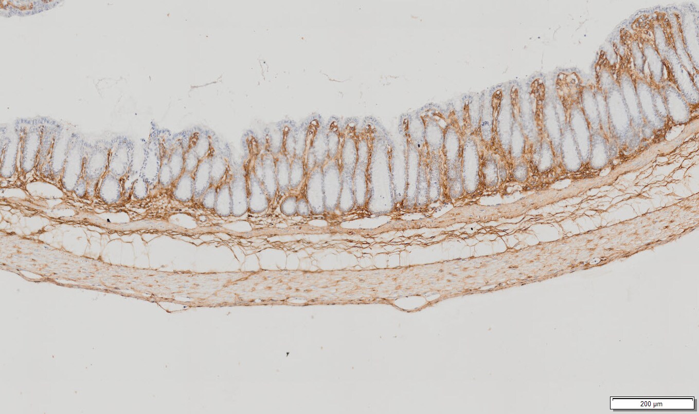
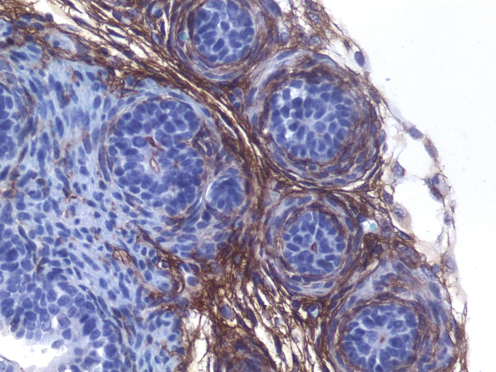
Detection of Mouse Decorin by Immunohistochemistry
Increased CSF-decorin in three different mouse models of A beta pathology. a, b Immunostaining and quantification of decorin in the ChP. Scale bars, 100 µm. (n = 4). c Double immunostaining of DCN and PV or d SRIF in mouse hippocampus. e Quantification of decorin-positive cell-type distribution. Scale bars, 500 µm. (n = 3). f, g Immunostaining and quantification of decorin in the hippocampus. Scale bars, 500 µm. (n = 4). h Mouse CSF-decorin levels in three months old (n = 5), i 13 months old (n = 4–5), j 18 months old (n = 3–5) mice were measured by ELISA and quantified. k CSF-decorin levels in AppNL-F/NL-F mice of different ages were measured and quantified. (n = 3–5). l Decorin levels in A beta 42 treated mouse primary neurons and (m) were quantified (n = 4). n Quantification of decorin levels in conditioned media. (n = 8). Data in (b, g–k) were analyzed by one-way ANOVA followed by Dunnett’s multiple comparisons test. Data in (m, n) were analyzed by student’s t-test. Data in (e) were analyzed by two-way ANOVA followed by Tukey’s post hoc test. Data are represented as mean ± SEM; *p < 0.05, **p < 0.01, ***p < 0.001, ****p < 0.0001. DCN decorin, PV parvalbumin, SRIF somatotropin release-inhibiting factor, PN pyramidal neurons, ns not significant Image collected and cropped by CiteAb from the following open publication (https://pubmed.ncbi.nlm.nih.gov/35787306), licensed under a CC-BY license. Not internally tested by R&D Systems.
Mouse Decorin ELISA Standard Curve
Recombinant Mouse Decorin (Catalog # 1060-DE) was serially diluted and captured by Goat Anti-Mouse Decorin Antigen Affinity-purified Polyclonal Antibody (Catalog # AF1060) coated on a Clear Polystyrene Microplate (Catalog # DY990). Goat Anti-Mouse Decorin Antigen Affinity-purified Polyclonal Antibody (Catalog # AF1060) was biotinylated and incubated with the protein captured on the plate. Detection of the standard curve was achieved by incubating Streptavidin-HRP (Catalog # DY998)
Mouse Decorin ELISA Standard Curve
Recombinant Mouse Decorin (Catalog # 1060-DE) was serially diluted and captured by Goat Anti-Mouse Decorin Antigen Affinity-purified Polyclonal Antibody (Catalog # AF1060) coated on a Clear Polystyrene Microplate (Catalog # DY990). Goat Anti-Mouse Decorin Antigen Affinity-purified Polyclonal Antibody (Catalog # AF1060) was biotinylated and incubated with the protein captured on the plate. Detection of the standard curve was achieved by incubating Streptavidin-HRP (Catalog # DY998)Applications for Mouse Decorin Antibody
Application
Recommended Usage
Immunohistochemistry
5-15 µg/mL
Sample: Immersion fixed frozen sections of mouse embryo (13 d.p.c., dorsal root ganglion)
Sample: Immersion fixed frozen sections of mouse embryo (13 d.p.c., dorsal root ganglion)
Simple Western
10 µg/mL
Sample: Mouse adipose tissue and mouse colon tissue
Sample: Mouse adipose tissue and mouse colon tissue
Western Blot
0.5 µg/mL
Sample: Mouse adipose tissue and mouse colon tissue
Sample: Mouse adipose tissue and mouse colon tissue
Mouse Decorin Sandwich Immunoassay
Please Note: Optimal dilutions of this antibody should be experimentally determined.
Reviewed Applications
Read 6 reviews rated 4.8 using AF1060 in the following applications:
Formulation, Preparation, and Storage
Purification
Antigen Affinity-purified
Reconstitution
Reconstitute at 0.2 mg/mL in sterile PBS. For liquid material, refer to CoA for concentration.
Loading...
Formulation
Lyophilized from a 0.2 μm filtered solution in PBS with Trehalose. *Small pack size (SP) is supplied either lyophilized or as a 0.2 µm filtered solution in PBS.
Shipping
Lyophilized product is shipped at ambient temperature. Liquid small pack size (-SP) is shipped with polar packs. Upon receipt, store immediately at the temperature recommended below.
Stability & Storage
Use a manual defrost freezer and avoid repeated freeze-thaw cycles.
- 12 months from date of receipt, -20 to -70 °C as supplied.
- 1 month, 2 to 8 °C under sterile conditions after reconstitution.
- 6 months, -20 to -70 °C under sterile conditions after reconstitution.
Calculators
Background: Decorin
References
- Iozzo, R.V. (1998) Annu. Rev. Biochem. 67:609.
- Tufvesson, E. and G. Westergren-Thorsson (2003) J. Cell Science 116:4857.
Alternate Names
DCN, DSPG2, PG-II, PGS2, SLRR1B
Gene Symbol
DCN
UniProt
Additional Decorin Products
Product Documents for Mouse Decorin Antibody
Certificate of Analysis
To download a Certificate of Analysis, please enter a lot or batch number in the search box below.
Note: Certificate of Analysis not available for kit components.
Product Specific Notices for Mouse Decorin Antibody
For research use only
Related Research Areas
Citations for Mouse Decorin Antibody
 Powered by Bioz
Customer Reviews for Mouse Decorin Antibody (6)
4.8 out of 5
6 Customer Ratings
Have you used Mouse Decorin Antibody?
Submit a review and receive an Amazon gift card!
$25/€18/£15/$25CAN/¥2500 Yen for a review with an image
$10/€7/£6/$10CAN/¥1110 Yen for a review without an image
Submit a review
Customer Images
Showing
1
-
5 of
6 reviews
Showing All
Filter By:
-
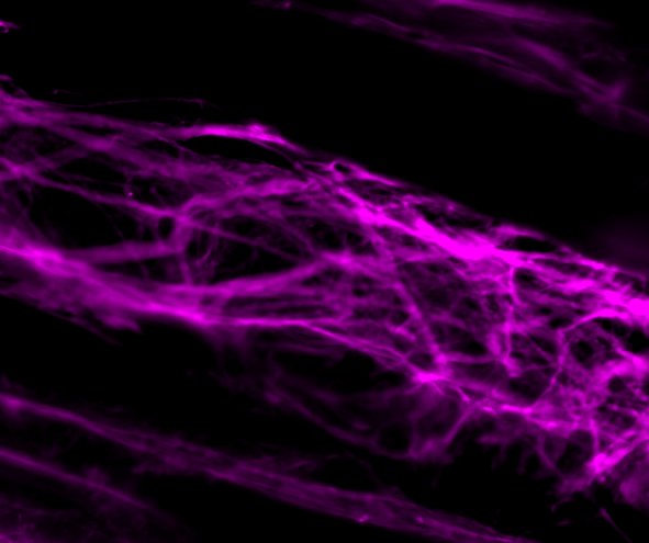
Application: Immunohistochemistry-FrozenSample Tested: skeletal muscleVerified Customer | Posted 10/28/2024Decorin on murine skeletal muscle longitudinal section.30 micrometer sections of skeletal muscle Floating immunofluorescence 2% normal donkey serum + 2% BSA - 3h room temp blocking 1:500 dilution in PBS 0.3M glycin + 0.3% triton x100 + 1% BSA Overnight incubation at room temp Secondary 1:1000 - 2h room temp.

-
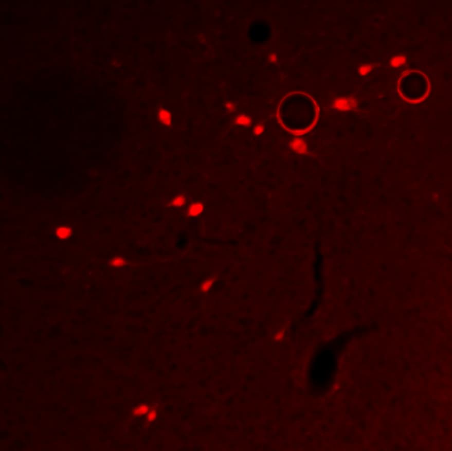
Application: Immunocytochemistry/ImmunofluorescenceSample Tested: Adult brainSpecies: Rat and MouseVerified Customer | Posted 04/10/20184C overnight, very strong and specific
-
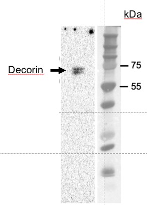
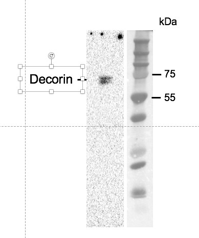
Application: Western BlotSample Tested: Liver tissueSpecies: MouseVerified Customer | Posted 03/20/2018Primary antibody was diluted in 5% milk/TBST at 1:1000. Blot was incubated in cold room overnight.
-
Application: Western BlotSample Tested: mouse liver lysateSpecies: MouseVerified Customer | Posted 09/08/2017A total of 80ug of mouse liver protein lysate was loaded.Primary antibody was diluted in 5% milk/TBST at 1:1000. Blot was incubated in cold room overnight.
-
Application: ImmunohistochemistrySample Tested: Colon cancer tissue and Colon tissueSpecies: MouseVerified Customer | Posted 05/18/2017
-
Application: ImmunohistochemistrySample Tested: Prostate tissueSpecies: MouseVerified Customer | Posted 05/19/2016
There are no reviews that match your criteria.
Protocols
Find general support by application which include: protocols, troubleshooting, illustrated assays, videos and webinars.
- Antigen Retrieval Protocol (PIER)
- Antigen Retrieval for Frozen Sections Protocol
- Appropriate Fixation of IHC/ICC Samples
- Cellular Response to Hypoxia Protocols
- Chromogenic IHC Staining of Formalin-Fixed Paraffin-Embedded (FFPE) Tissue Protocol
- Chromogenic Immunohistochemistry Staining of Frozen Tissue
- ClariTSA™ Fluorophore Kits
- Detection & Visualization of Antibody Binding
- Fluorescent IHC Staining of Frozen Tissue Protocol
- Graphic Protocol for Heat-induced Epitope Retrieval
- Graphic Protocol for the Preparation and Fluorescent IHC Staining of Frozen Tissue Sections
- Graphic Protocol for the Preparation and Fluorescent IHC Staining of Paraffin-embedded Tissue Sections
- Graphic Protocol for the Preparation of Gelatin-coated Slides for Histological Tissue Sections
- IHC Sample Preparation (Frozen sections vs Paraffin)
- Immunofluorescent IHC Staining of Formalin-Fixed Paraffin-Embedded (FFPE) Tissue Protocol
- Immunohistochemistry (IHC) and Immunocytochemistry (ICC) Protocols
- Immunohistochemistry Frozen Troubleshooting
- Immunohistochemistry Paraffin Troubleshooting
- Preparing Samples for IHC/ICC Experiments
- Preventing Non-Specific Staining (Non-Specific Binding)
- Primary Antibody Selection & Optimization
- Protocol for Heat-Induced Epitope Retrieval (HIER)
- Protocol for Making a 4% Formaldehyde Solution in PBS
- Protocol for VisUCyte™ HRP Polymer Detection Reagent
- Protocol for the Preparation & Fixation of Cells on Coverslips
- Protocol for the Preparation and Chromogenic IHC Staining of Frozen Tissue Sections
- Protocol for the Preparation and Chromogenic IHC Staining of Frozen Tissue Sections - Graphic
- Protocol for the Preparation and Chromogenic IHC Staining of Paraffin-embedded Tissue Sections
- Protocol for the Preparation and Chromogenic IHC Staining of Paraffin-embedded Tissue Sections - Graphic
- Protocol for the Preparation and Fluorescent IHC Staining of Frozen Tissue Sections
- Protocol for the Preparation and Fluorescent IHC Staining of Paraffin-embedded Tissue Sections
- Protocol for the Preparation of Gelatin-coated Slides for Histological Tissue Sections
- R&D Systems Quality Control Western Blot Protocol
- TUNEL and Active Caspase-3 Detection by IHC/ICC Protocol
- The Importance of IHC/ICC Controls
- Troubleshooting Guide: Immunohistochemistry
- Troubleshooting Guide: Western Blot Figures
- Western Blot Conditions
- Western Blot Protocol
- Western Blot Protocol for Cell Lysates
- Western Blot Troubleshooting
- Western Blot Troubleshooting Guide
- View all Protocols, Troubleshooting, Illustrated assays and Webinars
Loading...
Associated Pathways

