Gastrokine 1 (GKN1; also CA11 and AMP-18) is a 17-20 kDa member of the Gastrokine protein family. It has limited expression, being restricted to mucous secreting pyloric antrum epithelial cells. Gastrokine 1 appears to promote epithelial cell proliferation and migration, and induce the formation of tight junctions between epithelial cells. By contrast, gastrokine 1 induces Fas expression in tumor cells, resulting in apoptosis. Mature mouse gastrokine 1 is 166 amino acids (aa) in length (aa 36-201 of SwissProt accession Q9CR36 or aa 19-184 of accession NP_079742). Based on the SwissProt sequence, it possesses one BRICHOS domain (aa 71-165) that contains a mitogenic sequence (aa 113-131). There is one potential alternative start site at Met18. Over aa 38-201 (accession Q9CR36), mouse gastrokine 1 shares 65% and 92% aa sequence identity with human and rat gastrokine 1, respectively.
Mouse Gastrokine 1 Antibody (759821)
R&D Systems | Catalog # MAB7287

Key Product Details
Species Reactivity
Validated:
Mouse
Cited:
Mouse
Applications
Validated:
Immunohistochemistry, Western Blot
Cited:
Immunohistochemistry
Label
Unconjugated
Antibody Source
Monoclonal Rat IgG2A Clone # 759821
Loading...
Product Specifications
Immunogen
E. coli-derived recombinant mouse Gastrokine 1
Tyr38-Tyr201
Accession # NP_079742
Tyr38-Tyr201
Accession # NP_079742
Specificity
Detects mouse Gastrokine 1 in direct ELISAs and mouse and rat Gastrokine 1 in Western blots. In Western blots, approximately 10% cross‑reactivity with recombinant human (rh) Gastrokine 1 and no cross-reactivity with rhBlottin is observed.
Clonality
Monoclonal
Host
Rat
Isotype
IgG2A
Scientific Data Images for Mouse Gastrokine 1 Antibody (759821)
Detection of Mouse and Rat Gastrokine 1 by Western Blot.
Western blot shows lysates of mouse stomach tissue and rat stomach tissue. PVDF membrane was probed with 1 µg/mL of Rat Anti-Mouse Gastrokine 1 Monoclonal Antibody (Catalog # MAB7287) followed by HRP-conjugated Anti-Rat IgG Secondary Antibody (Catalog # HAF005). A specific band was detected for Gastrokine 1 at approximately 17 kDa (as indicated). This experiment was conducted under reducing conditions and using Immunoblot Buffer Group 1.
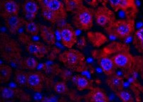
Gastrokine 1 in Mouse Intestine.
Gastrokine 1 was detected in perfusion fixed frozen sections of mouse intestine using Rat Anti-Mouse Gastrokine 1 Monoclonal Antibody (Catalog # MAB7287) at 25 µg/mL overnight at 4 °C. Tissue was stained using the NorthernLights™ 557-conjugated Anti-Rat IgG Secondary Antibody (red; Catalog # NL013) and counterstained with DAPI (blue). Specific staining was localized to gastric glands. View our protocol for Fluorescent IHC Staining of Frozen Tissue Sections.Applications for Mouse Gastrokine 1 Antibody (759821)
Application
Recommended Usage
Immunohistochemistry
8-25 µg/mL
Sample: Perfusion fixed frozen sections of mouse intestine
Sample: Perfusion fixed frozen sections of mouse intestine
Western Blot
1 µg/mL
Sample: Mouse stomach tissue and rat stomach tissue
Sample: Mouse stomach tissue and rat stomach tissue
Reviewed Applications
Read 1 review rated 5 using MAB7287 in the following applications:
Formulation, Preparation, and Storage
Purification
Protein A or G purified from hybridoma culture supernatant
Reconstitution
Sterile PBS to a final concentration of 0.5 mg/mL. For liquid material, refer to CoA for concentration.
Loading...
Formulation
Lyophilized from a 0.2 μm filtered solution in PBS with Trehalose. *Small pack size (SP) is supplied either lyophilized or as a 0.2 µm filtered solution in PBS.
Shipping
Lyophilized product is shipped at ambient temperature. Liquid small pack size (-SP) is shipped with polar packs. Upon receipt, store immediately at the temperature recommended below.
Stability & Storage
Use a manual defrost freezer and avoid repeated freeze-thaw cycles.
- 12 months from date of receipt, -20 to -70 °C as supplied.
- 1 month, 2 to 8 °C under sterile conditions after reconstitution.
- 6 months, -20 to -70 °C under sterile conditions after reconstitution.
Calculators
Background: Gastrokine 1
Long Name
Take out Column D name
Alternate Names
AMP18, BRICD1, CA11, FOV, Foveolin, GKN1
Gene Symbol
GKN1
UniProt
Additional Gastrokine 1 Products
Product Documents for Mouse Gastrokine 1 Antibody (759821)
Certificate of Analysis
To download a Certificate of Analysis, please enter a lot or batch number in the search box below.
Note: Certificate of Analysis not available for kit components.
Product Specific Notices for Mouse Gastrokine 1 Antibody (759821)
For research use only
Related Research Areas
Citations for Mouse Gastrokine 1 Antibody (759821)
 Powered by Bioz
Customer Reviews for Mouse Gastrokine 1 Antibody (759821) (1)
5 out of 5
1 Customer Rating
Have you used Mouse Gastrokine 1 Antibody (759821)?
Submit a review and receive an Amazon gift card!
$25/€18/£15/$25CAN/¥2500 Yen for a review with an image
$10/€7/£6/$10CAN/¥1110 Yen for a review without an image
Submit a review
Customer Images
Showing
1
-
1 of
1 review
Showing All
Filter By:
-
Application: ImmunohistochemistrySample Tested: IntestineSpecies: MouseVerified Customer | Posted 03/06/2022

There are no reviews that match your criteria.
Protocols
Find general support by application which include: protocols, troubleshooting, illustrated assays, videos and webinars.
- Antigen Retrieval Protocol (PIER)
- Antigen Retrieval for Frozen Sections Protocol
- Appropriate Fixation of IHC/ICC Samples
- Cellular Response to Hypoxia Protocols
- Chromogenic IHC Staining of Formalin-Fixed Paraffin-Embedded (FFPE) Tissue Protocol
- Chromogenic Immunohistochemistry Staining of Frozen Tissue
- ClariTSA™ Fluorophore Kits
- Detection & Visualization of Antibody Binding
- Fluorescent IHC Staining of Frozen Tissue Protocol
- Graphic Protocol for Heat-induced Epitope Retrieval
- Graphic Protocol for the Preparation and Fluorescent IHC Staining of Frozen Tissue Sections
- Graphic Protocol for the Preparation and Fluorescent IHC Staining of Paraffin-embedded Tissue Sections
- Graphic Protocol for the Preparation of Gelatin-coated Slides for Histological Tissue Sections
- IHC Sample Preparation (Frozen sections vs Paraffin)
- Immunofluorescent IHC Staining of Formalin-Fixed Paraffin-Embedded (FFPE) Tissue Protocol
- Immunohistochemistry (IHC) and Immunocytochemistry (ICC) Protocols
- Immunohistochemistry Frozen Troubleshooting
- Immunohistochemistry Paraffin Troubleshooting
- Preparing Samples for IHC/ICC Experiments
- Preventing Non-Specific Staining (Non-Specific Binding)
- Primary Antibody Selection & Optimization
- Protocol for Heat-Induced Epitope Retrieval (HIER)
- Protocol for Making a 4% Formaldehyde Solution in PBS
- Protocol for VisUCyte™ HRP Polymer Detection Reagent
- Protocol for the Preparation & Fixation of Cells on Coverslips
- Protocol for the Preparation and Chromogenic IHC Staining of Frozen Tissue Sections
- Protocol for the Preparation and Chromogenic IHC Staining of Frozen Tissue Sections - Graphic
- Protocol for the Preparation and Chromogenic IHC Staining of Paraffin-embedded Tissue Sections
- Protocol for the Preparation and Chromogenic IHC Staining of Paraffin-embedded Tissue Sections - Graphic
- Protocol for the Preparation and Fluorescent IHC Staining of Frozen Tissue Sections
- Protocol for the Preparation and Fluorescent IHC Staining of Paraffin-embedded Tissue Sections
- Protocol for the Preparation of Gelatin-coated Slides for Histological Tissue Sections
- R&D Systems Quality Control Western Blot Protocol
- TUNEL and Active Caspase-3 Detection by IHC/ICC Protocol
- The Importance of IHC/ICC Controls
- Troubleshooting Guide: Immunohistochemistry
- Troubleshooting Guide: Western Blot Figures
- Western Blot Conditions
- Western Blot Protocol
- Western Blot Protocol for Cell Lysates
- Western Blot Troubleshooting
- Western Blot Troubleshooting Guide
- View all Protocols, Troubleshooting, Illustrated assays and Webinars
Loading...