Insulin-like growth factor I, also known as somatomedin C, is the dominant effector of growth hormone and is structurally homologous to proinsulin. Mouse IGF-I is synthesized as two precursor isoforms with alternate N- and C-terminal propeptides (1). These isoforms are differentially expressed by various tissues (1). The 7.6 kDa mature IGF-I is identical between isoforms and is generated by proteolytic removal of the N- and C-terminal regions. Mature mouse IGF-I shares 94% and 99% aa sequence identity with human and rat IGF-I, respectively (2), and exhibits cross-species activity. It shares 60% aa sequence identity with mature mouse IGF‑II. Circulating IGF-I is produced by hepatocytes, while local IGF-I is produced by many other tissues in which it has paracrine effects (1). IGF-I induces the proliferation, migration, and differentiation of a wide variety of cell types during development and postnatally (3). IGF-I regulates glucose and fatty acid metabolism, steroid hormone activity, and cartilage and bone metabolism (4-7). It plays an important role in muscle regeneration and tumor progression (1, 8). IGF-I binds IGF-I R, IGF-II R, and the insulin receptor, although its effects are mediated primarily by IGF-I R (9). IGF-I association with IGF binding proteins increases its plasma half-life and modulates its interactions with receptors (10).

Key Product Details
Species Reactivity
Validated:
Mouse
Cited:
Human, Mouse, Rat, Transgenic Mouse
Applications
Validated:
Immunohistochemistry, Western Blot, Neutralization
Cited:
Immunohistochemistry, Immunohistochemistry-Paraffin, Immunohistochemistry-Frozen, Western Blot, Neutralization, Immunocytochemistry, In vivo assay
Label
Unconjugated
Antibody Source
Polyclonal Goat IgG
Loading...
Product Specifications
Immunogen
E. coli-derived recombinant mouse IGF-I/IGF-1
Gl33-Ala102
Accession # Q8CAR0
Gl33-Ala102
Accession # Q8CAR0
Specificity
Detects mouse IGF-I/IGF-1 in direct ELISAs and Western blots. In direct ELISAs, approximately 15% cross-reactivity with recombinant human IGF-I/IGF-1 is observed, and less than 2% cross-reactivity with recombinant mouse IGF-II/IGF-2 is observed.
Clonality
Polyclonal
Host
Goat
Isotype
IgG
Endotoxin Level
<0.10 EU per 1 μg of the antibody by the LAL method.
Scientific Data Images for Mouse IGF-I/IGF-1 Antibody
Cell Proliferation Induced by IGF-I/IGF-1 and Neutralization by Mouse IGF-I/IGF-1 Antibody.
Recombinant Mouse IGF-I/IGF-1 (Catalog # 791-MG) stimulates proliferation in the MCF-7 human breast cancer cell line in a dose-dependent manner (orange line). Proliferation elicited by Recombinant Mouse IGF-I/IGF-1 (15 ng/mL) is neutralized (green line) by increasing concen-trations of Goat Anti-Mouse IGF-I/IGF-1 Antigen Affinity-purified Polyclonal Antibody (Catalog # AF791). The ND50 is typically 0.2-1.0 µg/mL.
IGF-I/IGF-1 in Mouse Embryo.
IGF-I/IGF-1 was detected in immersion fixed frozen sections of mouse embryo (13 d.p.c.) using Goat Anti-Mouse IGF-I/IGF-1 Antigen Affinity-purified Polyclonal Antibody (Catalog # AF791) at 15 µg/mL overnight at 4 °C. Tissue was stained using the Anti-Goat HRP-DAB Cell & Tissue Staining Kit (brown; Catalog # CTS008) and counterstained with hematoxylin (blue). Lower panel shows a lack of labeling when primary antibodies are omitted and tissue is stained only with secondary antibody followed by incubation with detection reagents. Specific staining was localized to developing brain and muscle cells. View our protocol for Chromogenic IHC Staining of Frozen Tissue Sections.
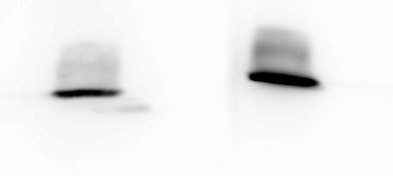
Detection of Mouse IGF-I/IGF-1 by Western Blot
Activation of IGF signaling pathways 36 and 48 h after i.p. injection of paraquat. (A) WB detection of IGF-I from blood and of key signal transduction proteins in IGF pathways (P-tyrosine-activated forms and total protein) from lung tissue. Gel loading was controlled by vinculin. (B) Quantification of WB by chemiluminescence. Phosphotyrosine-IGF-1R (P-IGF-1R) and P-IRS-1 were detected using a phosphotyrosine-specific antibody after IP. Note that increase in IGF-1R abundance over time was very similar whether detected from IP samples or by direct WB. Tests were performed in 11- to 13-week-old mice. N = 3 per group; mean ± SEM, expressed in arbitrary units. *P < 0.05, Mann–Whitney U-test. Image collected and cropped by CiteAb from the following open publication (https://pubmed.ncbi.nlm.nih.gov/23898955), licensed under a CC-BY license. Not internally tested by R&D Systems.
Mouse / Rat IGF-I ELISA Standard Curve
Recombinant Mouse IGF-I/IGF-1 (Catalog # 791-MG) was serially diluted and captured by Hamster Anti-Mouse IGF-I/IGF-1 Monoclonal Antibody (Catalog # MAB791) coated on a Clear Polystyrene Microplate (Catalog # DY990). Goat Anti-Mouse IGF-I/IGF-1 Antigen Affinity-purified Polyclonal Antibody (Catalog # AF791) was biotinylated and incubated with the protein captured on the plate. Detection of the standard curve was achieved by incubating Streptavidin-HRP (Catalog # DY998)Applications for Mouse IGF-I/IGF-1 Antibody
Application
Recommended Usage
Immunohistochemistry
5-15 µg/mL
Sample: Immersion fixed frozen sections of mouse embryo (E13)
Sample: Immersion fixed frozen sections of mouse embryo (E13)
Western Blot
0.1 µg/mL
Sample: Recombinant Mouse IGF-I/IGF-1 (Catalog # 791-MG)
Sample: Recombinant Mouse IGF-I/IGF-1 (Catalog # 791-MG)
Neutralization
Measured by its ability to neutralize IGF-I/IGF-1-induced proliferation in the MCF‑7 human breast cancer cell line. Karey, K. P. et al. (1988) Cancer Research 48:4083. The Neutralization Dose (ND50) is typically 0.2-1.0 µg/mL in the presence of 15 ng/mL Recombinant Mouse IGF-I/IGF-1.
Reviewed Applications
Read 2 reviews rated 4 using AF791 in the following applications:
Formulation, Preparation, and Storage
Purification
Antigen Affinity-purified
Reconstitution
Reconstitute at 0.2 mg/mL in sterile PBS. For liquid material, refer to CoA for concentration.
Loading...
Formulation
Lyophilized from a 0.2 μm filtered solution in PBS with Trehalose. *Small pack size (SP) is supplied either lyophilized or as a 0.2 µm filtered solution in PBS.
Shipping
Lyophilized product is shipped at ambient temperature. Liquid small pack size (-SP) is shipped with polar packs. Upon receipt, store immediately at the temperature recommended below.
Stability & Storage
Use a manual defrost freezer and avoid repeated freeze-thaw cycles.
- 12 months from date of receipt, -20 to -70 °C as supplied.
- 1 month, 2 to 8 °C under sterile conditions after reconstitution.
- 6 months, -20 to -70 °C under sterile conditions after reconstitution.
Calculators
Background: IGF-I/IGF-1
References
- Philippou, A. et al. (2007) In Vivo 21:45.
- Bell, G.I. et al. (1986) Nucleic Acids Res. 14:7873.
- Guvakova, M.A. (2007) Int. J. Biochem. Cell Biol. 39:890.
- Clemmons, D.R. (2006) Curr. Opin. Pharmacol. 6:620.
- Bluher, S. et al. (2005) Best Pract. Res. Clin. Endocrinol. Metab. 19:577.
- Garcia-Segura, L.M. et al. (2006) Neuroendocrinology 84:275.
- Malemud, C.J. (2007) Clin. Chim. Acta 375:10.
- Samani, A.A. et al. (2007) Endocrine Rev. 28:20.
- LeRoith, D. and S. Yakar (2007) Nat. Clin. Pract. Endocrinol. Metab. 3:302.
- Denley, A. et al. (2005) Cytokine Growth Factor Rev. 16:421.
Long Name
Insulin-like Growth Factor I/Insulin-like Growth Factor 1
Alternate Names
IGF-1, IGF1, IGFI, Somatomedin A, Somatomedin C
Gene Symbol
IGF1
UniProt
Additional IGF-I/IGF-1 Products
Product Documents for Mouse IGF-I/IGF-1 Antibody
Certificate of Analysis
To download a Certificate of Analysis, please enter a lot or batch number in the search box below.
Note: Certificate of Analysis not available for kit components.
Product Specific Notices for Mouse IGF-I/IGF-1 Antibody
For research use only
Citations for Mouse IGF-I/IGF-1 Antibody
 Powered by Bioz
Customer Reviews for Mouse IGF-I/IGF-1 Antibody (2)
4 out of 5
2 Customer Ratings
Have you used Mouse IGF-I/IGF-1 Antibody?
Submit a review and receive an Amazon gift card!
$25/€18/£15/$25CAN/¥2500 Yen for a review with an image
$10/€7/£6/$10CAN/¥1110 Yen for a review without an image
Submit a review
Customer Images
Showing
1
-
2 of
2 reviews
Showing All
Filter By:
-
Application: Western BlotSample Tested: Purified proteinSpecies: MouseVerified Customer | Posted 04/18/2019Could not detect IGF-1 when I used liver samples from mice.

-
Application: Western BlotSample Tested: See PMID 23407451Species: MouseVerified Customer | Posted 01/09/2015
There are no reviews that match your criteria.
Protocols
Find general support by application which include: protocols, troubleshooting, illustrated assays, videos and webinars.
- Antigen Retrieval Protocol (PIER)
- Antigen Retrieval for Frozen Sections Protocol
- Appropriate Fixation of IHC/ICC Samples
- Cellular Response to Hypoxia Protocols
- Chromogenic IHC Staining of Formalin-Fixed Paraffin-Embedded (FFPE) Tissue Protocol
- Chromogenic Immunohistochemistry Staining of Frozen Tissue
- ClariTSA™ Fluorophore Kits
- Detection & Visualization of Antibody Binding
- Fluorescent IHC Staining of Frozen Tissue Protocol
- Graphic Protocol for Heat-induced Epitope Retrieval
- Graphic Protocol for the Preparation and Fluorescent IHC Staining of Frozen Tissue Sections
- Graphic Protocol for the Preparation and Fluorescent IHC Staining of Paraffin-embedded Tissue Sections
- Graphic Protocol for the Preparation of Gelatin-coated Slides for Histological Tissue Sections
- IHC Sample Preparation (Frozen sections vs Paraffin)
- Immunofluorescent IHC Staining of Formalin-Fixed Paraffin-Embedded (FFPE) Tissue Protocol
- Immunohistochemistry (IHC) and Immunocytochemistry (ICC) Protocols
- Immunohistochemistry Frozen Troubleshooting
- Immunohistochemistry Paraffin Troubleshooting
- Preparing Samples for IHC/ICC Experiments
- Preventing Non-Specific Staining (Non-Specific Binding)
- Primary Antibody Selection & Optimization
- Protocol for Heat-Induced Epitope Retrieval (HIER)
- Protocol for Making a 4% Formaldehyde Solution in PBS
- Protocol for VisUCyte™ HRP Polymer Detection Reagent
- Protocol for the Preparation & Fixation of Cells on Coverslips
- Protocol for the Preparation and Chromogenic IHC Staining of Frozen Tissue Sections
- Protocol for the Preparation and Chromogenic IHC Staining of Frozen Tissue Sections - Graphic
- Protocol for the Preparation and Chromogenic IHC Staining of Paraffin-embedded Tissue Sections
- Protocol for the Preparation and Chromogenic IHC Staining of Paraffin-embedded Tissue Sections - Graphic
- Protocol for the Preparation and Fluorescent IHC Staining of Frozen Tissue Sections
- Protocol for the Preparation and Fluorescent IHC Staining of Paraffin-embedded Tissue Sections
- Protocol for the Preparation of Gelatin-coated Slides for Histological Tissue Sections
- R&D Systems Quality Control Western Blot Protocol
- TUNEL and Active Caspase-3 Detection by IHC/ICC Protocol
- The Importance of IHC/ICC Controls
- Troubleshooting Guide: Immunohistochemistry
- Troubleshooting Guide: Western Blot Figures
- Western Blot Conditions
- Western Blot Protocol
- Western Blot Protocol for Cell Lysates
- Western Blot Troubleshooting
- Western Blot Troubleshooting Guide
- View all Protocols, Troubleshooting, Illustrated assays and Webinars


